
Paulodora sinensis Wang & Zhong, 2018
|
publication ID |
https://doi.org/10.11646/zootaxa.4446.1.3 |
|
publication LSID |
lsid:zoobank.org:pub:AC2EA154-7E31-4E91-AF4A-9C95FE504123 |
|
DOI |
https://doi.org/10.5281/zenodo.5958668 |
|
persistent identifier |
https://treatment.plazi.org/id/7E20083B-FFD5-0A45-FF74-C1BDC83BFCE8 |
|
treatment provided by |
Plazi |
|
scientific name |
Paulodora sinensis Wang & Zhong |
| status |
sp. nov. |
Paulodora sinensis Wang & Zhong n. sp.
( Figs. 1–3 View FIGURE 1 View FIGURE 2 View FIGURE 3 )
Material examined. Holotype: PLA-Po041, whole-mounted specimen. Paratypes: PLA-Po042–043, wholemounted specimens. Other specimens: PLA-Po045, mounted slides of stylet; PLA-Po044-1~044-4, longitudinal serially-sectioned specimens. Specimens were collected in the intertidal Zone (water temperature: 20–21?; salinity: 19–20‰), Eastern ShenZhen City, Guangdong Province, China ( 22°28′N, 114°32′E) by Y.T.L in May 2017. All specimens were preserved in IZCAS GoogleMaps .
Etymology. The species is named because it was firstly discovered in China.
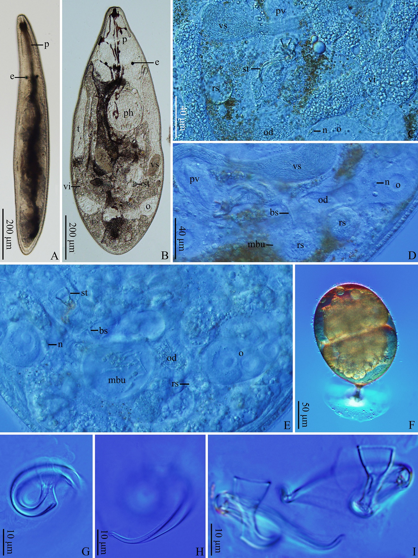
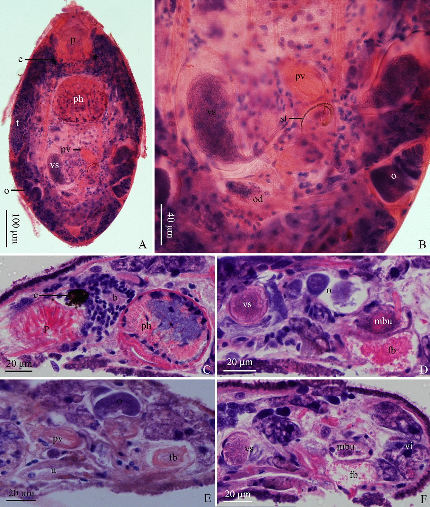
Description: The mature individual is 850–1,130 µm in length and 140–160 µm in width (n=3) ( Fig. 1A View FIGURE 1 ). A light-yellow longitudinal stripe is situated dorsally along the centerline of the body. The proboscis is 120–130 µm in length and 55–65 µm in basal diameter (n=3) ( Fig. 1A–B View FIGURE 1 , 2A & C View FIGURE 2 , 3A View FIGURE 3 ). The paired black eyes (16–18 µm in diameter) are situated posterior to the proboscis and the distance between two eyes is 48–55 µm (n=3) ( Fig. 1A–B View FIGURE 1 , 2A & C View FIGURE 2 , 3A View FIGURE 3 ). The brain is situated ventrally and posterior to the eyes ( Fig. 2C View FIGURE 2 , 3A View FIGURE 3 ). The pharynx (125–135 µm in diameter, n=3) is located at 40% of the body ( Fig. 1B View FIGURE 1 , 2A & C View FIGURE 2 , 3A View FIGURE 3 ).
The paired rod-like testes (150–190 µm in length, 30–40 µm in width, n=3) are located at middle part of the body ( Fig. 1B View FIGURE 1 , 2A View FIGURE 2 , 3A View FIGURE 3 ). Two baseball-bat shaped seminal vesicles (100–120 µm in length, n=3) are also located at the middle of the body ( Fig. 1B–D View FIGURE 1 , 2A–B, D & F View FIGURE 2 , 3A–B View FIGURE 3 ) and fuse at their distal ends, where the ejaculatory duct is formed. The ejaculatory duct runs next to the prostatic vesicle and enters the stylet from its opening. The prostatic vesicle is oblong-shaped (61–72 µm in length, 26–32 µm in width, n=3), of which the distal part is connected to the base of stylet and is partially inserted into it ( Fig. 1C–D View FIGURE 1 , 2A–B & E View FIGURE 2 , 3A–B View FIGURE 3 ). The double-walled stylet (95–130 µm in length, n=3) is located at male atrium ( Fig. 1B–C E & G–I View FIGURE 1 , 2B & E View FIGURE 2 , 3A–D View FIGURE 3 ). The base of stylet is funnel-shaped (10–12 µm in diameter, n=3), while the rest of the stylet twists into spiral shape. The outer stylet forms a minor fold proximally, while the inner stylet (7–9 µm in basal diameter) bends along the outer stylet distally. The bursa stalk is slender (~30 µm in length), and connects the male atrium to the male bursa ( Fig. 1D–E View FIGURE 1 , 3B View FIGURE 3 ). The globular male bursa, with maximal diameter up to 90 µm, is located at dorsal side ( Fig. 1D–E View FIGURE 1 , 2D & F View FIGURE 2 , 3A–B View FIGURE 3 ).
Two rod-like vitellaria are located dorsally at both sides of the body, extending from the pharynx to the posterior end of the body ( Fig. 1B View FIGURE 1 , 2F View FIGURE 2 , 3A View FIGURE 3 ). The paired kidney-shaped ovaries (100–110 µm in length, n=3) ( Fig. 1B–E View FIGURE 1 , 2A–B & D–E View FIGURE 2 , 3A–B View FIGURE 3 ) are connected to oviducts, at the junction with a hard “noZZle” ( Fig. 1C–E View FIGURE 1 , 3B View FIGURE 3 ) structure. The female duct is formed posterior to the fusion of two oviducts and is filled with sperm. In some of the individuals, a globular seminal receptacle can be observed form their oviducts (25–31 µm in diameter, n=3) ( Fig. 1D–E View FIGURE 1 , 3B View FIGURE 3 ). The uterus connects to the common genital atrium anteriorly ( Fig. 2E View FIGURE 2 , 3A–B View FIGURE 3 ). A female bursa is located posterior to the uterus, and connects to the common genital atrium ( Fig. 2D–F View FIGURE 2 , 3A–B View FIGURE 3 ). The egg (180 µm in length) is oval-shaped and has a stalk ( Fig. 1F View FIGURE 1 ).
Remarks. To date, there are 16 Paulodora species ( Paulodora sinensis n. sp. included) recorded globally. There are three species, including P. dolichocephala (Pereyaslawsewa, 1892) Karling, 1956 , P. matarazzoi Marcus, 1948 , and P. watsoni Artois & Tessens, 2008 , similar to Paulodora sinensis n. sp., which all have an elongated stylet with a large twist. Among them, the stylets of three species form a fold proximally: P. dolichocephala (Pereyaslawsewa, 1892) Karling, 1956 , P. watsoni Artois & Tessens, 2008 and Paulodora sinensis n. sp.. In P. matarazzoi , its stylet lacks the proximal fold and is in open circle shape, which is exclusive in this species. In P. watsoni , its stylet twists at around 270°, continues more or less straight for some distance and twists again 90° distally ( Artois & Tessens, 2008). In addition, the stylet of P. watsoni is longer than that of P. sinensis n. sp. ( Tab. 2). In P. dolichocephala , the stylet has highest degree of twist (> 500°) and its outer stylet forms a large fold proximally. In P. sinensis n. sp., its stylet forms a minor fold proximally and twists at around 360°. In summary, the morphological characteristics of P. sinensis n. sp. are distinct from those of the above similar species. As such, it is evident that Paulodora sinensis n. sp. described in this study is a new species within the genus Paulodora .

| IZCAS |
Institute of Zoology, Chinese Academy of Sciences |
No known copyright restrictions apply. See Agosti, D., Egloff, W., 2009. Taxonomic information exchange and copyright: the Plazi approach. BMC Research Notes 2009, 2:53 for further explanation.
|
Kingdom |
|
|
Phylum |
|
|
Class |
|
|
Order |
|
|
SubOrder |
Kalyptorhynchia |
|
Family |
|
|
Genus |