
Zinophora slotowi, Redman & Hamer & Barraclough, 2003
|
publication ID |
https://doi.org/ 10.5281/zenodo.7666308 |
|
persistent identifier |
https://treatment.plazi.org/id/7E3D87A0-8823-8020-FE39-BCCB0062F3AE |
|
treatment provided by |
Felipe |
|
scientific name |
Zinophora slotowi |
| status |
sp. nov. |
Zinophora slotowi View in CoL sp. n. Redman
Figs 3 View Fig , 175–183 View Figs 175–183
Type material (examined): Holotype: SOUTH AFRICA: Eastern Cape: 1ơ, Mt. Ayliff [3029CD], on road, xi.1961, R. F. Lawrence ( NMSA 8209 About NMSA ) . Paratype: 1ơ, same data as holotype .
Etymology: This species is named for Prof. Robert Slotow (University of Natal), in recognition of his interest in millipede biogeography, and his contribution of numerous specimens for this study.
Diagnosis: Telopodite with one long, narrow, apically tapered and slightly curved femoral spine partially concealed under posterior telocoxal fold ( Figs 176, 179, 180 View Figs 175–183 ). Distally, medial margin of anterior telocoxal fold extended to form a large, aborally directed, flattened process with two to four teeth/telocoxal spines apically (spine plate) ( Figs 176, 177, 179 View Figs 175–183 ).
Description:
Dimensions: Males, n = 3. Body width 6.0, 6.0–7.5; collum width 6.3, 6.0–7.5; body length 70.0, 65.0–75.0; leg length 4.0, 4.0–4.7; antenna length 5.0, 5.0–5.0.
Number of segments: 43, 43–45.
Colour: Head, clypeus, and collum brown. Prozonites and mesozonites orange, metazonites brown. Pre-anal ring and anal valve orange. Tip of caudal spine dark brown. Legs and antennae brown.
First ozopore: Segment 5.
Collum: One submarginal groove, anterior corner rounded, forming an angle of about 90˚ ( Fig. 182 View Figs 175–183 ).
Gonopods: One long, narrow and slightly curved femoral spine emerging from flexure of telopodite ( Fig. 180 View Figs 175–183 ), this partially concealed under posterior telocoxal fold ( Figs 176, 179 View Figs 175–183 ). Pectinophore curved towards thumb. Thumb a concave/saucer-like laminate plate widening apically, dentate along distal margin and curved towards pectinophore. Second lamella distally widened ( Figs 178, 181 View Figs 175–183 ). Distally, medial margin of anterior telocoxal fold extended to form a spine plate, a prominent, aborally directed, flattened, broad, plate-like protrusion with two to four teeth/telocoxal spines apically, positioned one above the other ( Figs 177, 179 View Figs 175–183 ). Anterior margin of posterior telocoxal fold sloping diagonally, not concealing telocoxal spines nor overlapping opposite posterior telocoxal fold ( Figs 175, 176 View Figs 175–183 ). Lateral margin of posterior telocoxal fold without distinct projection. Pre-anal ring: Caudal spine extending beyond posterior margin of anal valve and upturned distally.
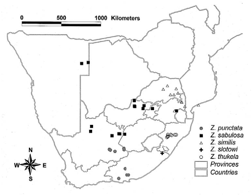
Distribution: The species is known from one locality, namely Mt Ayliff in the Eastern Cape ( Fig. 3 View Fig ).
Remarks: Lawrence initially labelled the type material as Z. diplodonta , but this was because he had examined only the specimen with two spines on the telocoxal spine-plate, without dissecting out the gonopods. Z. diplodonta is distributed in the northern parts of the subregion, and the Eastern Cape locality falls far out of this range.
There is a degree of variation in the structure of the gonopods. One specimen has two telocoxal spines ( Fig. 177 View Figs 175–183 ) and the other has four ( Fig. 179 View Figs 175–183 ). In both cases one spine is further split into two, i.e. apically bifid.

No known copyright restrictions apply. See Agosti, D., Egloff, W., 2009. Taxonomic information exchange and copyright: the Plazi approach. BMC Research Notes 2009, 2:53 for further explanation.