
Calappa karenae, Ng, Peter K. L. & Lai, Joelle C. Y., 2012
|
publication ID |
https://doi.org/10.5281/zenodo.211197 |
|
DOI |
https://doi.org/10.5281/zenodo.6170014 |
|
persistent identifier |
https://treatment.plazi.org/id/7F09EC44-FF9B-A405-FF3F-D456FA4DAA9D |
|
treatment provided by |
Plazi |
|
scientific name |
Calappa karenae |
| status |
sp. nov. |
Calappa karenae View in CoL sp. nov.
( Figs. 1–5 View FIGURE 1 View FIGURE 2 View FIGURE 3 View FIGURE 4 View FIGURE 5 )
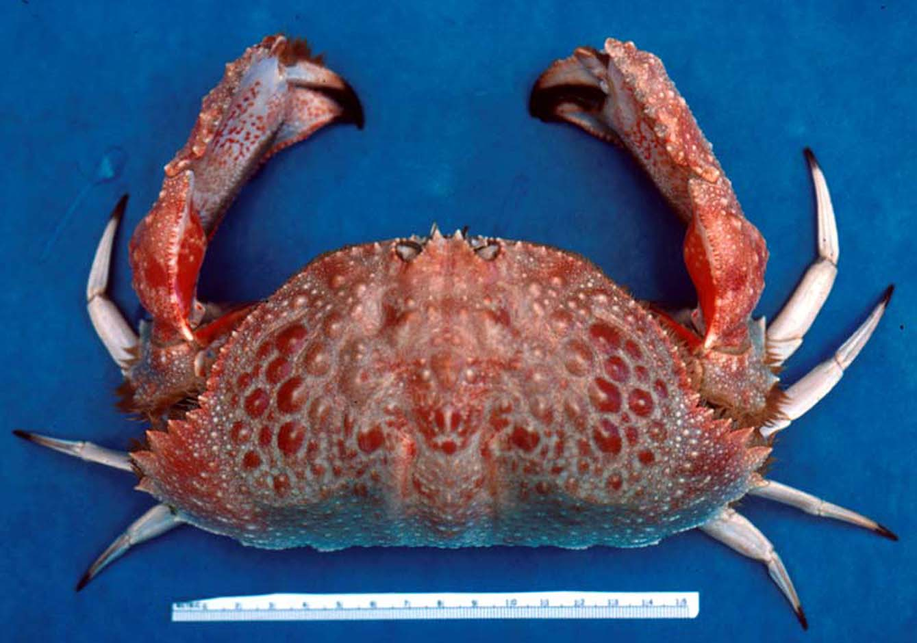
Calappa View in CoL sp. 1 ( aff. sebastiani Galil, 1997 )— Paulay et al. 2003: 496 [not Calappa sebastiani Galil, 1997 ] Material examined. Holotype: male (180.4 by 95.9 mm) (USNM 1150290 ), Haputo, Guam, in crab-trap, 100 fathoms (= 183 m), coll. E. Smith & L. Eldredge, 10 April 1986. Paratype: 1 male (CL 89.6 mm, damaged carapace; dimensions estimated from Fig. 1 View FIGURE 1 , 197.0 mm by 95.0 mm) (USNM 1150291 ), same data as holotype.
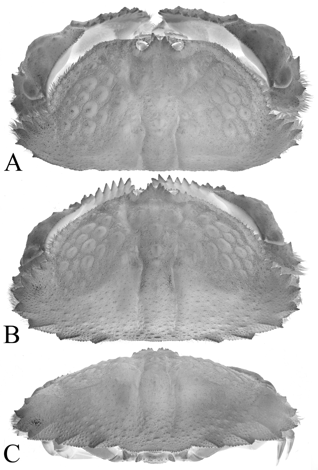
Diagnosis. Carapace much broader than long, width to length ratio 1.88–2.07. Anterior two-thirds of dorsal surface with large, round submammillate tubercles; posterior one-third with numerous low small granules ( Fig. 1 View FIGURE 1 , 2 View FIGURE 2 ). Pterygostomian lobe with broad inner oblique longitudinal groove ( Fig. 4 View FIGURE 4 D). Posterolateral margin of clypeiform expansion with 8 triangular teeth ( Figs. 1 View FIGURE 1 , 2 View FIGURE 2 ). Outer margin of merus of cheliped with 4 lobes ( Fig. 4 View FIGURE 4 C). Chela with 6 high, lamelliform teeth on dorsal margin ( Figs. 3 View FIGURE 3 A, 4A, B). Male thoracic sternites 4–6 relatively broad ( Fig. 3 View FIGURE 3 B, C). Male abdomen relatively broad; somite 6 subrectangular, slightly wider than long ( Fig. 3 View FIGURE 3 B, C). G1 C-shaped; tip truncated ( Fig. 5 View FIGURE 5 B, C, D, E).
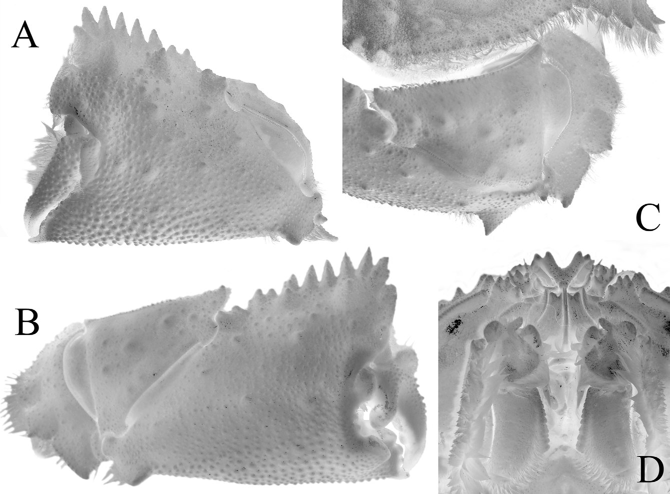
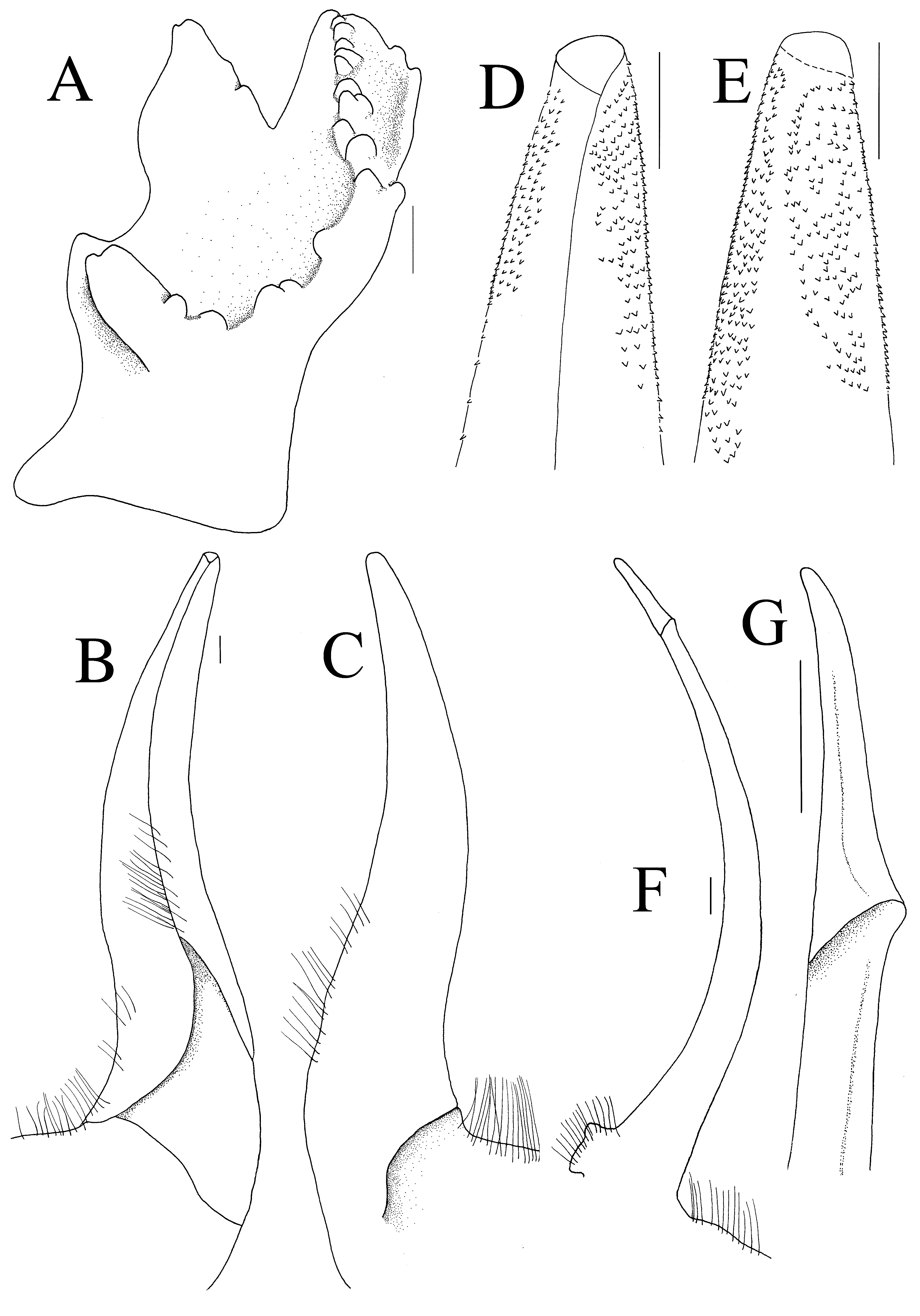
Description. Carapace transversely ovate, much broader than long, width to length ratio 1.88 ( holotype), 2.07 (estimated from photograph of paratype when intact) ( Figs. 1 View FIGURE 1 , 2 View FIGURE 2 ). Dorsal surface convex, anterior two-thirds covered with large, round submammillate tubercles, resembling low pustules, surface between tubercles finely granular; posterior third with numerous low small granules, short transverse striae, becoming more prominent, relatively larger towards posterior margin ( Figs. 1 View FIGURE 1 , 2 View FIGURE 2 ). Gastric region convex, with median part highest point of carapace, surface with low granules, tubercles; longitudinal gastro-cardiac grooves relatively deep, extending to just before posterior margin ( Figs. 1 View FIGURE 1 , 2 View FIGURE 2 ). Frontal margin distinctly bifurcated, with deep, broad U-shaped median cleft separating triangular teeth, separated from supraorbital margin by broad cleft ( Fig. 2 View FIGURE 2 A). Supraorbital margin uneven, granular, with 2 narrow longitudinal fissures on outer half ( Fig. 2 View FIGURE 2 A). Eyes folded obliquely, external orbital tooth low, tubercular ( Fig. 3 View FIGURE 3 A). Suborbital margin broad, dentiform, appearing sublobiform, with 3 low but distinct teeth, separated from external orbital angle by deep fissure ( Fig. 3 View FIGURE 3 A). Pterygostomian lobe at end of posterior margin of epistome with relatively broad inner oblique longitudinal groove ( Fig. 3 View FIGURE 3 A). Basal antennal article subquadrate, cusp-like, with transverse median ridge; proximal margin with prominent granules, distal margin deeply notched ( Figs. 3 View FIGURE 3 A, 5A). Longitudinal epistomial septum appears anteriorly complete ( Figs. 3 View FIGURE 3 A, 4D). Proepistome rounded ( Fig. 3 View FIGURE 3 A). Third maxilliped with margins of merus densely setose, distal margin cleft, anterolateral angle auriculiform ( Fig. 4 View FIGURE 4 D). Anterolateral margin arcuate, with 12–15 blunt, rounded unevenly sized tubercles, first tubercle distinct, subsequent ones very low, not easily discernible; last 5 or 6 progressively larger, more dentiform. Posterolateral margin of clypeiform expansion well developed, margins densely setose but not obscuring margin, with 8 triangular teeth, each tooth with denticulate margins, longitudinal median ridge (very strong in last 3 posterior teeth); first 4 teeth symmetrical, directed obliquely anteriorly; fifth tooth slightly asymmetrical, posterior part slightly longer, directed laterally; sixth tooth largest, broadest, asymmetrical, inner margin twice length of outer margin, directed posteriorly; last 2 teeth progressively smaller; posterior carapace margin gently concave, granular, not flanked by teeth but low lobes ( Figs. 1 View FIGURE 1 , 2 View FIGURE 2 ).
Chelipeds asymmetrical, right larger ( Figs. 1 View FIGURE 1 , 2 View FIGURE 2 A, B, 3A). Outer margin of merus with 4 lobes, first lobe very low, distalmost largest, sharpest; tip of lobes 3, 4 with sharp tubercle distally ( Fig. 4 View FIGURE 4 C). Carpus subtriangular, outer surface granular with scattered low tubercles ( Fig. 4 View FIGURE 4 B, C). Chela high, dorsal margin with prominent crest, with 6 high, lamelliform teeth, first smaller than second, directed slightly anteriorly; next 5 teeth vertical, acutely triangular, progressively smaller; base of crest with 2 small teeth; outer surface of manus with numerous granules, more prominent becoming denser, more prominent towards ventral margin with scattered large rounded tubercles; granules relatively more prominent in smaller chela; ventral margin lined with prominent row of granules, with prominent triangular tooth on proximal edge of ventral margin ( Figs. 3 View FIGURE 3 A, 4A-C). Dactylus of major chela with distinct basal curved cutting tooth, rest of margin with several low teeth; pollex with 3 molariform teeth, first tooth largest, subsequent teeth small. Dactylus of minor chela slender, curved, proximal two-thirds granular, cutting margin lined with denticles; pollex relatively stouter, surface granular, proximal cutting margin lined with denticles, distal part more blade-like ( Fig. 4 View FIGURE 4 A, B). Ambulatory legs slender, surfaces smooth; first leg longest; merus, carpus, propodus laterally flattened; dactylus styliform, gently curved ( Fig. 1 View FIGURE 1 ).
Thoracic sternum narrow; sternites 1, 2 completely fused; separated from sternite 3 by convex shallow groove; sternites 3, 4 fused but median sutures still visible, medially interrupted with longitudinal depression ( Fig. 3 View FIGURE 3 B); male thoracic sternites 4–6 relatively low, broad, surface covered with low, flattened granules ( Fig. 3 View FIGURE 3 B, C). Sternoabdominal cavity almost reaching thoracic suture 3/4 ( Fig. 3 View FIGURE 3 B).
Abdomen relatively broad; somite 1 longitudinally narrow with lateral parts expanded, anterior margin distinctly granular; somite 2 with lateral parts expanded to form subauriculiform process, surface prominently granular; somites 3–5 fused but median sutures just visible as shallow broad grooves, present as clefts laterally; somite 3 sub-rectangular, outer margin appears lobiform, granular, scalloped; somite 4 trapezoidal, lateral margins sinuous; somite 5 rectangular, lateral margins concave, distal margin granular; somite 6 subrectangular, slightly wider than long, lateral margins concave; telson acutely triangular, converging gradually to sharp tip; lateral margins gently concave ( Fig. 3 View FIGURE 3 B, C).
G1 relatively stout, slightly sinuous ( Fig. 5 View FIGURE 5 B, C); tip truncated, distal surface lined with numerous minute spinules ( Fig. 5 View FIGURE 5 D, E). G2 subequal in length to G1, slender, curved, distal tip rounded ( Fig. 5 View FIGURE 5 F, G).
Colour. On the basis of the colour photograph by Roy Kropp ( Fig. 1 View FIGURE 1 ), the live colouration of C. karenae is striking. It ranges from orange-red anteriorly to a light salmon pink in the posterior part of the carapace, with the round, submammillate tubercles in the middle third of the carapace scarlet red and bright yellow at the apex, resembling small pustules. Tips of movable fingers are dark brown, as are the tips of propodus on its ambulatory legs. The inner surface of the cheliped carpus is red, and the inner surface of its manus bears numerous small red spots. The preserved specimens have lost their colour and are now uniformly beige.
Etymology. The authors are delighted to dedicate this distinctive new species to Ms Karen Reed, a custodian to the priceless crustacean collections at USNM, who helped us eventually locate these elusive specimens.
Remarks. A label associated with the present specimens, written in 1979 by the late Austin Williams notes that the species “keys to hepatica in Sakai 1976 but cannot be that”. The general features of C. karenae sp. nov. superficially resemble C. hepatica ( Linnaeus, 1758) , but the differences are marked. Calappa hepatica has a green to purplish-grey carapace, never red. The front does not project so far anteriorly, there are no mammary-like tubercles on the carapace, and most importantly, the carapace is much less broad, with the width to length ratio of about 1.6 to 1.7 (see Galil 1997). The basal article of the antenna is also completely different ( Galil 1997: Fig. 14a). The carapace of C. karenae sp. nov. is wider than any other Calappa species in the Indo-West Pacific: C. calappa ( Linnaeus, 1758) the next widest species, has a width to length ratio of 1.7 to 1.8 ( Galil 1997). The carapace width to length ratios of the holotype and paratype of C. karenae sp. nov. is 1.88 and 2.07. respectively Calappa japonica Ortmann, 1892 has been reported to attain a larger carapace length (see Lai & Ng 2006; Ng et al. 2011) but its width to length ratio is approximately 1.4 in adult males.
Calappa karenae View in CoL sp. nov. was originally identified as C. sebastieni Galil, 1997 View in CoL (labelled as “ Calappa aff. sebastieni View in CoL ” in Paulay et al. 2003) on the basis of the photograph taken by Roy Kropp, but they are clearly different species, with C. sebastieni View in CoL possessing a very different rostrum ( Galil 1997: fig. 20f) and a proportionately narrower carapace.
The species perhaps closest to C. karenae View in CoL sp. nov. is the poorly known C. yamasitae Sakai, 1980 View in CoL , described from two specimens ( holotype male 110.0 × 66.0 mm and paratype female 100.0 × 62.0 mm) from Mie Prefecture in Japan. Unfortunately, comparisons between the two species can only be made based on Sakai’s publication, as the whereabouts of the only two known specimens of C. yamasitae View in CoL is unknown. They are neither present in the various Japanese museums where Sakai’s material are known to be deposited, nor in Senckenberg Museum in Frankfurt where a good part of his collection was posthumously donated. The specimens may be lost, mislabeled, or misplaced in one of these museums. Sakai provided a colour photograph of C. yamasitae View in CoL ( Sakai 1980: frontispiece fig. 2) although we do not know if it is an accurate representation of its live colour. The holotype as figured by Sakai (1980: frontispiece Fig. 2 View FIGURE 2 ) had a carapace that was reddish-orange, with the large tubercles relatively darker in colour. The colouration, presence of mammary-type tubercles on carapace and general form of the chela are shared characters in both species. Their carapace proportions, however, are different; the width to length ratio is 1.6 for C. yamasitae View in CoL whereas it is 1.88 in the holotype of C. karenae View in CoL sp. nov. ( 2.07 in the paratype). While this disparity may be due to the much larger sizes of our specimens (CW 110.0 mm, C. yamasitae View in CoL vs. 180.4 mm, C. karenae View in CoL ), it seems rather unlikely. The carapace width to length ratios of adult Calappa View in CoL species do not usually vary by more than 0.2 even when considering changes associated with size are considered (unpublished data; based on 10 C. calappa View in CoL adult males with CW greater than 100.0 mm).
From the colour photograph of C. yamasitae View in CoL ( Sakai 1980: frontispiece fig. 2), this species also appears to possess a more depressed hepatic (similar to C. gallus View in CoL ) region than C. karenae View in CoL sp. nov. The curvatures of the antero-lateral margins of both species are different. It is more when compared with convex in C. yamasitae View in CoL than C. karenae View in CoL sp. nov., although this is likely due to the relative width differences between the two species. The lateral spines along the clypeiform expansions on the posterolateral margin of the carapace points laterally in C. yamisitae but anteriorly in C. karenae View in CoL sp. nov.
The dorsal crest of the chela was also described as having 9 or 10 long teeth in C. yamasitae View in CoL (see Sakai 1980: 7), but there are only six such teeth in C. karenae View in CoL sp. nov., and even counting the two low teeth on the proximal part of the palm, there are a total of eight ( Fig. 4 View FIGURE 4 A, B). The teeth of the dorsal crest are more acute in C. karenae View in CoL sp. nov.; they are higher and have narrower bases than those seen in C. yamasitae View in CoL . The front also does not protrude very much in C. yamasitae View in CoL (see Sakai 1980: frontispiece fig. 2, text fig. 2a) while it does so prominently in C. karenae View in CoL sp. nov. ( Figs. 1 View FIGURE 1 , 2 View FIGURE 2 A, B). Sakai (1980: 6) described the front of C. yamasitae View in CoL as “consisting of two median obtuse teeth, which are separated medially by a U-shaped sinus”. The frontal margin of C. karenae View in CoL comprises two relatively sharp teeth, separated by a deeper U-shaped sulcus instead.
More importantly, the male abdominal somite 6 of C. yamasitae View in CoL is more transversely rectangular, with the telson evenly triangular, and the lateral margins of both structures are convex ( Sakai 1980: text fig. 3b). In contrast, the male somite 6 is quadrate, with curved, concave lateral margins and the telson more elongated, with straight lateral margins in C. karenae View in CoL sp. nov. ( Fig. 3 View FIGURE 3 B). The G1 is also relatively more slender in C. karenae View in CoL sp. nov. ( Fig. 5 View FIGURE 5 B, C), with the median and basal parts proportionately stouter in C. yamasitae View in CoL ( Sakai 1980: text fig. 3c)
The new species also resembles species in the C. japonica Ortmann, 1892 View in CoL , group, which includes C. exanthematosa Alcock & Anderson, 1894 View in CoL , and C. africana Lai & Ng, 2006 View in CoL (see Ng et al. 2011), especially in the general form of the mammary-like granules on the carapace. Other than the different colour patterns, C. karenae View in CoL sp. nov. can be separated from C. japonica View in CoL , C. exanthematosa View in CoL and C. africana View in CoL by its much broader carapace, more deeply clefted distal margin of the first maxilliped endopod, the rounded rather than triangular and compressed proepistome, and proportionately broader groove on the pterygostomian lobe (cf. Lai & Ng 2006; Ng et al. 2011).
The use of deep-water traps in Guam and the rest of the Mariana Is have already uncovered many new and/or rare species of homolids, poupiniids, inachids and mathildellids ( Eldredge 1980; Williams & Moffitt 1991; Ng & Wang 2002; Crosnier & Ng 2004). The present discovery of Calappa karenae View in CoL sp. nov. attests to the value of using trapping in areas with deep steep cliffs which cannot be sampled by trawls or dredges (see Mendoza et al. 2010).

No known copyright restrictions apply. See Agosti, D., Egloff, W., 2009. Taxonomic information exchange and copyright: the Plazi approach. BMC Research Notes 2009, 2:53 for further explanation.
|
Kingdom |
|
|
Phylum |
|
|
Class |
|
|
Order |
|
|
InfraOrder |
Brachyura |
|
Family |
|
|
Genus |
Calappa karenae
| Ng, Peter K. L. & Lai, Joelle C. Y. 2012 |
C. africana
| Lai & Ng 2006 |
aff. sebastiani
| Galil 1997 |
Calappa sebastiani
| Galil 1997 |
C. sebastieni
| Galil 1997 |
C. yamasitae
| Sakai 1980 |
C. exanthematosa
| Alcock & Anderson 1894 |
C. japonica
| Ortmann 1892 |