
Paranursallia spinosa n.
|
publication ID |
https://doi.org/ 10.5252/g2015n2a3 |
|
publication LSID |
urn:lsid:zoobank.org:pub:ADA93E56-4694-4ED6-A95C-99675FE54FEF |
|
persistent identifier |
https://treatment.plazi.org/id/C49AED7E-3471-4FCE-906D-1FFDFEF82D27 |
|
taxon LSID |
lsid:zoobank.org:act:C49AED7E-3471-4FCE-906D-1FFDFEF82D27 |
|
treatment provided by |
Felipe |
|
scientific name |
Paranursallia spinosa n. |
| status |
n. |
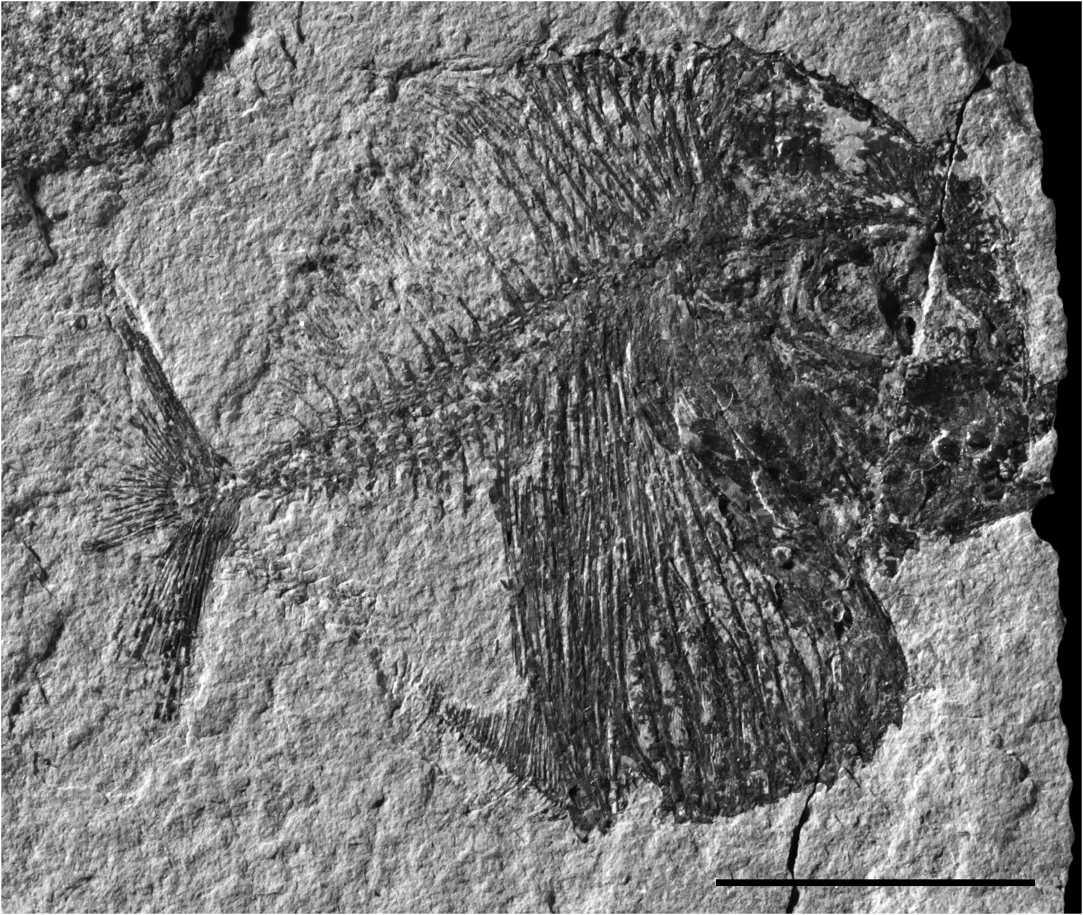
HOLOTYPE. — Sample MNHN.F.PSA214, a complete specimen in right view ( Fig. 3 View FIG ). Total length: 31 mm.
FORMATION AND LOCALITY. — Marine Cenomanian of Dir Oulad Yahia, Jebel Bargou, North Tunisia.
ETYMOLOGY. — From the Latin spinosus, -a, -um, spiny, in reference to the spiny upper margin of the dermosupraoccital in the new species.
DIAGNOSIS. — Paranursallia n. gen. with a series of small spines on the dermosupraoccipital upper margin. Frontal reaching the parietal. 8 scutes in the dorsal ridge. 8 precloacal and 2 postcloacal scutes in the ventral keel. About 30 principal rays in the caudal fin. HOLOTYPE MORPHOMETRIC DATA
In percentage (%) of the standard length (26 mm):
Length of the head (opercle included) .............................. 47.7% Depth of the head (in the occipital region) ....................... 62.1%
Maximal depth of the body .............................................. 93.9% Prepelvic length ................................................................ 47.0% Predorsal length ................................................................ 66.7% Preanal length ................................................................... 57.6%
NSP
OSTEOLOGY
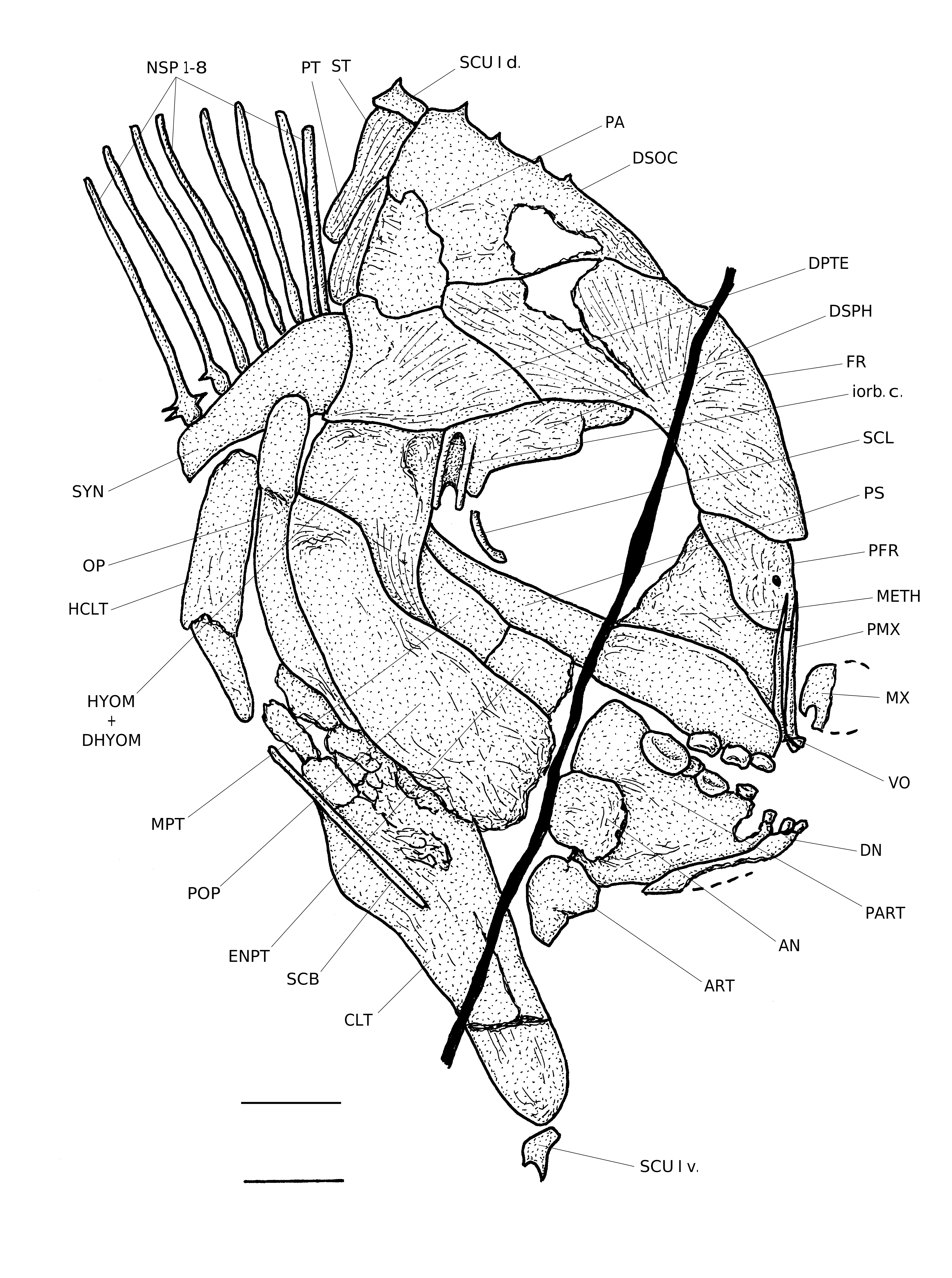
The skull ( Fig. 4 View FIG )
The head is large in comparison to the body size.The skull is deeper than long, with a rounded frontal border. The orbit is large and the snout very short, with its anterior border vertically oriented. The dermal bones are slightly ornamented with some thin ridges. The endocranial bones are not visible, except the mesethmoid. The mandibular lower margin and the cleithral anterior margin meet at almost a right angle, forming a large “V”-shaped notch at the junction between the head and the abdomen.
The mesethmoid is bulky but very short. The bone is partly covered by a pair of broad but short prefrontals. The vomer is massive. Only three vomerian teeth are preserved. They are molariform and belong to the right lateral row.
The skull roof is formed by the dermosupraoccipital and the paired frontals,parietals and dermopterotics.The frontal is long, broad and curved.Posteriorly,it sutures not only with the dermosupraoccipital and the dermopterotic but also reaches the rather small parietal.The dermosupraoccipital is a large bone.Its dorsal margin bears four small spines. There is no temporal fenestra and no brush-like process on the parietal. The autosphenotic is entirely hidden by the hyomandibula and the dermosphenotic. The sensory canals on the skull roof are not visible.
The parasphenoid is short, straight and toothless. No trace of the other sphenoid bones is visible in the orbit.
A part of the metapterygoid and of the entopterygoid appears between the preopercle and the parasphenoid. The quadrate and the symplectic are not preserved.
The long and very thin premaxilla bears two incisiform teeth. Only a small fragment of the maxilla is preserved. The mandible is triangular in shape and as deep as long. The dentary is reduced to its ventral branch. It bears two incisiform teeth. Five molariform teeth are visible on the upper margin of the large preaticular. Their size increases from before to behind. The angular and articular are well developed.
A very large dermosphenotic forms the upper border of the orbit. It bears the top of the infraorbital sensory canal. No other bone of the orbital ring is preserved. A fragment of a sclerotic bone is visible just above the parasphenoid.
The preopercle is much larger than the exposed part of the fused hyomandibula and dermohyomandibula. The opercle is long and narrow. No trace branchiostegal rays is visible.
The hyoid bar and the branchial skeleton are unknown.
The girdles ( Figs 4 View FIG ; 8 View FIG )
The posttemporal is a small narrow bone pressed against the posterior margin of the parietal. The hypercleithrum (= supracleithrum) is a deep bone. The ventral branch of the cleithrum is long but rather narrow (cf. Nursall 1996: fig. 11a). Some very small fragments of the pectoral fin are present but the number of rays is not determinable.
A few short and very thin pelvic rays are preserved in the cloacal vestibule. The pelvic bones are not visible.
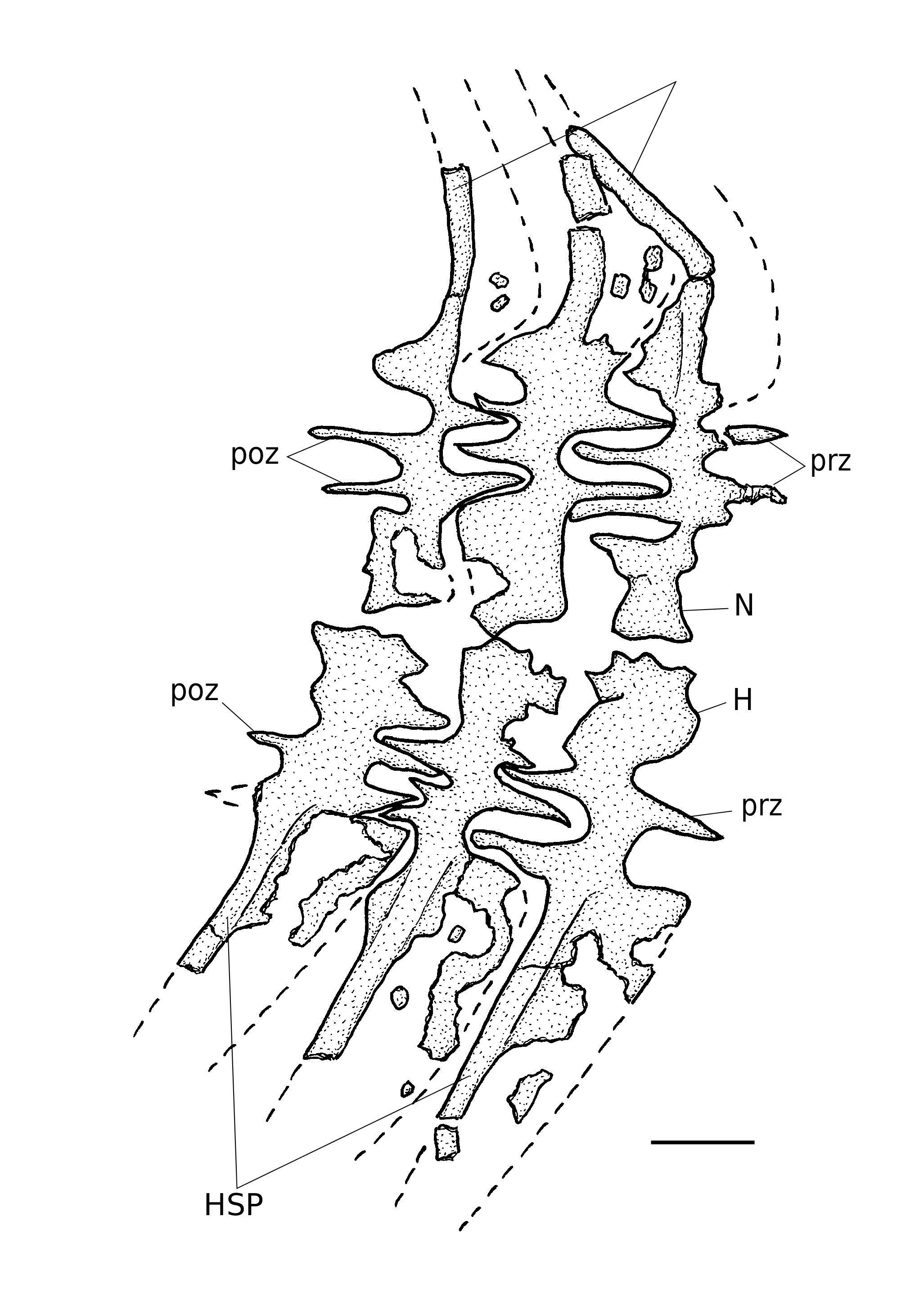
The axial skeleton ( Fig. 5 View FIG )
Starting from the caudal region, the vertebral axis progressively elevates to reach anteriorly the level of the orbit dorsal border. The vertebrae are constituted by dorsal and ventral arcocentra. No chordacentrum or autocentrum is present. The neural and haemal arches do not completely surround the notochord. The first neural arches are fused together. They form a large synarcual articulated to the rear of the skull and probably including the two exocciptals. There are 27 neural spines before the epichordal series and 14 haemal spines before the hypochordal elements. Most neural and haemal spines bear an anterior sagittal thin bony wing. The anteriormost 8 neural spines are autogenous, devoid of anterior sagittal flange and rest on the synarcual. The two last neural spines and the four last haemal spines before the caudal skeleton are much shorter than the preceding ones. Posteriorly to the synarcual, the neural arches are interlocked together by two pre- and two postzygapophyses. The same system exists on the haemal arches in caudal region of the fish. A few ribs are visible between the scales in the abdominal region but their exact number is not known. The postcoelomic bone is a long and rather thin bone reaching both the vertebral axis and the ventral margin of the fish.
The dorsal and anal fin ( Figs 3 View FIG ; 8 View FIG )
The dorsal and anal fins are badly preserved. The dorsal fin shape is unknown. The anal fin shape is strip-like and corresponds to the A 2 type of Poyato-Ariza & Wenz (2002: fig. 34). A few rays and 63 complete or fragmentary pterygiophores are visible in the dorsal fin but the last ones are missing. The total number of dorsal pterygiophores must be about 70. Some rays and the first 33 pterygiophores of the anal fin are preserved. The total length of the anal fin basis represents 13.2 mm and the 33 preserved pterygiophores cover 5.9mm of this length. We can thus estimate that the complete anal fin was supported by 73 pterygiophores.
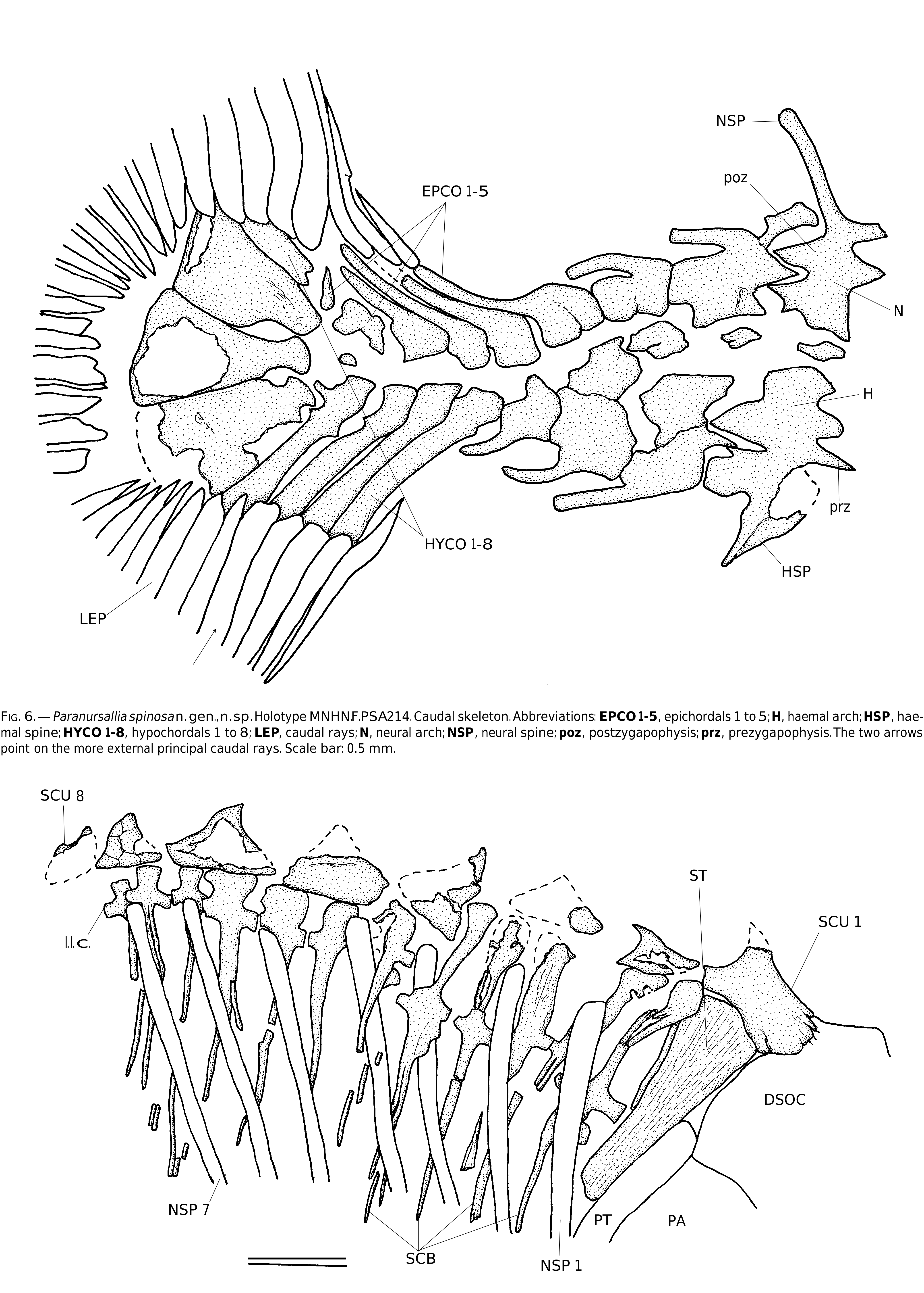
The caudal skeleton ( Fig. 6 View FIG )
The caudal peduncle is short, with reduced neural and haemal spines. The caudal endoskeleton contains 5 epichordal and 8 hypochordal elements. The neural arches of the epichordal series bear long but thin neural spines, except the fifth one that is short and broken away from its neural arch. The first four hypochordal elements are well developed, all together long and rather broad. The fifth, sixth and seventh hypochordals are hypertrophied. These three elements have approximately the same width. The eighth hypochordal is not enlarged. No urodermal is visible but this apparent absence is perhaps due to the taphonomic events.
The caudal fin is of the vertical type (Poyato-Ariza & Wenz 2002: fig. 36 F). There are 29 principal rays, 3 dorsal and 4 ventral procurrent rays.
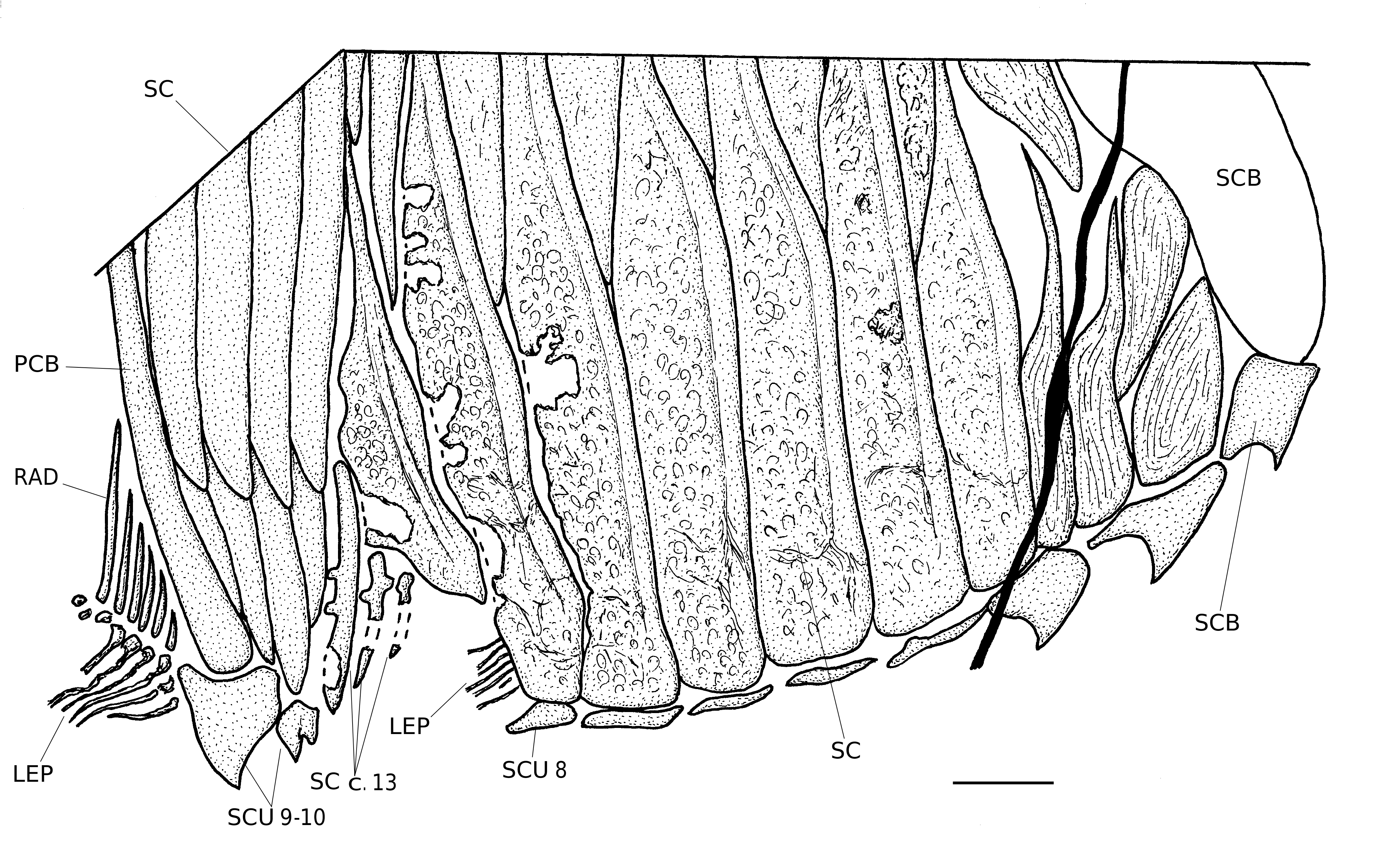
Squamation ( Figs 7; 8 View FIG )
There are flank scales only in the abdominal region of the body. In the ventralmost area of the situs viscerum, the scales are complete, slightly ornamented and articulated together. There are 9 rows of these large and broad body scales before the cloaca and 4 rows of narrower scales behind the cloaca. One smaller scale overhangs the cloaca and three small scales are visible in the cloacal vestibule. No bifid cloacal scale is present. The other body scales are reduced to scale bars.
The dorsal ridge is formed by a series of 8 scutes, each of them bearing one median spine. Only a very small part of the eighth element is preserved. The first dorsal scute is larger than the others. It is articulated with the dermosupraoccipital and rests on the dorsal margin of the supratemporal. Two scale bars are associated with each other dorsal scute. These dorsal scale bars bear a small transverse tube for the lateral line sensory canal.
The ventral keel contains 10 scutes, 8 before and 2 behind the cloaca. The first three and the two postcloacal scutes bear a median spine. The first ventral scute is located just below the cleithrum and the last one below the postcoelomic bone.
| MNHN |
Museum National d'Histoire Naturelle |
No known copyright restrictions apply. See Agosti, D., Egloff, W., 2009. Taxonomic information exchange and copyright: the Plazi approach. BMC Research Notes 2009, 2:53 for further explanation.