
Archiconchoecia (Archiconchoecia) chavturi, Kornicker, Louis S. & Harrison-Nelson, Elizabeth, 2005
|
publication ID |
https://doi.org/ 10.5281/zenodo.170303 |
|
DOI |
https://doi.org/10.5281/zenodo.6269095 |
|
persistent identifier |
https://treatment.plazi.org/id/805787E8-BE29-FFC7-FE83-A91829C5FD96 |
|
treatment provided by |
Plazi |
|
scientific name |
Archiconchoecia (Archiconchoecia) chavturi |
| status |
sp. nov. |
Archiconchoecia (Archiconchoecia) chavturi View in CoL , new species
Figs. 1–8 View FIGURE 1 View FIGURE 5 View FIGURE 8
Etymology. The species is named in honor of V. G. Chavtur, eminent specialist in the systematics and ecology of Ostracoda.
Holotype. USNM 1078583, adult female, carapace in alcohol, appendages on slide. Paratypes. None.
Type Locality. Tica Site, East Pacific Rise, 9°50.447'N, 104°17.493'W, 2500 m bottom depth, temperature up to 13°C. Alvin dive number 3851, sample number TC2b. Sample taken with a Bushmaster jr. from Riftia pachyptila aggregations with white and straight growing tubes.
Description of adult female ( Figs. 1–8 View FIGURE 1 View FIGURE 5 View FIGURE 8 ). Carapace oval in lateral view with greatest height just posterior to midlength ( Figs. 1 View FIGURE 1 A, 2A). In dorsal view valves widest near midlength (Fig. 2C). A small glandular opening on posterior edge of each valve dorsal to midheight; gland slightly lower on right valve ( Fig. 1 View FIGURE 1 D). Glands present within infold of ventral, anteroventral, and posteroventral margins of each valve ( Figs. 1 View FIGURE 1 B,C, 2D). Rostrum and incisure of each valve small ( Fig. 1 View FIGURE 1 A–C,E). Right valve with indentation in dorsal margin near midlength prior to removal of body ( Fig. 1 View FIGURE 1 A), but straight after removal of body ( Fig. 1 View FIGURE 1 B). Posterior angular hinge structure on each valve well developed (Figs. 2B, 3A). Surface of valves without either striations or reticulations (possibly lost during preservation).
Central Adductor Muscle Scars: Ends of 5–8 muscles detached from valve in muscle area visible through valve and may have been attached to valve (Fig. 2C). Five scars forming Jshape present at base of usual location of central adductor attachments ( Fig. 1 View FIGURE 1 B,C).
Carapace Size: Carapace length 0.60 mm, height 0.40 mm.
First Antenna: Interpreted to have 6 articles, but sutures not present between articles 1 and 2, and 2 and 3. Separation of articles 1 and 2 indicated by step in dorsal margin, and separation of articles 2 and 3 indicated by termination of internal muscle (Fig. 3D). Article 2 with slender terminal dorsal bristle with indistinct marginal spines (Figs. 2F, 3B–D). Article 5 with 4 long filamentlike bristles. Article 6 with 2 long filamentlike bristles (Figs. 2F, 3D) (1 filamentlike bristle displaced on left limb (dashed in Fig. 3D)). Black proximal spots on limbs (Figs. 2F, 3B,D).
Second Antenna (Figs. 2A,E, 3B,C, 4): Protopod: ventral margin with small bare process between bases of exopod and endopod, closer to endopod (Fig. 4B,C); protopod of left limb with about 12 small black pigment spots. Exopod of right limb with 8 articles separated by sutures (exopod of left limb incomplete): article 1 with small dorsal spines (on right limb only) and short, slender, bare, medial terminal bristle (Fig. 4A,B); article 2 of right limb without bristle (all bristles broken off on left limb); natatory bristles on articles 3–7 with bases medial; article 8 with 2 short bristles (Fig. 4A,D). Endopod: article 1 without processes mammillaria; ventral margin with minute digitations and spines; dorsal margin with a and bbristles (indistinct spines observed only on one bristle) (Fig. 4B,C). Articles 2 and 3 fused, with total of 5 filamentlike bristles of about same length; tips of filaments tapered. (The bristle of the 2nd exopodial article of the right limb was observed to be absent prior to dissection of the specimen, but whether or not it had broken off is unknown. Although 9 exopodial articles are the usual number, only 8 could be detected on the right limb when viewed under oil immersion with X100 objective lens.).
FIGURE 2. Archiconchoecia chavturi , new species, adult female, holotype: A, compete specimen from left side; B, dorsal part of complete specimen from left side; C, dorsal view of complete specimen, anterior to right, (Bellonci Organ projecting at right; black dots on 1st antennae); D, inside edge of ventral margin of left valve showing glandular openings; E, anterior of left valve showing elongate Bellonci Organ and part of left 2nd antenna; F, dorsal distal view of right 1st antenna (nabs). (nabs = not all bristles shown.)
FIGURE 3. Archiconchoecia chavturi , new species, adult female, holotype: A, ventral view of dorsal posterior end of carapace with valve partly open; B, anterior of body from right side showing Bellonci Organ and parts of right 1st and 2nd antennae; C, anterior of body from left side showing Bellonci Organ, parts of left 1st and 2nd antenna and two processes on anterior of body; D, Bellonci Organ and left 1st antenna, lv.
(lv = lateral view.)
FIGURE 4. Archiconchoecia chavturi , new species, adult female, holotype: A, B, part right 2nd antenna, mv; C, D, endopod and exopod left 2nd antenna, lv (nabs).
(lv=lateral view; mv = medial view; nabs = not all bristles shown.)
Mandible ( Figs. 5 View FIGURE 5 , 6A): Coxa: anterior margin with distal triangular process; terminal edge with about 6 triangular teeth lateral and partly anterior to dense rows of minute triangular teeth (number of rows difficult to resolve) ( Fig. 5 View FIGURE 5 A,D,E). Basis endite: distal edge with 6 small triangular teeth (posterior tooth slightly larger than others) and 1 larger tooth lateral to anterior triangular tooth; tubeshaped bristle and pointed bristle on edge proximal to posterior triangular tooth; a few hairs along edge proximal to pointed bristle; 1 long bristle on anterior margin of endite proximal to teeth ( Fig. 5 View FIGURE 5 A,C,D); lateral side of endite distal to stout internal muscle with 2 long proximal and 2 short distal bristles ( Fig. 5 View FIGURE 5 C). Dorsal edge of basis with 1 or 2 short bristles; medial side of basis near dorsal edge with 2 exopodial bristles (length uncertain) (Fig. 6A). Endopod ( Figs. 5 View FIGURE 5 A,B, 6A): article 1 with 4 bristles (3 ventral, 1 dorsal); article 2 with 6 bristles (4 ventral, 2 dorsal); article 3 with 6 bristles (4 ventral, 2 terminal (dorsal terminal bristle clawlike)). (Several bristles on endopod broke off during dissection; these are dashed in Figs. 5 View FIGURE 5 A, 6A.) Limb without black spots.
Maxilla (Maxillula) (Fig. 6A–C): Precoxa endite with 5 stout bristles and 1 tubular bristle; coxa endite with 4 stout bristles and 1 tubular bristle. Basis with long spinous ventral bristle. Endopod: article 1 with 8 bristles; article 2 with bristles missing (bristles broken off during dissection).
Fifth Limb (Fig. 6A,D): Epipod (not shown in fig.) with 3 sets of plumose bristles, each set with about 5 bristles. Coxa with 3 endites: endite I with 2 short bristles (longest with marginal hairs); endite II with 2 long hirsute bristles; endite III with 2 claws and 4 bristles (3 long hirsute). Basis with 5 bristles (1 long hirsute) on or near ventral margin, and 1 long dorsal exopodial bristle. Endopod: article 1 with 2 or 3 bristles (1 or 2 ventral, 1 dorsal) near midlength; article 2 short with 3 terminal bristles.
Sixth Limb (Figs. 6E,F, 7A): Epipod with 3 sets of plumose bristles, each set with 4 to 6 bristles (Figs. 6E, 7A). Coxa without bristles. Basis with 1 distal ventral bristle, 1 dorsal bristle near midlength, and 1 terminal dorsal exopodial bristle (not observed on left limb). Endopod: article 1 with 1 dorsal bristle near midlength; article 2 with dorsal bristle near midlength and none or 1 ventral bristle near midlength; short 3rd article with 3 terminal bristles (middle bristle longest, ventral bristle shortest).
Seventh Limb (Figs. 6G, 7A): Short with 2 terminal bristles.
Furca (Fig. 7A,B,E,F): Each lamella with 6 claws (most claws missing from specimen when received), and 1 unpaired bristle.
Bellonci Organ (Figs. 2E, 3B–D): Elongate, reaching bend at tip of 1st antenna, with rounded tip bearing 2 minute spines.
Lips (Fig. 7C): With marginal spines.
Anterior of Body (Figs. 3C, 7C,D): With rounded process on each side.
Posterior of Body (Fig. 7A,B,E,F): Without spines or segments.
Spermatheca (Figs. 7A,B, 8): Sac with 2 lobes located adjacent to furca; right lobe larger; both lobes of studied specimen contain stringlike sperm; more sperm present inside right lobe.
FIGURE 6. Archiconchoecia chavturi , new species, adult female, holotype: A, anterior of body showing left mandible, maxilla, and 5th limb, lv (nabs); B, C, maxillae (nabs); D, 5th limb; E, F, 6th limbs (nabs); G, 7th limb. (lv = lateral view; nabs = not all bristles shown.)
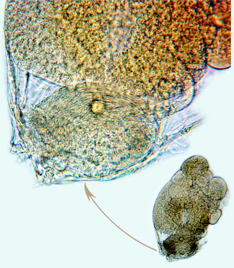
Eggs: Studied specimen with about 15 eggs of different diameters (Fig. 7A,B); largest egg closest to spermatheca.
Gut Content: Abundant appendage fragments, bristles, and bristlelike claws.
FIGURE 7. Archiconchoecia chavturi , new species, adult female, holotype: A, posterior of body from right side showing eggs, spermatheca, 6th and 7th limbs, and furca (not all furcal claws shown); B, eggs, spermatheca containing threadlike sperm, and proximal part of furca; C, posterior view of anterior of body compressed under cover slip showing upper and lower lips and two anterior processes; D, anterior view of body showing two anterior processes; E, F, right and left furcal lamellae (dashed claws missing from specimen).
Comparisons. In the “Key to Species of Subgenus Archiconchoecia (Archiconchoecia) (Adult Female and Male)” presented by Chavtur and Stovbun (2003:145), the female in the present collection keyed out to A. (A.) propinqua Chavtur and Stovbun, 2003 . A difference between that species and A. (A.) chavturi is that the Bellonci Organ of A. (A.) chavturi has a rounded tip with 2 minute spines, whereas on A. (A.) propinqua the tip is bare and pointed; also, the organ extends farther over the bend at the distal end of the 1st antenna than that of A. (A.) chavturi (compare Fig. 3B with Chavtur and Stovbun, 2003: fig. 4D). In addition, the posterior shell glands appear to be much broader on A. (A.) propinqua than the glands of A. (A.) chavturi (compare Fig. 3A with Chavtur and Stovbun, 2003: Fig. 6A. B).
Taxonomic note. According to Diebel (1962:240), type specimens of Archiconchoecia striata Müller, 1894 , type species of the genus, are at the Museum for Naturkunde, Berlin. In answer to a letter from the senior author asking to borrow a specimen, Dr. Charles Oliver Coleman, Curator of Crustacea at the museum, informed us (Email, 16 Nov 2004) that the material had been borrowed in 1962 and not returned, and that the museum was unable to recover the material after death of the borrower, and that the specimens are considered lost.


| USNM |
Smithsonian Institution, National Museum of Natural History |
No known copyright restrictions apply. See Agosti, D., Egloff, W., 2009. Taxonomic information exchange and copyright: the Plazi approach. BMC Research Notes 2009, 2:53 for further explanation.