
Sphaerodoropsis heteropapillata Hartmann-Schröder, 1987
|
publication ID |
https://doi.org/ 10.11646/zootaxa.4000.2.3 |
|
publication LSID |
lsid:zoobank.org:pub:7EDEDAEE-642C-4F9D-A04D-141815D73343 |
|
DOI |
https://doi.org/10.5281/zenodo.5667467 |
|
persistent identifier |
https://treatment.plazi.org/id/81007D79-8D55-257F-FF0F-FA07FC731DA2 |
|
treatment provided by |
Plazi |
|
scientific name |
Sphaerodoropsis heteropapillata Hartmann-Schröder, 1987 |
| status |
|
Sphaerodoropsis heteropapillata Hartmann-Schröder, 1987 View in CoL new rank
Figs 4 View FIGURE 4 M–N, 5I, 11
Sphaerodoropsis multipapillata heteropapillata Hartmann-Schröder, 1987: 50 View in CoL –51, Figs 32–36.
Material examined. Holotype ZMH P.18875 Point Lonsdale, Geelong, Victoria, Australia, Abrasion terrace near lighthouse, on coralline algae, 24 Dec 1975.
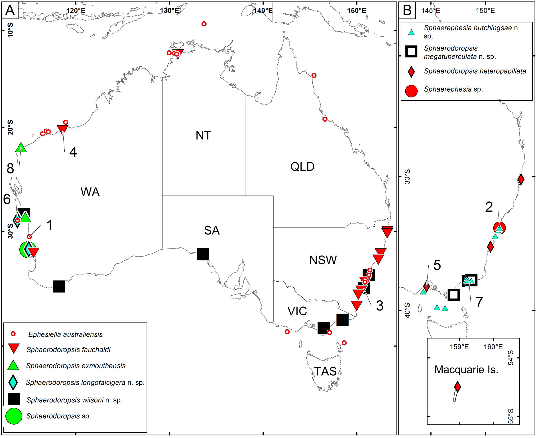
Additional material. New South Wales: AM W.42683 (1 spec.), Split Solitary Island, 30° 14' S, 153° 10' 48" E, 15 m, 7 Mar 1991, mixed red algae; AM W.42686 (4 specs, 2 on SEM stubs), South Solitary Island, 30° 12' 07" S, 153° 15' 59" E, 14.5 m, 1 May 2005, coarse sediment; AM W.42684 (1 spec.), Burrill Rock, Ulladulla, 35° 23' 25" S, 150° 28' 11" E, 10 m, 1 May 1997, Ecklonia holdfasts. Tasmania: AM W.42678 (2 specs), Maquarie Island, 54° 30' S, 158° 57' E, 15 m, 19 Oct 1983, algae. Victoria: NMV F.90823 (2 specs), Beware reef, Cape Conran, 37° 49' 20'' S, 148° 47' 23'' E, 5 m, 15 Apr 1998, subtidal rocky reefs.
Comparative material. Sphaerodoridium multipapillata Hartmann-Schröder, 1974 , holotype, ZMH P.14336.
Diagnosis. Ellipsoid body. Dorsum with more than 50 spherical sessile tubercles in two sizes in about five transversal rows per segment, in an irregular pattern. Ventrum with 50–60 spherical papillae in two sizes in 5 irregular transversal rows. Two parapodial papillae, one each on anterior and posterior surfaces. Parapodia with 6– 10 chaetae per fascicle, blades as long as wide. Chaetae semi-compound, with shaft distally widened and blades short and wide (as long as broad), both provided of stout spines.
Re-description. Measurements and general morphology. Female with eggs. Body ellipsoid, measuring 2.8 mm long, 1.0 mm wide, with 19 chaetigers, anterior end bluntly rounded ( Fig. 11 View FIGURE 11 B–C), slightly narrowing along posterior segments; convex dorsum ( Fig. 11 View FIGURE 11 A) and flat ventrum ( Fig. 11 View FIGURE 11 B). Segmentation inconspicuous ( Fig. 11 View FIGURE 11 B–D), tegument wrinkled ( Fig. 11 View FIGURE 11 A–B). Preserved specimen yellowish, lacking pigmentation.
Head. Indistinguishable rounded appendages, similar to surrounding papillae ( Fig. 11 View FIGURE 11 B–C).
Tubercles. Spherical and sessile tubercles dissimilar in size present on dorsum in 5–7 irregular transversal rows with more than 50 tubercles in each chaetiger ( Figs 4 View FIGURE 4 M, 11A, C–D). Ventral surface with sessile and spherical papillae dissimilar in size, with 50–60 papillae per chaetiger in five irregular transversal rows, ventral papillae somewhat smaller than dorsal tubercles ( Figs 4 View FIGURE 4 N, 11B, D). Body epithelium with microscopic oblong granules ( Fig. 11 View FIGURE 11 F).
Parapodia. Parapodia sub-conical, increasing in size towards chaetiger 3, around 1–2 times longer than wide in mid-chaetigers ( Fig. 11 View FIGURE 11 E, G–H), and decreasing slightly towards posterior chaetigers. Acicular lobe anterior to chaetae fascicle, ellipsoid, 2–3 times longer than wide, projecting longer than ventral cirrus ( Fig. 11 View FIGURE 11 G–H), with pores on distal end ( Fig. 11 View FIGURE 11 H). Ventral cirri ellipsoid ( Fig. 11 View FIGURE 11 G–H). Parapodial papillae spherical, one at base of anterior surface ( Figs 5 View FIGURE 5 I, 11G), and one at base of posterior surface ( Figs 5 View FIGURE 5 I, 10H); similar throughout chaetigers. Chaetae. Chaetal fascicles arranged in a curved transverse row behind acicular lobe, numbering 6–10 chaetae per fascicle ( Figs 5 View FIGURE 5 I, 11E, G–H). All chaetae semi-compound ( Fig. 11 View FIGURE 11 J–M), appearing sometimes as simple and suggesting a fusion of shaft and blades ( Fig. 11 View FIGURE 11 L–M). Blades as long as wide ( Fig. 11 View FIGURE 11 J–M); with an outer distal thin spine in some chaetae ( Fig. 11 View FIGURE 11 L–M). Shaft with widened distal end with saw-toothed spinulation ( Fig. 11 View FIGURE 11 L–M), continuing along most of blade length.
Pygidium . Pygidium terminal, with inconspicuous spherical anal cirri similar in shape as surrounding papillae ( Fig. 11 View FIGURE 11 I).
Internal features. One pair of eyes as sickle-shaped dark brown or black spots deeply embedded below integument in chaetigers 2–3. Muscular pharynx not observed.
Reproductive features. Holotype and other two females with large eggs measuring approximately 130 µm. ‘Copulatory organs’ not observed in any specimen.
Variation. Specimens examined are all ellipsoid, ranging 1.0– 2.3 mm long, 0.3–0.7 mm wide, with 14–20 chaetigers and resemble the holotype in the short and inconspicuous prostomial appendages similar in shape to surrounding papillae and the shape and size of parapodia. In the original description four parapodial papillae were reported, only two have been observed after reexamination of the holotype and in additional material. Parapodial papillae are difficult to observe, and also judgment of their position as they are placed at the base of parapodia. Definition of chaetae is ambiguous. Most chaetae are simple when observed in the scanning electron microscope, although some give the impression of being semi-compound ( Fig. 11 View FIGURE 11 J–M), and even break at the joint between shaft and blade ( Fig. 11 View FIGURE 11 M). In the light microscope chaetae have a clear appearance of being compound ( Fig. 11 View FIGURE 11 J– K), as they were described in the original description ( Hartmann-Schröder 1987: Fig. 36).
Remarks. Sphaerodoropsis heteropapillata is characterized by having relatively large dorsal tubercles of a range of sizes and by bearing semi-compound chaetae. In addition, relatively large papillae are present on the ventral side. Dorsal and ventral tubercles are arranged in irregular transversal rows being difficult to outline, especially when specimens are contracted. It was included within the informal Sphaerodoropsis Group 4 (sensu Borowski 1994) gathering species with ‘macrotubercles’ scattered in 3–4 transversal rows, together with S. multipapillata (Hartmann-Schröder, 1974) , from Tanzania. Both species also share the similar type of wide semicompound chaetae ( Hartmann-Schröder 1974a, 1987). Sphaerodoropsis heteropapillata (which only the holotype had been reported to date) was originally described as a subspecies of S. multipapillata . The geographic distance suggests it is unlikely the two represent separated populations of the same species. There are also clear differences between the species. S phaerodoropsis heteropapillata bears parapodial papillae while in S. multipapillata parapodia lack papillae ( Hartmann-Schröder 1974a, 1987; this study). Moreover, in S. heteropapillata dorsal tubercles clearly have different sizes, while in S. multipapillata papillae are of similar size. This warrants elevation of the subspecies to rank of species.
S phaerodoropsis heteropapillata also resembles a recently described species from Lizard Island, Great Barrier Reef, Sphaerodoropsis plurituberculata Capa and Rouse, 2015. Both species bear several more or less clearly longitudinal rows of spherical and sessile tubercles, variable in size, arranged in several transverse rows per segment, over dorsum and ventrum; around six semi-compound chaetae per parapodium with distally enlarged shaft and short blades, serrated, with distal long spines on the edge opposite to serration and shaft with conspicuous spinulation continuing along most of blade. Differences between these two Australian species lay in the relative length of the head appendages, number and arrangement of dorsal and ventral tubercles.
Sphaerodoropsis plurituberculata is most similar to S. heteropapillata View in CoL because both species are covered with dissimilar dorsal tubercles, while in S. multipapillata View in CoL these are of similar size ( Hartmann-Schröder 1974a; Hartmann-Schröder 1987; Capa & Rouse 2015). Differences between S. heteropapillata View in CoL and the new species include the length of the prostomial appendages (inconspicuous in S. heteropapillata View in CoL and digitiform in S. plurituberculata , at least when not contracted); the number of ventral papillae (around 50 per segment in S. heteropapillata View in CoL and 30 in S. plurituberculata ); and the number of parapodial papillae ( S. heteropapillata View in CoL bears one per parapodium: one at anterior surface while S. plurituberculata lacks parapodial papillae) ( Capa & Rouse 2015).
‘Copulatory organs’ have not been observed in S. heteropapillata View in CoL , but in Sphaerodoropsis plurituberculata male copulatory organ were described as enlarged, bottle-shaped and porous ventral cirrus and females presented an oval and flat tubercle with a porous surface also ventral to parapodia of chaetiger 6, in addition to the ventral cirrus ( Capa & Rouse 2015).
Type locality. Point Lonsdale, Geelong, Victoria.
Distribution. Victoria, New South Wales and the Sub-Antarctic Maquarie Island, from 0 to 15 m (Hartmann- Schröder 1987, Fig. 15 View FIGURE 15 ).
Habitat. Coarse sediment and among algae in subtidal rocky reefs.


No known copyright restrictions apply. See Agosti, D., Egloff, W., 2009. Taxonomic information exchange and copyright: the Plazi approach. BMC Research Notes 2009, 2:53 for further explanation.
|
Kingdom |
|
|
Phylum |
|
|
Class |
|
|
Order |
|
|
Family |
|
|
Genus |
Sphaerodoropsis heteropapillata Hartmann-Schröder, 1987
| Capa, Maria & Bakken, Torkild 2015 |
Sphaerodoropsis multipapillata heteropapillata Hartmann-Schröder, 1987 : 50
| Hartmann-Schroder 1987: 50 |