
Psilorrhynchus bifasciatus (Blanchard, 1844)
|
publication ID |
https://doi.org/ 10.37520/aemnp.2022.003 |
|
publication LSID |
lsid:zoobank.org:pub:B80C1377-88E3-4A2C-8413-3577781E6406 |
|
persistent identifier |
https://treatment.plazi.org/id/8254F800-1E22-DD3D-6874-C73DEEAAFB3D |
|
treatment provided by |
Felipe |
|
scientific name |
Psilorrhynchus bifasciatus (Blanchard, 1844) |
| status |
|
Psilorrhynchus bifasciatus (Blanchard, 1844)
( Figs 10–49 View Figs 10–13 View Figs 14–18 View Figs 19–35 View Figs 36–38 View Figs 39–49 )
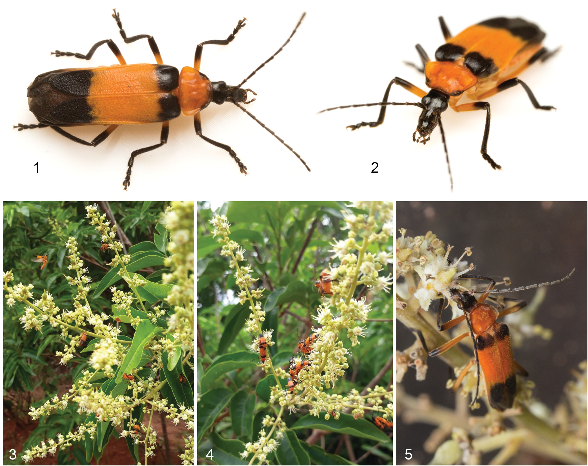
Material examined. BRAZIL: Mൺඍඈ Gඋඈඌඌඈ ൽඈ Sඎඅ: Selvíria, Fazenda Bovinocultura da UNESP, 20°20′28.18″S, 51°24′16.86″W, 22.x.2018 (on Matayba guianensis inflorescence), L. Migliore leg. (4 JJ, 4 ♀♀, MZSP 45585–45592, 5 E, 10 L1, 2 L2, 6 L3 MZSP Im. Col. 10369); Maciço do Urucum, 19°12′13.5″S 57°37′46.3″W, 12.–15.i.2019, winkler, M. A. Ulysséa leg. (1 L3, MZSP Im. Col. 10370).
Adult’s behaviour. Adults of Psilorrhynchus bifasciatus were abundant on the inflorescences of Matayba guianensis Aubl. ( Sapindaceae, Cupanieae ) ( Figs 3–5 View Figs 1–5 ). Around 15:00 h (local time), the hottest time of the day, hundreds of specimens were observed in each of two nearby flowering bushes. By 16:30 h they were much less abundant, with only a few tens of remaining specimens on each bush. The next morning no more specimens were found in neither bush.
While in the plant, they were very active, rapidly and ceaselessly foraging throughout the flowers by inserting the elongate head to reach the nectar disc located between petals and stamens. The pollen in the elongate and conspicuous stamens was apparently despised. During collection, the specimens were transferred to plastic vials containing branches and inflorescences of M. guianensis for transportation. Immediately after the transfer, the specimens searched for the flowers and carried on foraging.
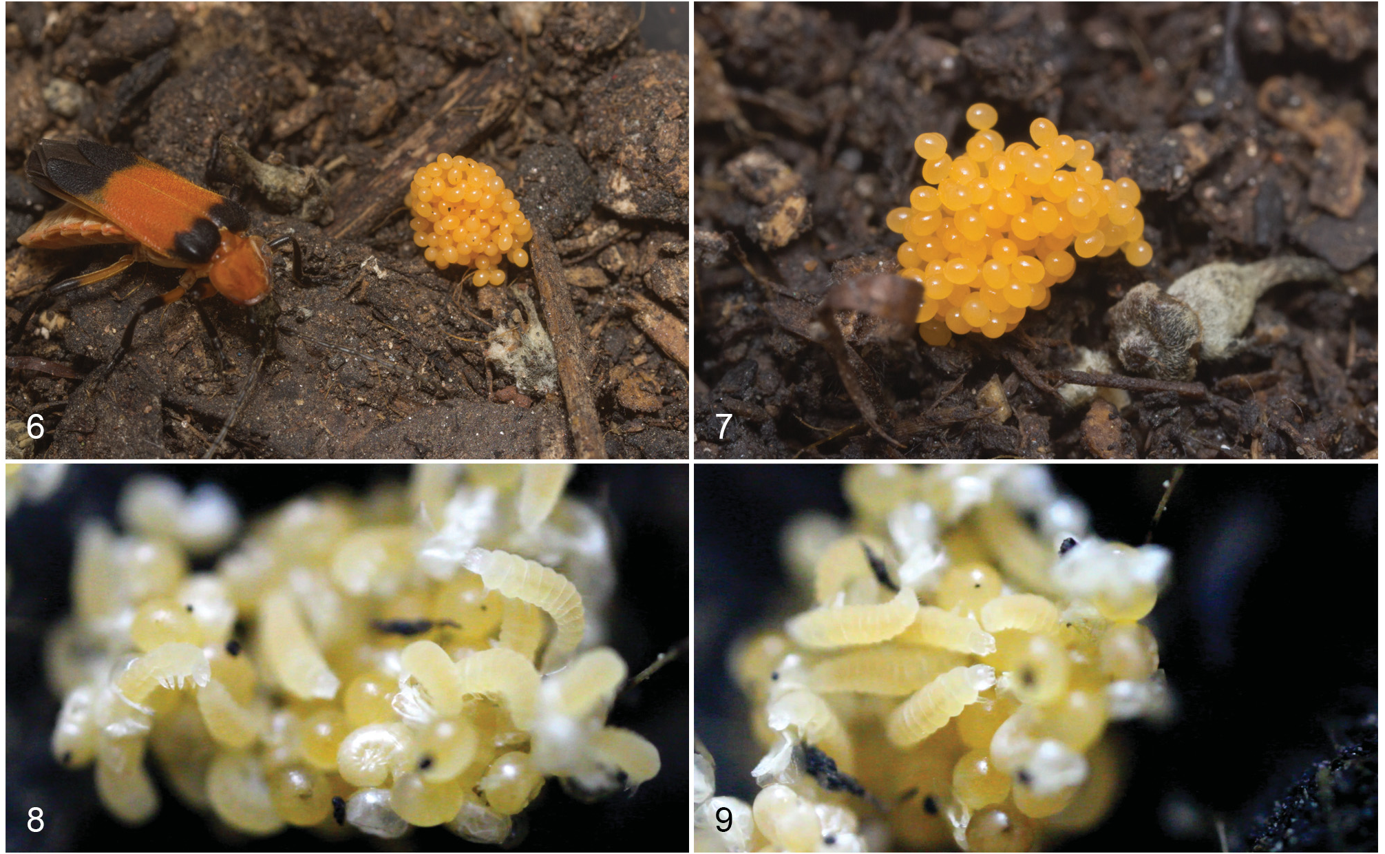
The copulation occurred on the plant while the females kept foraging. The time of each copulation and the possible change of couples could not be observed in situ. In the laboratory, the specimens continuously kept foraging in the offered inflorescences, although apparently, no more nectar leftovers were available therein. Occasional copulation occurred only during the following two days after collection.Afterwards, the females were kept in individual plastic containers for oviposition. Two females laid eggs 7 and 10 days after the last copulation. Each female laid large piles of orange eggs directly on the soil ( Figs 6–7 View Figs 6–9 ), but only one pile was fertilized. Day after day, the fertilized eggs got clearer, turning white and slightly translucent, whereas the unfertilized eggs kept the orange colour for several days until they perished.
Egg hatching took place 9 days after the oviposition ( Figs 8–9 View Figs 6–9 ). In the beginning, a few larvae hatched, and then 5–6 hours later, many other larvae started hatching simultaneously. The first instar larvae were moveable since the hatching, moving legs and mouthparts and curving the body, although having the apex of the abdomen still attached to the chorion. After detachment, the first exploratory surveys carried out by the larvae were within the egg pile, apparently feeding on the chorion remains. One day after hatching, the larvae started exploring the soil around the egg pile. The next day, most larvae were dispersed and mostly inside the soil. Several cases of cannibalism were observed, with various larvae preying on the same prey.
The larvae were reared in the laboratory for about 20 days, until the third instar, when they died. The larvae hardly ever accepted the offered food sources, consisting of Collembola, pieces of flies or other insects, or artificial fish ration.
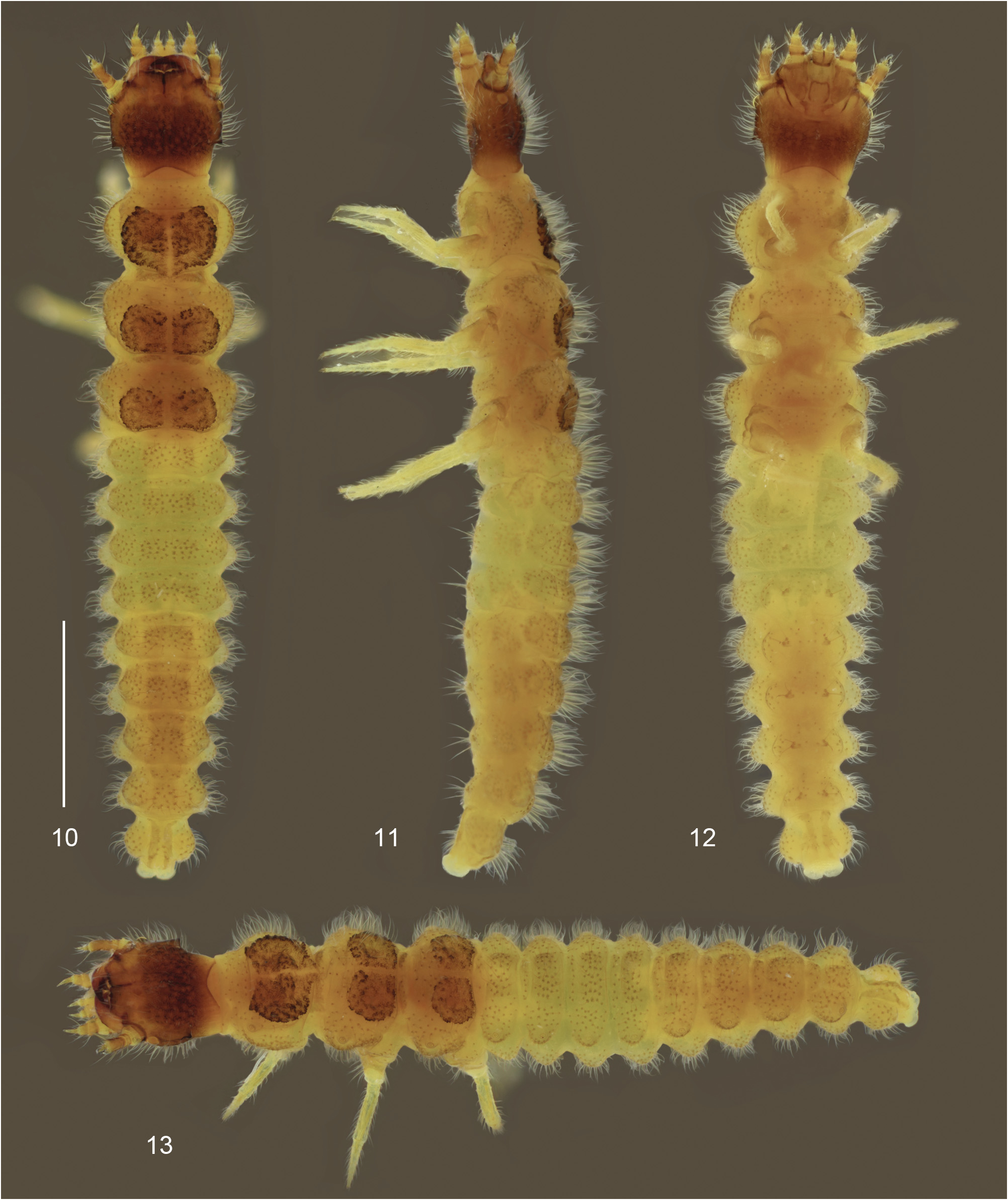
Description of immatures. Third instar larva ( Figs 10–35 View Figs 10–13 View Figs 14–18 View Figs 19–35 ). Colouration. Body mostly white to light grey, slightly translucent, except for light brown head, mouthparts and rough areas in pro-, meso- and metanotum (illustrated larva stained with iodine).
Pubescence. Very long, thick and sparse; setae erect, inserted in dark punctures; secondary layer of very short, thin and dense pubescence; abdominal segments II–VIII with pairs of tufts of long setae ventrally.
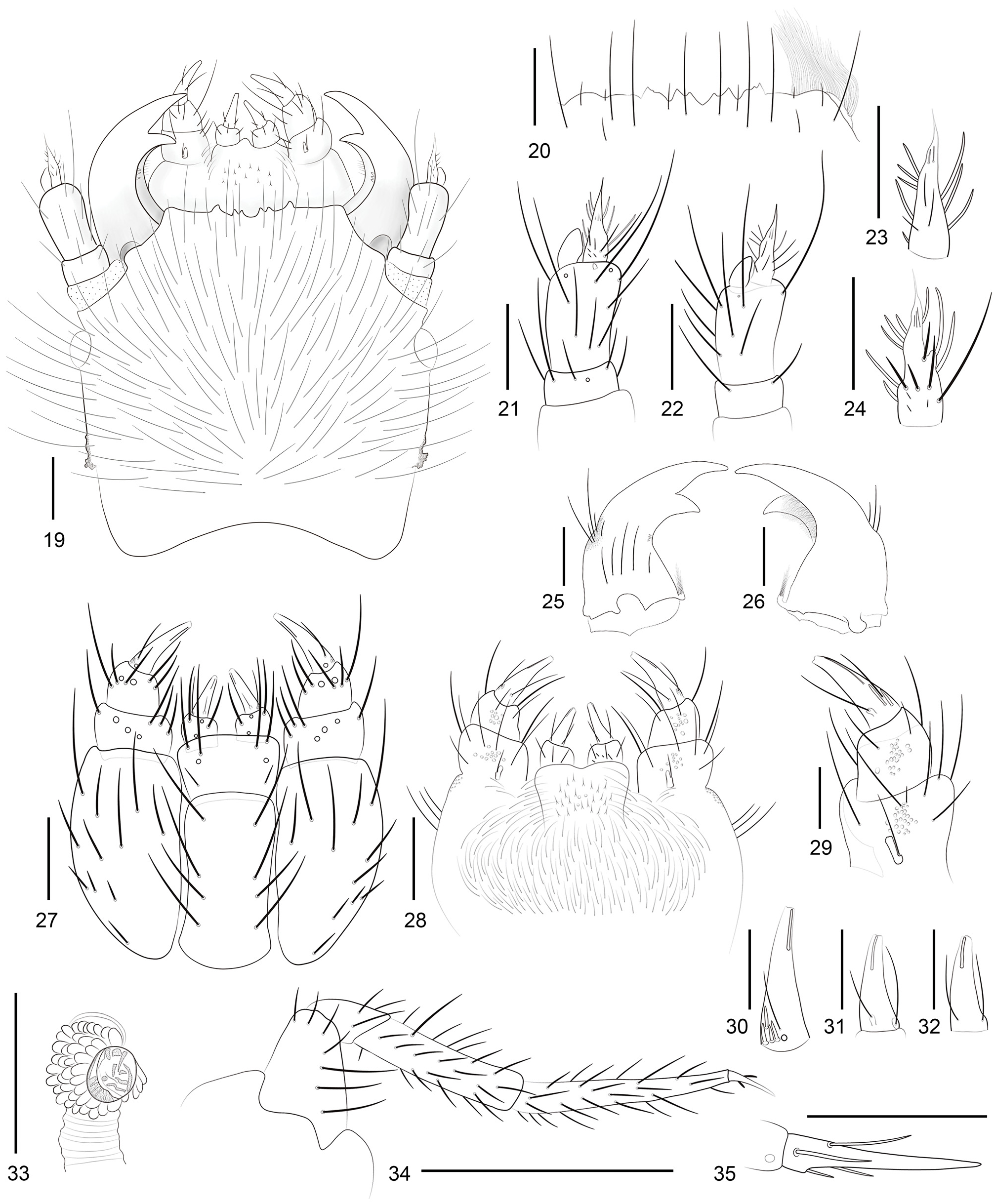
Structure. Head ( Figs 10–12 View Figs 10–13 , 19 View Figs 19–35 ) wider than long, not retracted into prothorax; prognathous, flattened dorsoventrally, vertex convex; lateral margins arched, slightly narrowed posteriorly, each side with prominent sclerotised irregular process near middle, forming straight parallel flaps; posterior edges of flaps forming irregular nodules; head constricted behind flaps, arched and slightly convergent posteriorly; posterior margin of head notched dorsally and ventrally; occipital foramen very wide; epicranial and gular sutures absent. Head with two distinct regions: anterior third of dorsal surface and nasale smooth, with sparse setae; posterior two thirds and ventral surface strongly rough, stronger medially in dorsal surface, covered with long setae and dense short pubescence; anterior and posterior regions feebly delimited by sinuous margins. One large stemma on each side, behind antennae. Nasale ( Fig. 20 View Figs 19–35 ) prominent, nearly as long as paranasal lobes, with median incision and three irregular teeth on each side, and nearly straight margin between teeth and paranasal lobe; long longitudinal dark line behind median incision; seven pairs of setae near anterior margin: three setae short and four setae long. Transverse, oblique, sclerotised plate ventrally, with deep irregular grooves on each side; median groove very wide with small rounded central protuberance; fringe of long, ramified setae, longer laterally, appearing dorsally below paranasal lobes. Antennae ( Figs 21–24 View Figs 19–35 ): antennomere I transverse with three setae and one campaniform sensillum near apex dorsally and two setae near apex ventrally; antennomere II elongate, with seven long setae, two campaniform sensilla and one sensorium dorsally and six setae and one campaniform sensillum ventrally, apex bearing antennomere III and one membranous, elongate sensorium; antennomere III elongate, narrowed apicad, shorter than II; in dorsal view, outer margin with two basal setae and group of three setae near apex; inner margin with three setae; dorsally, with group of three short setae near apex and three short setae near base, and five long setae distributed near median region; ventrally, two short setae near apex and two long setae in basal half. Mandibles ( Figs 25–26 View Figs 19–35 ) elongate, curved with well developed retinaculum; fringe of fine and long setae ventrally at base of subapical tooth; three tiny teeth dorsally at base of subapical tooth; dorsally, with 5–7 setae above acetabulum and setae of varied sizes near basal third laterally; penicillus formed by tuft of long setae. Maxillo-labial complex ( Figs 27–28 View Figs 19–35 ): stipes elongate; ventrally, with lateral margin rounded, with five long setae distributed on anterior half and six shorter on posterior half. Maxillary palpi ( Figs 29–30 View Figs 19–35 ) with three palpomeres: basal palpomere wider than long, slightly narrower than stipes at distal margin; ventrally, with three long setae near inner margin, one seta near outer margin, and three campaniform sensilla; dorsally, with five long setae; median palpomere slightly narrower than basal palpomere, narrowed apicad; with six setae and two campaniform sensilla ventrally, and two long setae dorsally; distal palpomere elongate, longer and narrower than median, gradually narrowed apicad, with one long seta and one campaniform sensillum near base ventrally, and one seta in groove near apex, outer margin with group of thick, short and decumbent setae near base; dorsally, with two long setae near base: one seta near outer margin and one pedunculate seta near inner margin; one long seta in groove near apex laterally. Galea minute, dorsal, with long lateral seta. Lacinia dorsal, formed by fringe of setae. Labium ( Figs 27–28 View Figs 19–35 ): postmentum elongate, rectangular with basal margin rounded and eight long setae; prementum wider than long, slightly narrowed basally, with anterior angles rounded, and with two long setae near each anterior angle, one short seta on each side near base and one campaniform sensillum on each side. Dorsal setae of labium wider than ventral ones. Labial palpi with two palpomeres: basal palpomere almost as wide as long, narrower at apex, with two long setae and two campaniform sensilla ventrally and with two long setae dorsally; distal palpomere ( Figs 31–32 View Figs 19–35 ) elongate, longer and narrower than basal palpomere, gradually narrowed apicad, ventrally and dorsally with two long setae on each side, near base, one of them pedunculate, and wide, decumbent and short setae near base laterally. Hypopharynx ( Fig. 28 View Figs 19–35 ) densely setose. Thorax ( Figs 10–14 View Figs 10–13 View Figs 14–18 ): pro-, meso- and metathorax wider than long, slightly wider than head, strongly constricted anteriorly; sides with broad and rounded ampullae. Prothorax with pair of large glandular pores near anterior corner, tergum with strong cordiform roughness; meso- and metathorax with glandular pores laterally, on lateral ampullae, notal roughness round and smaller. Narrow dorsal ampullae between segments. Mesothoracic spiracle ( Fig. 33 View Figs 19–35 ) circular, marginated by stout setae. Legs ( Fig. 34 View Figs 19–35 ) broadly separated, narrow, elongate and densely setose; pretarsus ( Fig. 35 View Figs 19–35 ) elongate with four short setae. Abdomen ( Figs 10–13 View Figs 10–13 , 17–18 View Figs 14–18 ): segments gradually tapering posteriorly, wide, band-like, longer posteriorly; segments I–VIII with rounded latero-dorsal ampullae bearing glandular openings, conical projecting lateral ampullae, and broad and narrow dorsal intersegmental ampullae; segment IX ( Figs 17–18 View Figs 14–18 ) narrowed anteriorly, sides broadly rounded and pair of glandular pores posteriorly on large latero-dorsal lobes; segment X membranous, bilobed; abdominal sternites I– VIII with long and thick ventral setae in pair of tufts per segment; segment IX with long ventral setae, more concentrated near apex. Abdominal spiracles small, rounded, located between lateral and dorso-lateral ampullae.
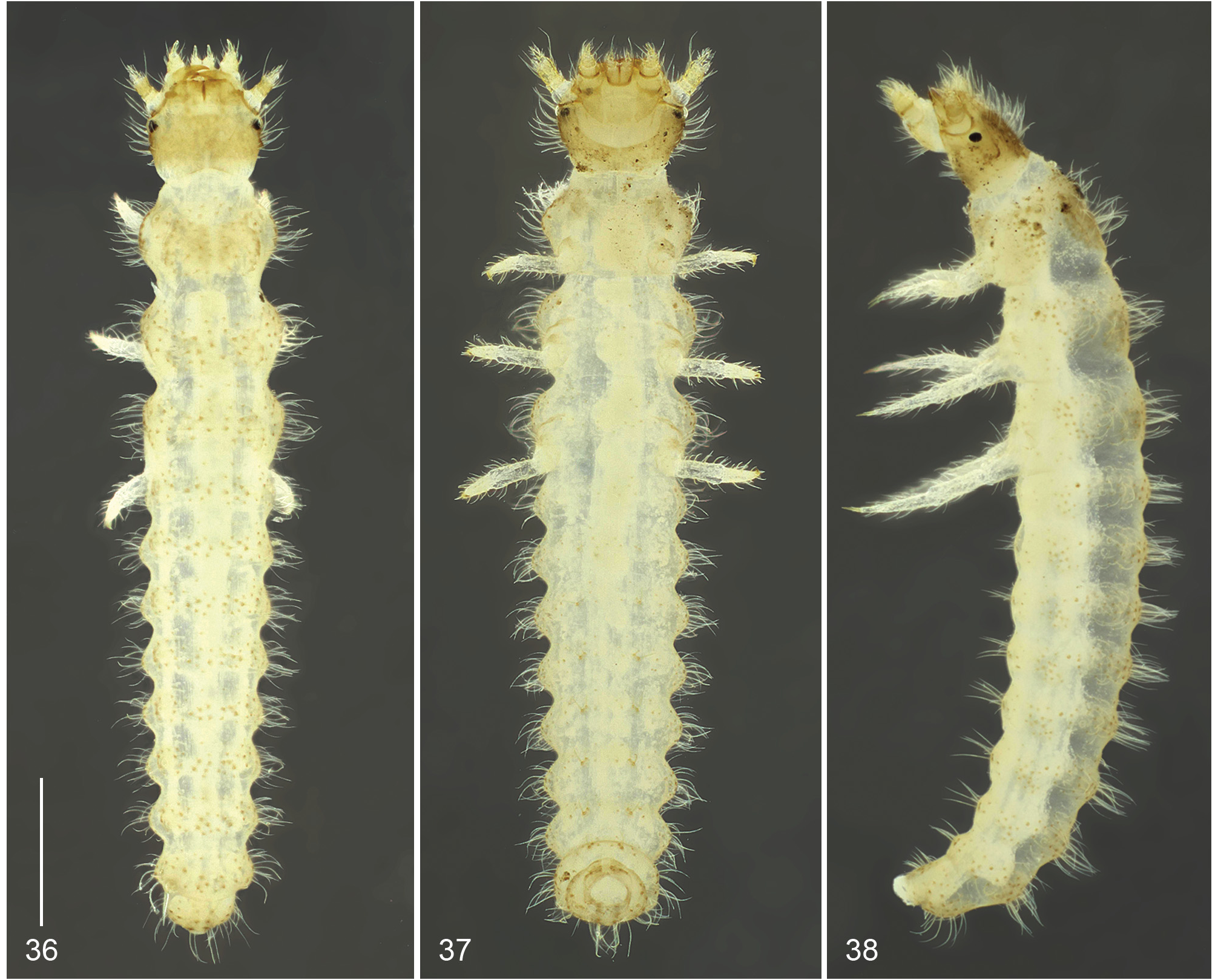
Second instar larva ( Figs 36–40 View Figs 36–38 View Figs 39–49 ). Colouration. Integument translucent, mostly whitish, except for head capsule, mouthparts, antennae, roughness of thorax, setae insertion and weakly sclerotised areas that are light brown.
Pubescence. Two layers of setae; body densely covered with very short and thin pubescence, giving the larva velvety aspect (seen under high magnification), and tufts of long and thick dorsal, lateral and ventral setae.
Structure. Head capsule wider than long; lateral margins slightly narrowed basally, each side with prominent sclerotized irregular process near middle; head densely covered with long and dense setae, except for narrow basal band. Stemmata prominent. Nasale with median incision and three asymmetrical and sharpened teeth on each side; two short and four long pairs of setae near anterior margin. Epipharynx with short fringe of ramified setae on each side seen from above. Thorax wider than long, slightly wider than head, each segment constricted anteriorly and posteriorly; sides with broad and rounded ampullae. Prothorax with pair of large glandular pores near anterior corner, tergum with weakly sclerotised area, rough; meso- and metathorax with glandular pores laterally, on lateral ampullae, tergal roughness weakly defined. Narrow dorsal ampullae between segments. Abdomen slightly tapering posteriorly, lateral ampullae rounded and laterally projected; dorsal and dorso-lateral ampullae weakly defined.
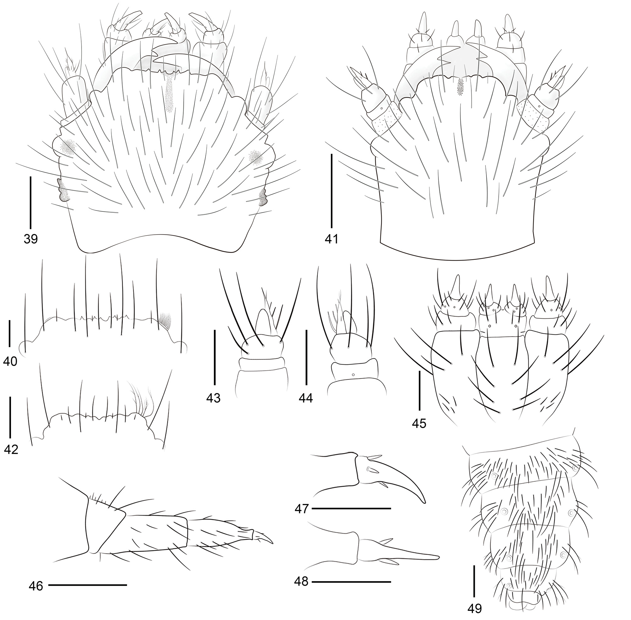
First instar larva ( Figs 41–49 View Figs 39–49 ). Colouration. Integument translucent, entirely whitish, except for light brown apex of mandibles.
Pubescence. body covered with very short and dense pubescence and medium length setae; each thoracic and abdominal segment with three pairs of dorsal, and two or three pairs of lateral long setae.
Structure. Head capsule ( Fig. 41 View Figs 39–49 ) almost as long as wide; lateral margins almost straight; stemmata prominent. Head covered with moderately dense pubescence of long setae, except in posterior fourth. Nasale ( Fig. 42 View Figs 39–49 ) irregular, slightly sinuous; each side with six pairs of setae near anterior margin and one at base dorsally; epipharynx with five long ramified setae on each side. Antennae ( Figs 43–44 View Figs 39–49 ) short and very wide; antennomere I transverse, band-like, with one campaniform sensillum dorsally; antennomere II wider than long, with three long dorsal setae and four ventral setae (some surpassing antennal length) and apex bearing antennomere III and one elongate, membranous sensorium; antennomere III elongate, gradually narrowed apicad, with four latero-internal setae and one dorsal seta. Maxillo-labial complex ( Fig. 45 View Figs 39–49 ): stipes elongate with eight setae, four basal setae shorter. Palpi very robust, except for distal palpomere; three palpomeres: basal palpomere wider than long, band-like with three long setae; median palpomere wider than long, strongly narrowed apicad, with eight setae and two campaniform sensilla; distal palpomere elongate and narrow, narrower apicad. Labium: postmentum elongate with eight long setae; prementum wider than long, bandlike, each side with one short and two long setae and one campaniform sensillum. Palpi with two palpomeres: basal palpomere very wide, band-like with three long setae and two campaniform sensilla; distal palpomere elongate and strongly narrower than basal palpomere, slightly narrowed apicad. Legs ( Fig. 46 View Figs 39–49 ) robust with apex of tibia abruptly narrowed, setae sparse; pretarsus ( Figs 47–48 View Figs 39–49 ) with very short and stout setae. Integument with setae moderately long and dense, more concentrated dorsally near middle of each tergite and with scattered longer setae; ramified setae on apex of abdomen ( Fig. 49 View Figs 39–49 ).
Eggs ( Figs 6–9 View Figs 6–9 ). Description. Eggs light orangish, progressively turning whitish when fertilized; slightly oval, surface smooth, without ornamentations; eggs adhering to each other, disposed in large masses on the soil.

| MZSP |
Sao Paulo, Museu de Zoologia da Universidade de Sao Paulo |
No known copyright restrictions apply. See Agosti, D., Egloff, W., 2009. Taxonomic information exchange and copyright: the Plazi approach. BMC Research Notes 2009, 2:53 for further explanation.
|
Kingdom |
|
|
Phylum |
|
|
Class |
|
|
Order |
|
|
Family |
|
|
Genus |