
Benedenia
|
publication ID |
https://doi.org/ 10.5281/zenodo.275591 |
|
DOI |
https://doi.org/10.5281/zenodo.6203158 |
|
persistent identifier |
https://treatment.plazi.org/id/82668798-FF87-FFE7-FF33-5E9FFA47FA45 |
|
treatment provided by |
Plazi |
|
scientific name |
Benedenia |
| status |
|
Key to the described species of Benedenia View in CoL
This key is accurate for the 25 currently described species of Benedenia . Numerous undescribed species of the genus exist and it is possible that many are likely to be incorrectly identified as described species if this key is used to identify them. Continual growth throughout their life, temperature dependent growth and host induced morphological variation in capsalids (see Discussion) are complicating factors. We strongly encourage the deposition of voucher specimens in recognised, curated museum collections of any capsalid taxa encountered ( Whittington 2004).
1a. Terminal part of penis adorned with sclerite or papillae ............................................................................................. 2
1b. Penis unadorned .......................................................................................................................................................... 4
2a. Penis with terminal sclerite ........................................................................................................................................ 3
2b. Penis with three terminal, conical papillae (see Fig. 24A of Whittington et al. 2001) ............................. B. jaliscana
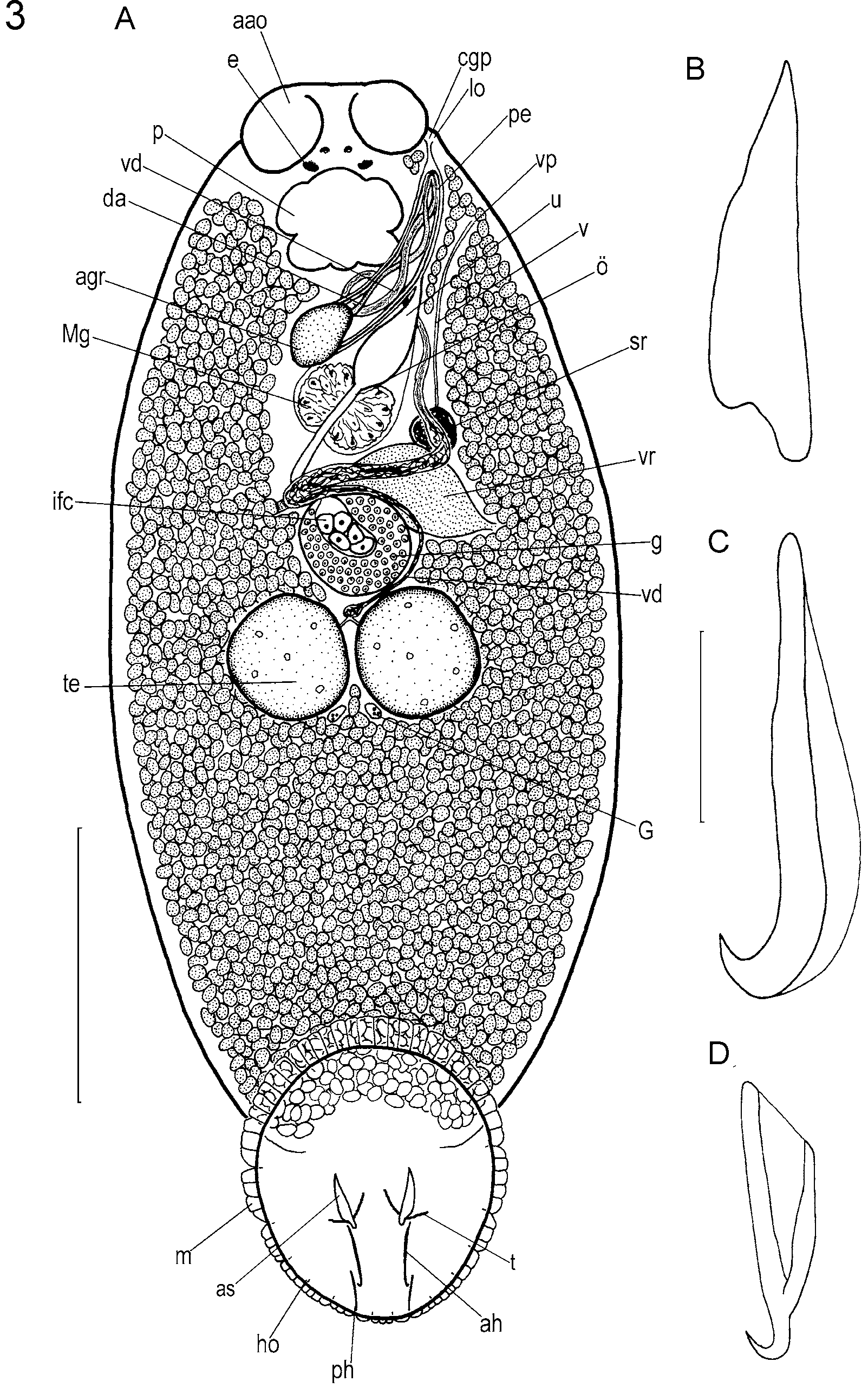
3a. Vaginal pore posterior to testes ( Figs. 1 View FIGURE 1 A, 2 in present study)....................................................................... B. ernsti View in CoL
3b. Vaginal pore anterior to testes, posterior to common genital pore................................................................ B. rohdei View in CoL
4a. Vaginal pore anterior to common genital pore (Fig. 10 of Whittington et al. 2001) ................................................. 5
4b. Vaginal pore adjacent or posterior to common genital pore but anterior to germarium (e.g. Fig. 12A-D of Whitting- ton et al. 2001) ............................................................................................................................................................ 7
5a. Anterior hamuli span almost half total length of haptor (Fig. 9A of Whittington et al. 2001); vaginal pore ventral .. ............................................................................................................................................................ B. acanthopagri View in CoL
5b. Anterior hamuli less than half total length of haptor (Fig. 9E, I of Whittington et al. 2001); vaginal pore dorsal .. 6
6a. Posterior hamuli invested in muscle fibres (Fig. 9H of Whittington et al. 2001); single lobe associated with com- mon genital pore and vaginal pore (Fig. 10B of Whittington et al. 2001) ...................................... B. anticavaginata View in CoL
6b. Posterior hamuli not invested in muscle fibres (Fig. 9L of Whittington et al. 2001); pair of lobes associated with common genital pore and single lobe associated with vaginal pore (Fig. 10C of Whittington et al. 2001). B. lutjani View in CoL
7a. Separate uterine, male and vaginal pores .................................................................................................................... 8
7b. Uterine and male copulatory organ pores common forming common genital pore and separate vaginal pore ......... 9
8a. Large (> 5 mm total length) species with accessory sclerites placed posteriorly on haptor (Fig. 4A of Whittington et al. 2001); long tapering penis .................................................................................................................... B. sciaenae View in CoL
8b. Small (<2.5 mm total length) species with broad anterior hamuli and accessory sclerites (Fig. 31A, B of Whitting- ton et al. 2001) .......................................................................................................................................... B. synagris View in CoL
9a. Vaginal pore in mid-body between common genital pore and germarium (Fig. 12A, B of Whittington et al. 2001). .......................................................................................................................................................................... B. ovata View in CoL
9b. Vaginal pore at a level between pharynx and common genital pore (e.g. Fig. 12B-D of Whittington et al. 2001). 10
10a. Penis with distinctive teat-shaped tip (Fig. 25A of Whittington et al. 2001) ........................................ B. monticellii View in CoL
10b. Penis without teat-shaped tip, but with blunt or tapering tip ................................................................................... 11
11a. Accessory sclerites with distal hooks or with three points ...................................................................................... 12
11b. Accessory sclerites with tapering, pointed distal tips ............................................................................................. 13
12a. Distal tips of accessory sclerites a recurved hook (Fig. 17A, B, E of Whittington et al. 2001) ................ B. elongata View in CoL
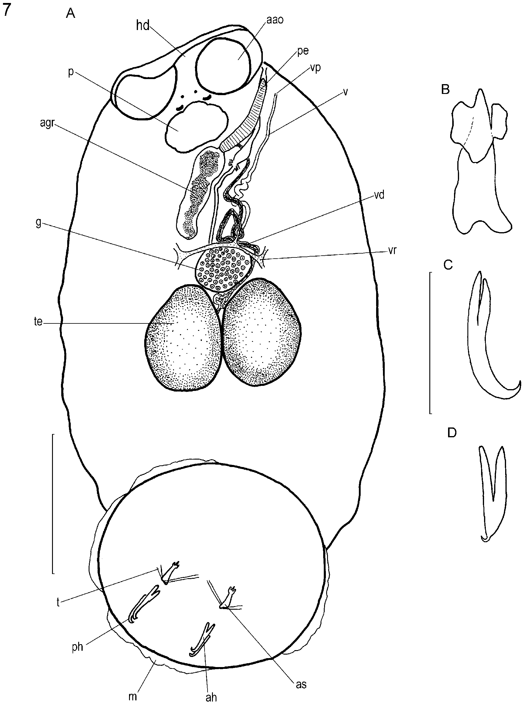
12b. Distal tips of accessory sclerites trident-like, with three points ( Fig. 7 View FIGURE 7 A, B of present study)............... B. ishikawae View in CoL
13a. Anterior hamuli with very broad roots; distal tips strongly recurved (Fig. 23B of Whittington et al. 2001) ............... ................................................................................................................................................................ B. innobilitata View in CoL
13b. Anterior hamuli without very broad roots................................................................................................................ 14
14a. Small lobe near vaginal pore (Fig. 18 of Whittington et al. 2001) ......................................................... B. epinepheli View in CoL
14b. Lobe near vaginal pore absent................................................................................................................................... 15
15a. Body broad, almost circular; anterior and posterior hamuli tiny relative to haptor (Fig. 29A, C, D of Whittington et al. 2001) .......................................................................................................................................................... B. sekii View in CoL
15b. Body elongate, comparatively narrow; anterior and posterior hamuli not tiny relative to haptor ............................ 16
16a. Anterior and posterior hamuli not overlapping ........................................................................................................ 17
16b. Anterior and posterior hamuli overlapping .............................................................................................................. 18
17a. Anterior hamuli recurved distally, with slender shafts (Fig. 21 of Whittington et al. 2001) ................... B. hendorffii View in CoL
17b. Anterior hamuli recurved distally with comparatively stout shafts (Fig. 30A, C, E, G of Whittington et al. 2001).... ..................................................................................................................................................................... B. seriolae View in CoL
18a. Anterior attachment organs strongly hooded (Fig. 22B of Whittington et al. 2001); wide vitellarium-free margin to body; medium-sized (2.5–7.5 mm total length) species ............................................................................ B. hoshinai View in CoL
18b. Anterior attachment organs weakly hooded or not hooded; vitellarium extends to near margin of body ................ 19
19a. Medium-large species (>2.5 mm total length, e.g. Fig. 3 View FIGURE 3 A of Whittington et al. 2001) with relatively large anterior attachment organs.................................................................................................................................... B. pompatica View in CoL
19b. Small-medium (<7.5 mm) species (e.g. Fig. 3 View FIGURE 3 C of Whittington et al. 2001) with relatively small anterior attachment organs ........................................................................................................................................................................ 20
20a. Distal tips of accessory sclerites located in posterior half of haptor (Fig. 16E of Whittington et al. 2001) ... B. scari View in CoL
20b. Distal tips of accessory sclerites located near centre of haptor ................................................................................ 21
21a. Penis broad proximally, with pointed or tapering tip ............................................................................................... 22
21b. Penis not broad proximally, with blunt tip (Fig. 15 of Whittington et al. 2001) ...................................................... 23
22a. Pair of prominent lobes near common genital pore; accessory sclerites with curved distal region; tip of penis pointed (Figs. 5A and 6A of present study) ............................................................................................. B. haywardi View in CoL
22b. No lobes near common genital pore; accessory sclerites straight (Figs. 16C and 18G of Whittington et al. 2001); tip of penis tapering but not pointed (Fig. 20 of Whittington et al. 2001) ................................................ B. hawaiiensis View in CoL
23a. Prominent wavy musculature near haptor margin; anterior and posterior hamuli of similar length (Figs. 13, 14, 16A, 19B, C of Whittington et al. 2001) .............................................................................................................. B. bodiani View in CoL
23b. Prominent wavy musculature absent from haptor margin ........................................................................................ 24
24a. Marginal valve comprising a single large lobe between positions of each hooklet (Fig. 16D of Whittington et al. 2001) ................................................................................................................................................................. B. lolo View in CoL
24b. Marginal valve comprising multiple lobes between positions of hooklets ( Fig. 3 View FIGURE 3 A of present study; adults very small (<1.5 mm in total length) (e.g. Fig. 3 View FIGURE 3 C of Whittington et al. 2001)..................................................... B. fieldsi View in CoL

No known copyright restrictions apply. See Agosti, D., Egloff, W., 2009. Taxonomic information exchange and copyright: the Plazi approach. BMC Research Notes 2009, 2:53 for further explanation.
|
Kingdom |
|
|
Phylum |
|
|
Class |
|
|
Order |
|
|
Family |
|
Kingdom |
|
|
Phylum |
|
|
Class |
|
|
Order |
|
|
Family |