
Donuea collustrata Bosselaers & Dierick
|
publication ID |
https://doi.org/ 10.5281/zenodo.293907 |
|
DOI |
https://doi.org/10.5281/zenodo.6201660 |
|
persistent identifier |
https://treatment.plazi.org/id/832C87E6-4C7F-FF86-7EBF-E985978EE80A |
|
treatment provided by |
Plazi |
|
scientific name |
Donuea collustrata Bosselaers & Dierick |
| status |
sp. nov. |
Donuea collustrata Bosselaers & Dierick sp. nov.
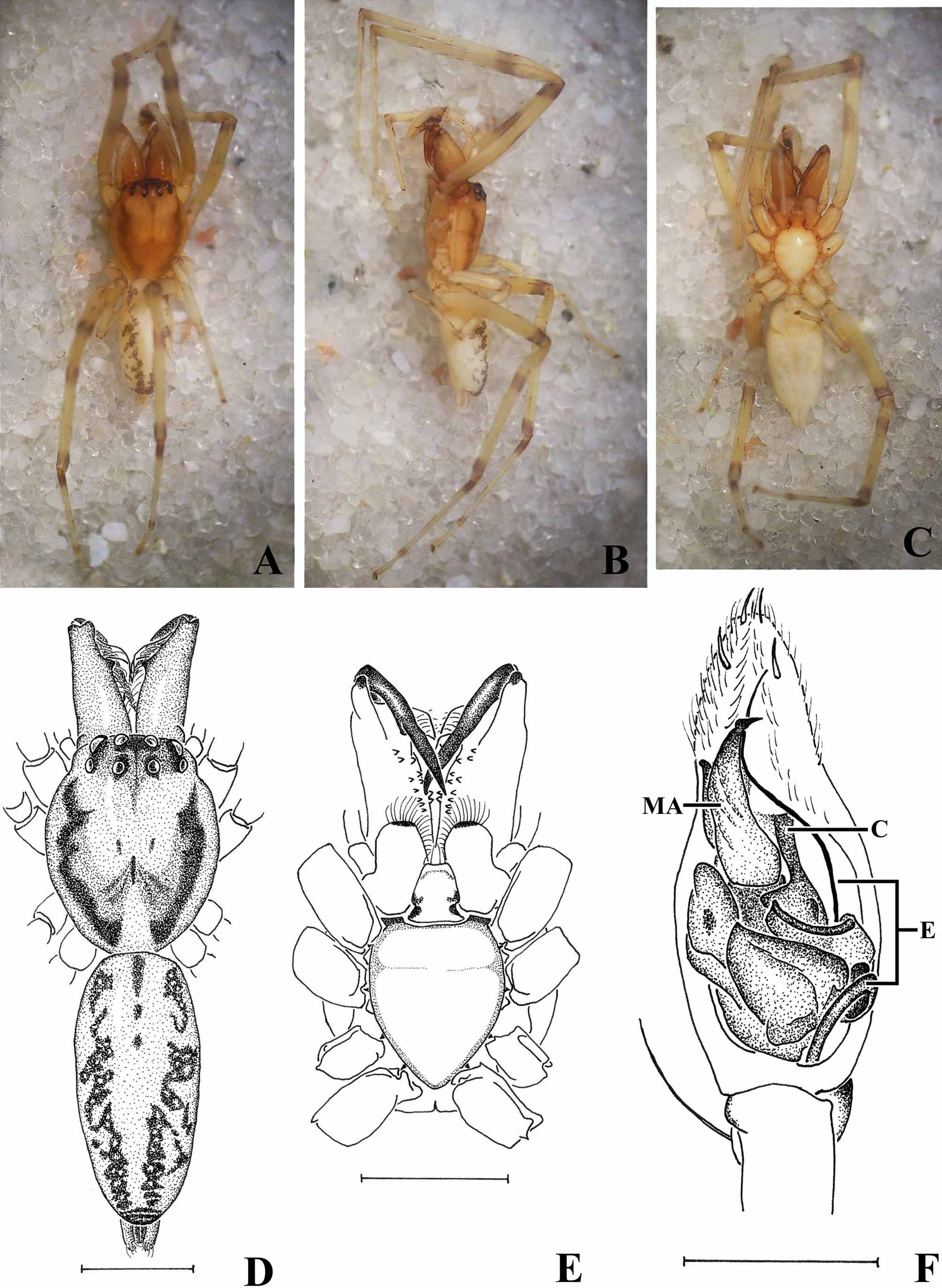
Figs. 1 A–E View FIGURE 1 A – E ; 2 A–F; 3 A–M; 4 A–I.
Type material. Holotype male: Madagascar, Foulpointe, forest on clay soil, beating vegetation, July 1995, A. Pauly leg. ( MRAC 206172). Paratype male, in copal from the Sambava area, NE Madagascar, MRAC.
Diagnosis. The species can be distinguished from the type species Donuea decorsei Simon, 1903 , by its bifid RTA as opposed to a simple one ( Simon 1903: 138), its large, bifurcated MA, a simple, pointed conductor situated ventrally instead of dorsally of MA and its shorter embolus which does not describe a circular loop before passing behind the tegulum ( Figs. 1 View FIGURE 1 A – E F, 4 C, F, I).
Description. Body length without chelicerae (holotype): 4.34, with chelicerae: 5.76. Carapace length: 1.81; w: 1.55, oval, yellow brown, darker in eye region, and with two brown lateral bands ( Fig. 1 A–B, D View FIGURE 1 A – E ). Fovea short and deep, pale brown, length 0.08, anterior end 1.14 from front. Eight eyes in two rows, ringed with black, AER w: 0.82, recurved from above, slightly procurved from front, PER w: 0.89, procurved from above, strongly procurved from front ( Figs. 1 A, D View FIGURE 1 A – E , 3 M View FIGURE 3 A – M ). MOQ depth: 0.32, anterior w: 0.37, posterior w: 0.42. All eyes subequal, ALE oval, others circular. All four median eyes with dark retina restricted to median 2/3. Anterior median eyes slightly closer to each other than to ALE. Eyes of PER equidistant, separated by about twice their diameter. ALE and PLE touching, on common tubercle. Clypeus vertical, 1/3 of diameter of AME. Chilum orange-brown, sclerotised, split ( Fig. 3 M View FIGURE 3 A – M ). Chelicerae large, yellow brown, elongated and pointing forward, parallel to the longitudinal axis of the body ( Figs. 1 A–D View FIGURE 1 A – E , 2 C, F View FIGURE 2 A – H , 3 A View FIGURE 3 A – M ). One large kneeshaped seta and a row of long, curved setae on ACR, cheliceral fangs brown. Six teeth on ACR, largest one closest to fang base, followed by an isolated very small tooth and two pairs of small teeth separated by a small distance. Seven teeth on PCR, a pair of medium sized teeth close to fang base, followed at some distance by five small teeth in a row. Sternum length: 1.05; w: 0.95, smooth, yellow, elongated and shield-shaped, with pale broad and blunt ICS between legs I–II and II-III, and thin, brown and pointed PCT facing all coxae ( Fig. 1 C, E View FIGURE 1 A – E ). Pleural bars ( Bosselaers & Jocqué 2002: 247, fig. 1P; Simon 1892: 11, fig. 29) yellow-brown continuous strips, weakly protruding between coxae. Labium brown, subtrapezoidal, as long as it is wide at its base, with white, thickened anterior rim. Endites subrectangular, with apical hair tuft and serrula, no oblique depression ( Fig. 1 C, E View FIGURE 1 A – E ). Abdomen cream, unicolorous ventrally and with two lateral, purple brown bands dorsally ( Fig. 1 A–B, D View FIGURE 1 A – E ). ALS contiguous, conical, with short, blunt apical segment. PMS very thin and slender, PLS subcylindrical, mottled with grey, with pale, rounded apical segment ( Figs. 1 B–D View FIGURE 1 A – E , 3 A View FIGURE 3 A – M ). Legs yellow, fe, ti and mt ringed with grey basally and/or terminally ( Figs. 1 A–C View FIGURE 1 A – E , 2 B–C, F View FIGURE 2 A – H ). No trochanter notch, no retrocoxal hymen ( Raven 1998; Bosselaers & Jocqué 2002), patellar indentation long and narrow, ¾ of pa length. Metatarsi III and IV with sparse ve terminal preening brush. Tarsi with two claws and dense claw tufts. Leg formula 1423. Leg spination ( Fig. 2 E View FIGURE 2 A – H ): fe: palp do 0-1-1 rlv 1-1-1-1-1-0; I pl 0-1-1-1 do 0-1-1-1; II pl 0- 0-0-1 do 0-1-1-1; III do 0-1-1-2; IV do 1-1-1-1-2-2; pa: palp pl 1; ti: palp pl 2-0-0; I pl 0-1-1 ve 2-2-2-2-2; II ve 2-2-2-2; III rl 0-1-0 plv 1-1-0; IV rl 0-1-1-0 plv 1-1-0 rlv 0-0-1; mt: I pl 1-0-0 ve 2-2-2; II ve 2-2-0; III pl 0- 1-0 rl 1-0-0 ve 2-0-0; IV rl 0-0-1 ve 2-0-2.
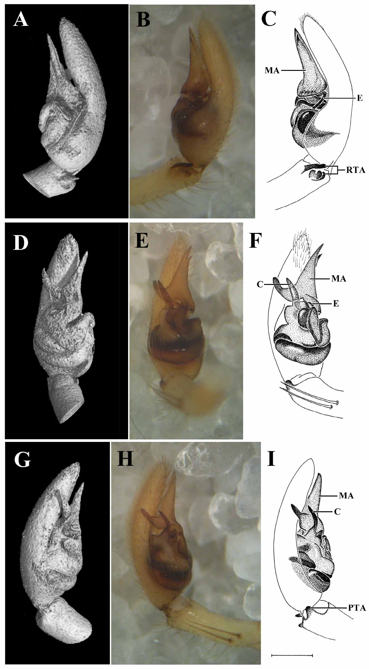
Male palp with a simple, blunt PTA ( Figs. 3 D, E, L View FIGURE 3 A – M , 4 F, I View FIGURE 4 A – I ) and a bifid RTA consisting of a long, pointed do part and a cup-shaped ve part ( Figs. 3 I, J, K, L View FIGURE 3 A – M , 4 A–C View FIGURE 4 A – I ). Palpal tarsus elongated and piriform, with a large, bifid, flag-shaped MA ( Figs. 3 C–F View FIGURE 3 A – M , 4 D–I View FIGURE 4 A – I ), a simple, blunt-tipped conductor situated ventrally of the pl branch of the MA ( Figs. 3 C–F, L View FIGURE 3 A – M , 4 D–I View FIGURE 4 A – I ) and a long embolus emerging from below the rl branch of the MA, proceeding basally and subsequently passing behind the tegulum ( Figs. 3 B–C, G–K View FIGURE 3 A – M , 4 A–C, F View FIGURE 4 A – I ).
Female. Unknown.
Etymology. The species epithet collustrata , irradiated, refers to the X-ray tomography technique used as an aid in the description of the present species.
Remarks. The specimen preserved in copal clearly is conspecific with the field-captured holotype. It has the same size (body length 4.21) and general morphology, and both specimens are identical with respect to a number of diagnostic details. Leg spination of the copal specimen is identical to the holotype although a few spines are missing, probably having been lost when the animal got caught in the sticky resin ( Figs. 2 E–F View FIGURE 2 A – H ). The copal specimen has lost most of its original colour, the abdomen being partly transparent and showing two dark spots in the anterior half, presumably corresponding with the book lungs. However, the leg annulations have been mostly preserved and coincide with those of the field-captured animal ( Figs. 1 A–C View FIGURE 1 A – E , 2 B, C, F View FIGURE 2 A – H ). Most significantly, palpal structure of both specimens is identical in minute detail, as can be proven by juxtaposing stereomicroscope observations of the field-captured holotype with X-ray micro-CT scans of the copal preserved specimen ( Fig. 4 A–I View FIGURE 4 A – I ).
The assignment of Donuea to Liocranidae is debatable, and it can indeed be argued that the genus would be better placed in Clubionidae . However, a formal transfer is outside the scope of the present contribution.


| MRAC |
Musée Royal de l’Afrique Centrale |
No known copyright restrictions apply. See Agosti, D., Egloff, W., 2009. Taxonomic information exchange and copyright: the Plazi approach. BMC Research Notes 2009, 2:53 for further explanation.
|
Kingdom |
|
|
Phylum |
|
|
Class |
|
|
Order |
|
|
Family |
|
|
Genus |