
Blennocampinae, Konow, 1890
|
publication ID |
https://doi.org/ 10.5733/afin.053.0204 |
|
DOI |
https://doi.org/10.5281/zenodo.7917759 |
|
persistent identifier |
https://treatment.plazi.org/id/836E1D24-BB3A-FF88-FEE2-06FBBA51FC0E |
|
treatment provided by |
Felipe |
|
scientific name |
Blennocampinae |
| status |
|
Key to genera of Afrotropical Blennocampinae
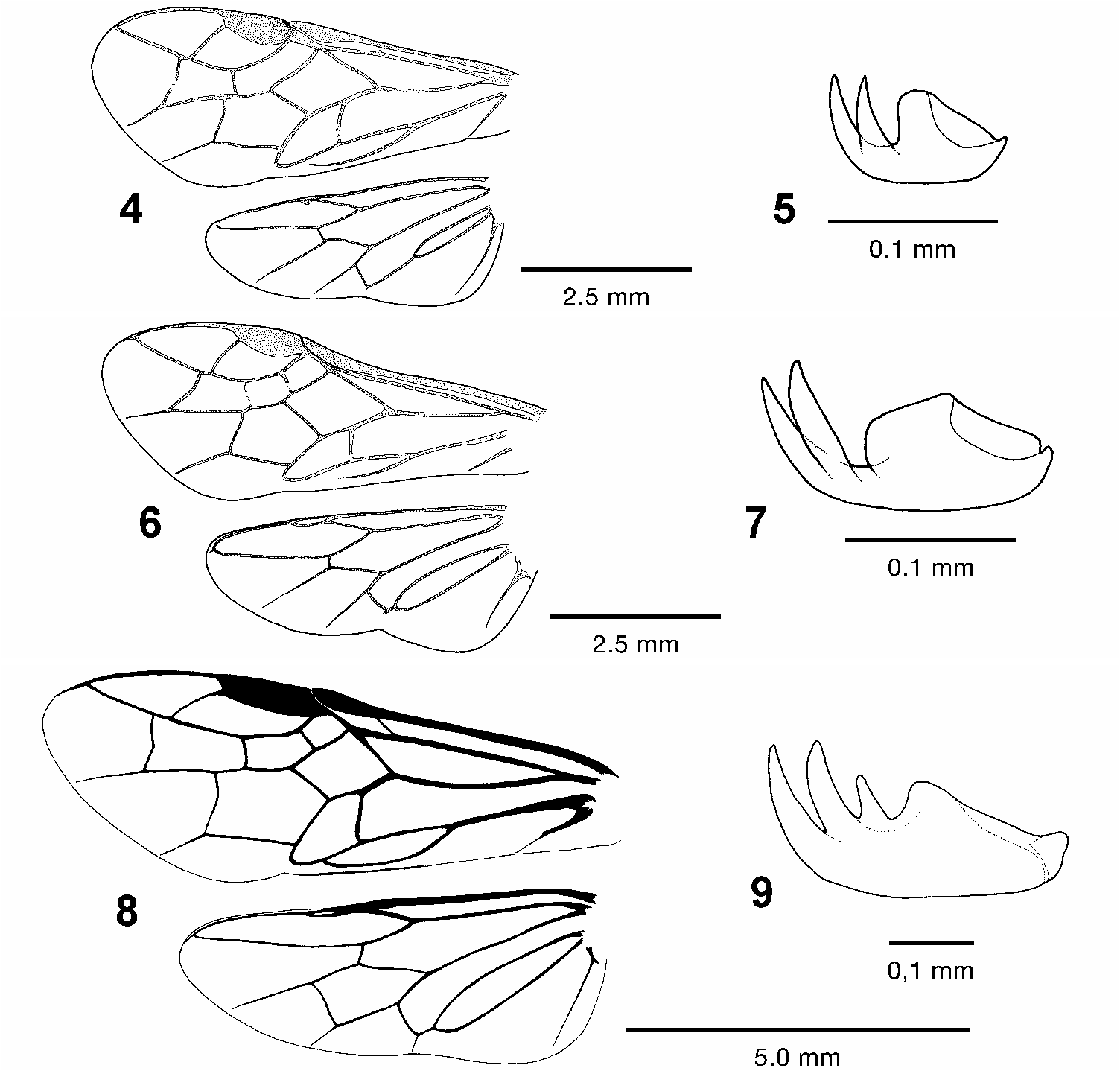
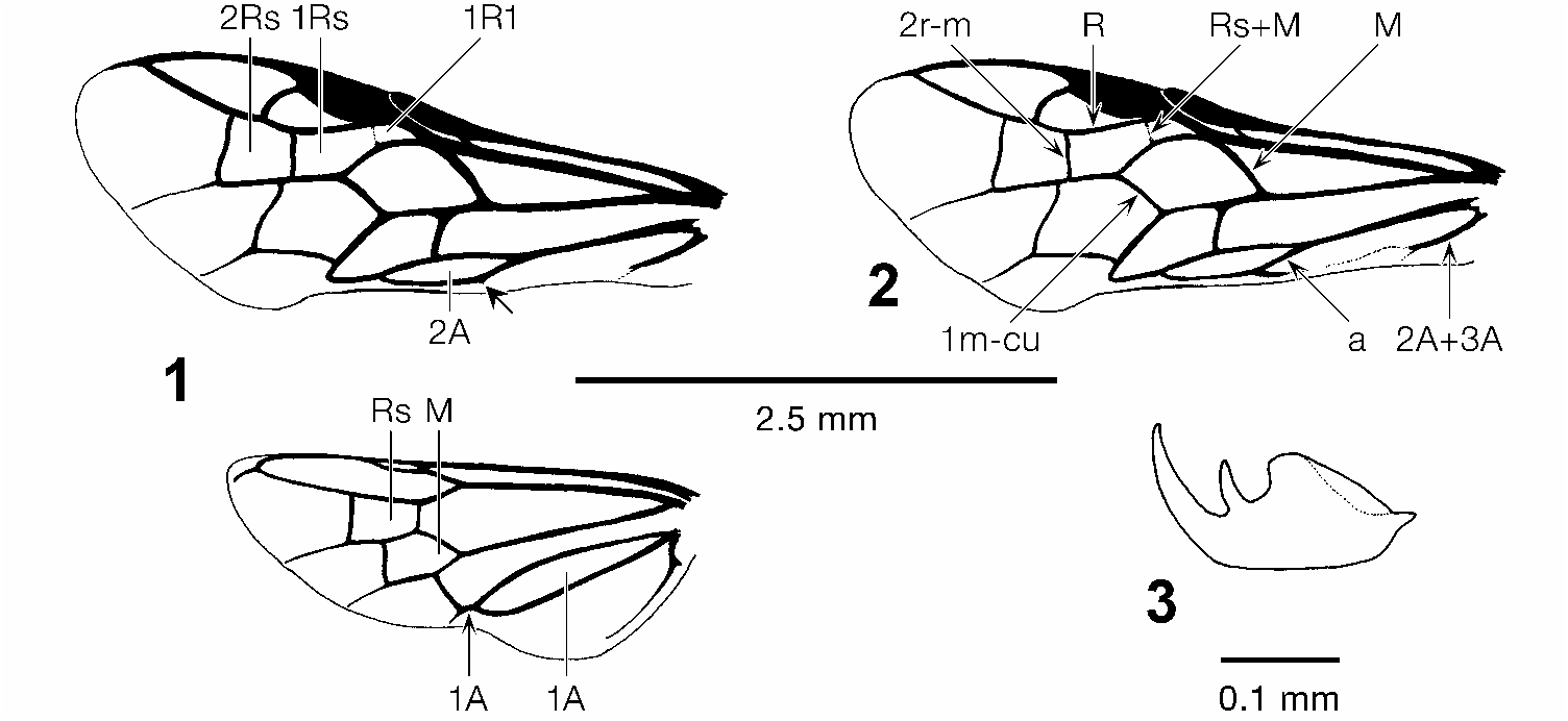
1 Hind wing with cell M ( Fig. 8 View Figs 4–9 ) or both Rs and M ( Fig. 1 View Figs 1–3 ) present..........................2
– Hind wing with cells Rs and M absent.....................................................................3
2 Hind wing only with M ( Fig. 8 View Figs 4–9 ); tarsal claws tridentate, with large basal lobe ( Fig.9 View Figs 4–9 ); mesepisternum without transverse groove or suture ..................... Trisodontophyes
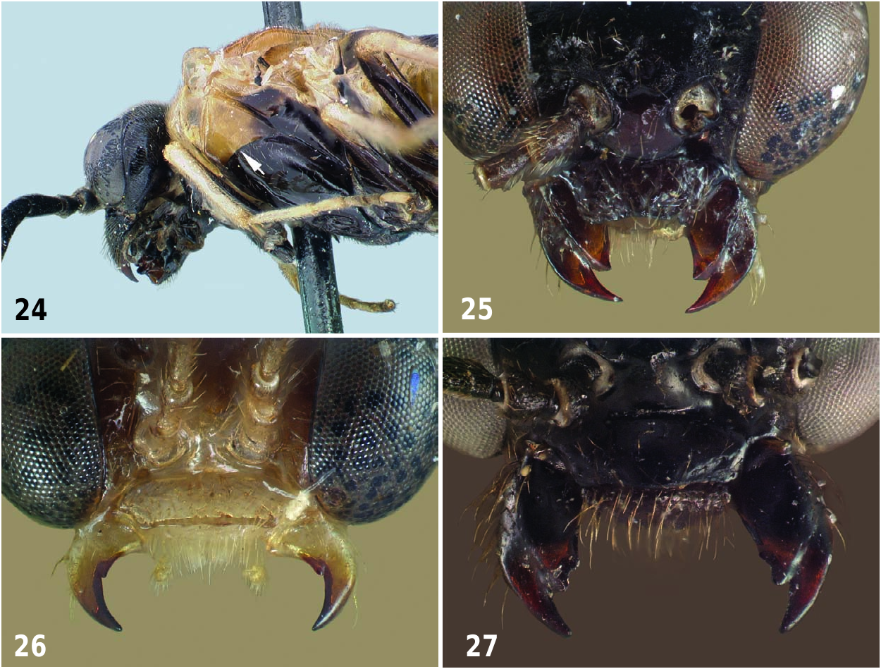
– Hind wing with both Rs and M both ( Fig. 1 View Figs 1–3 ); tarsal claws with one subapical tooth and enlarged basal lobe ( Fig. 3 View Figs 1–3 ); upper half of mesepisternum separated from lower by a transverse groove or suture ( Fig. 24 View Figs24–27 )..................................................... Distega
3 Fore wing with crossvein Rs+M absent, 1R1 and 1Rs are fused, thus there are only two cells ( Fig. 4 View Figs 4–9 ); anal cell of hind wing as long as anal vein 1A ( Fig. 4 View Figs 4–9 )................. ............................................................................................................. Aethiocampa
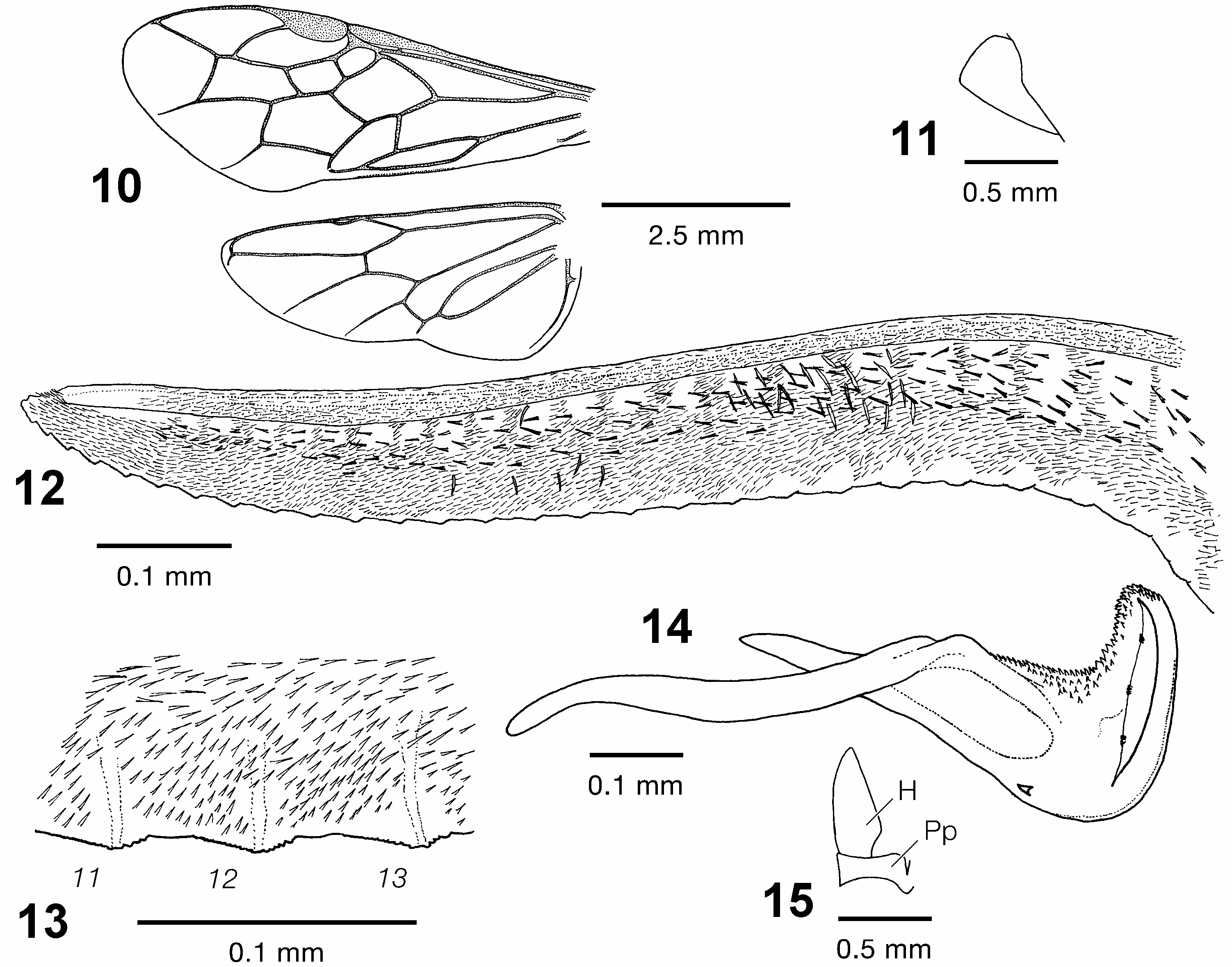
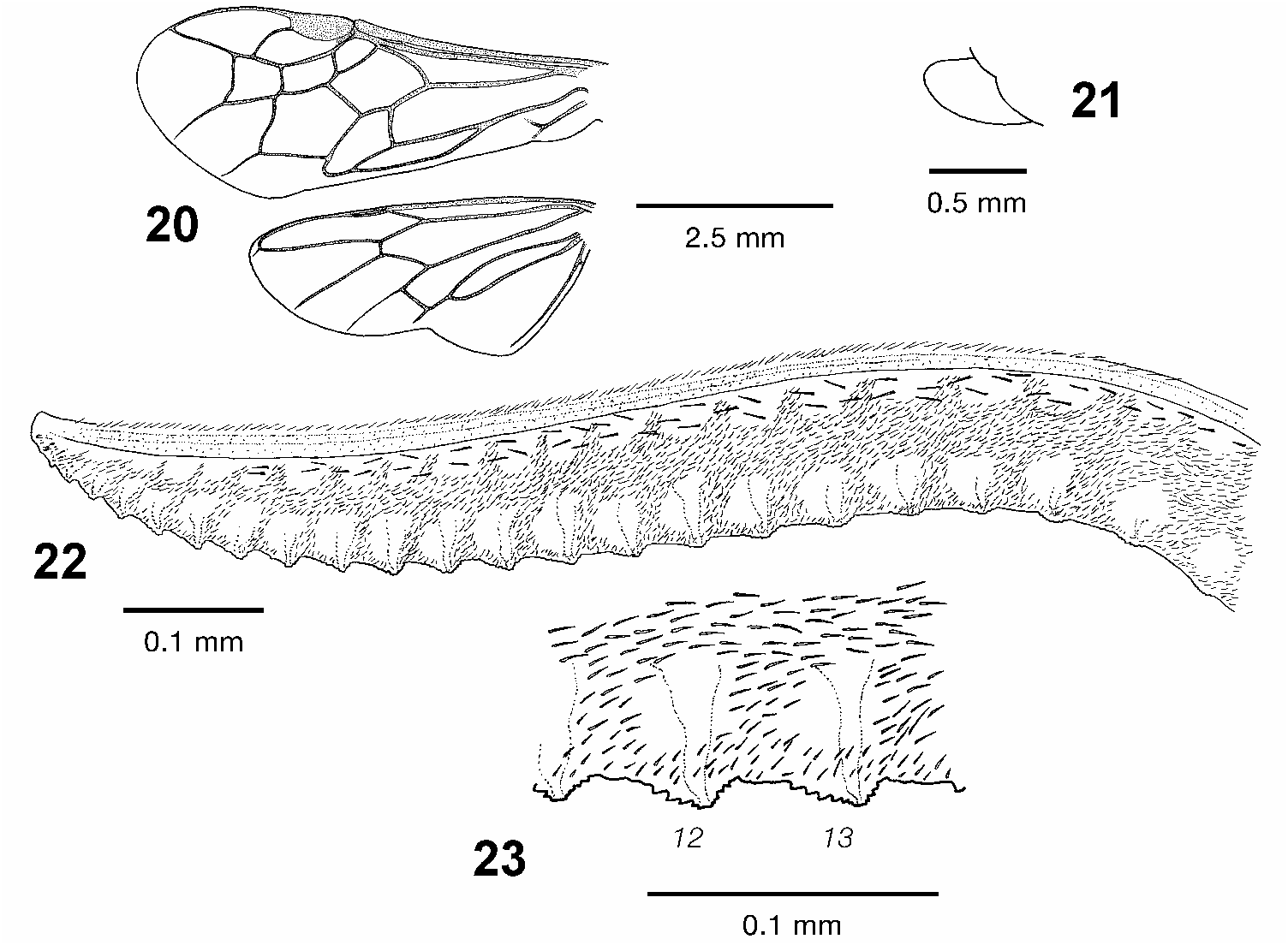
– Fore wing with crossvein Rs+M present, 1R1, 1Rs and 2Rs are present, thus there are three cells ( Figs 6 View Figs 4–9 , 10 View Figs 10–15 , 20 View Figs 20–23 ); anal cell of hind wing with conspicuously shorter petiole ( Figs 6 View Figs 4–9 , 10 View Figs 10–15 , 20 View Figs 20–23 )..............................................................................................4
4 Tarsal claws with conspicuously flattened basal lobe, about half as high as inner subapical tooth ( Fig. 7 View Figs 4–9 ); anal vein 2A of fore wing gradually obliterated apically, so anal cell (2A) open below; anal cell of hind wing very short, nearly impetiolate ( Fig. 6 View Figs 4–9 ) ..................................................................................................................... Tesslinia
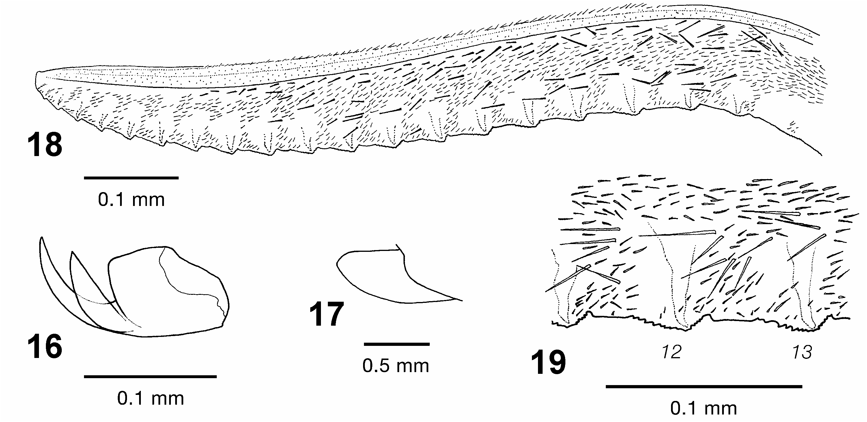
– Tarsal claws with enlarged basal lobe, nearly equal in height to inner subapical tooth ( Fig. 16 View Figs 16–19 ); anal vein 2A of fore wing completely developed, anal cell closed; anal cell of hind wing petiolate, about equal to width of anal cell ( Figs 10 View Figs 10–15 , 20 View Figs 20–23 )...................... ................................................................................................................ Durbadnus
No known copyright restrictions apply. See Agosti, D., Egloff, W., 2009. Taxonomic information exchange and copyright: the Plazi approach. BMC Research Notes 2009, 2:53 for further explanation.
|
Kingdom |
|
|
Phylum |
|
|
Class |
|
|
Order |
|
|
Family |