
Diadema antillarum (Philippi, 1845)
|
publication ID |
https://doi.org/ 10.11646/zootaxa.3636.1.6 |
|
publication LSID |
lsid:zoobank.org:pub:FF3A24CC-6545-4B77-83C5-2503143E7F16 |
|
DOI |
https://doi.org/10.5281/zenodo.6145096 |
|
persistent identifier |
https://treatment.plazi.org/id/84171C00-D225-F637-FC94-79A9166EFCAF |
|
treatment provided by |
Plazi |
|
scientific name |
Diadema antillarum (Philippi, 1845) |
| status |
|
Diadema antillarum (Philippi, 1845) View in CoL
Figs 1–4 View FIGURE 1 View FIGURE 2 View FIGURE 3 View FIGURE 4 , tables 1–3
Cidaris (Diadema) antillarum Philippi, 1845 . Arch. F. Naturg., 11 (1), p. 355.
Diadema antillarum, A. Agassiz 1863 . Bull. M.C.Z., 1, p.19. Nutting, 1895, Bull. Univ. Iowa, Lab. Nat. Hist. (3), p.224, unnumbered plate, fig.1 (young Diadema , labelled Aspidodiadema sp.)
Neotype diagnosis. Test and spines typically black with a red tinge. However, spines vary in colour, from white/ brown to black. Iridophores occur as a pentamerous ring around the apical disc. Arch-shaped depressions are found on the apical disc along the inner edges of genital plates only in juveniles and young adults. This character fades with age. In transverse section, ambulacral and interambulacral spines show an isosceles triangle-shaped solid wedges that constitute the shaft. These wedges typically number sixteen in ambulacral spines and twenty in the largest interambulacral spines. Only tridentate and triphyllous pedicellariae are present. Tridentate pedicellariae occur as two forms, one with broad valves and a narrow form. Both forms have moderately curved valves, and serrations along the edges of the valves.
Material examined. Neotype: (TFMCBMEQ/00237) in the ‘Museo de Ciencias Naturales de Tenerife’ (TFMC), Santa Cruz de Tenerife, Canary Islands, Spain.
Other material. One specimen of the Zoological Collection in the ‘Departamento de Biología Animal (Ciencias Marinas)’, Universidad de La Laguna, Tenerife, Canary Islands.
Etymology. The species name refers to the species occurrence in The Antilles Islands.
Ecology. Diadema antillarum is a key herbivore on Caribbean reefs. Until 1983 this species was abundant on Caribbean coral reefs and in seagrass beds. In 1983, a non-identified pathogenic infection resulted in a mass die-off of this species which reduced population sizes by more than 97% in the Caribbean Sea and western Atlantic (Lessios 1984b, 1988). Such population have failed to recover to pre-die-off numbers (Lessios 2005). Following the mass mortality of D. antillarum there was an immediate increase in algal growth, particularly in areas where herbivorous fish had been reduced in numbers through intense fishing pressure. In such regions, a reduction of algal-free areas suitable for coral settlement has been reported, and is believed to be responsible for the reduction in coral-cover (Lessios, 1988).
Diadema antillarum spawns around the time of the new moon, typically on the first two days of the first lunar quarter (Lessios, 1984a). Settlement times and levels of recruitment of D. antillarum have been found to vary in different localities (Lewis 1966; Bauer 1976; Lessios 1981, 1984b).
Distribution. Diadema antillarum is found off the coasts of the tropical western Atlantic Ocean, including the Caribbean Sea, tropical coasts of South America down to Brazil, and from Bermuda to Florida. It is typically found in shallow waters on coral reefs, but has been reported from depths of 70 m (Mortensen, 1940).
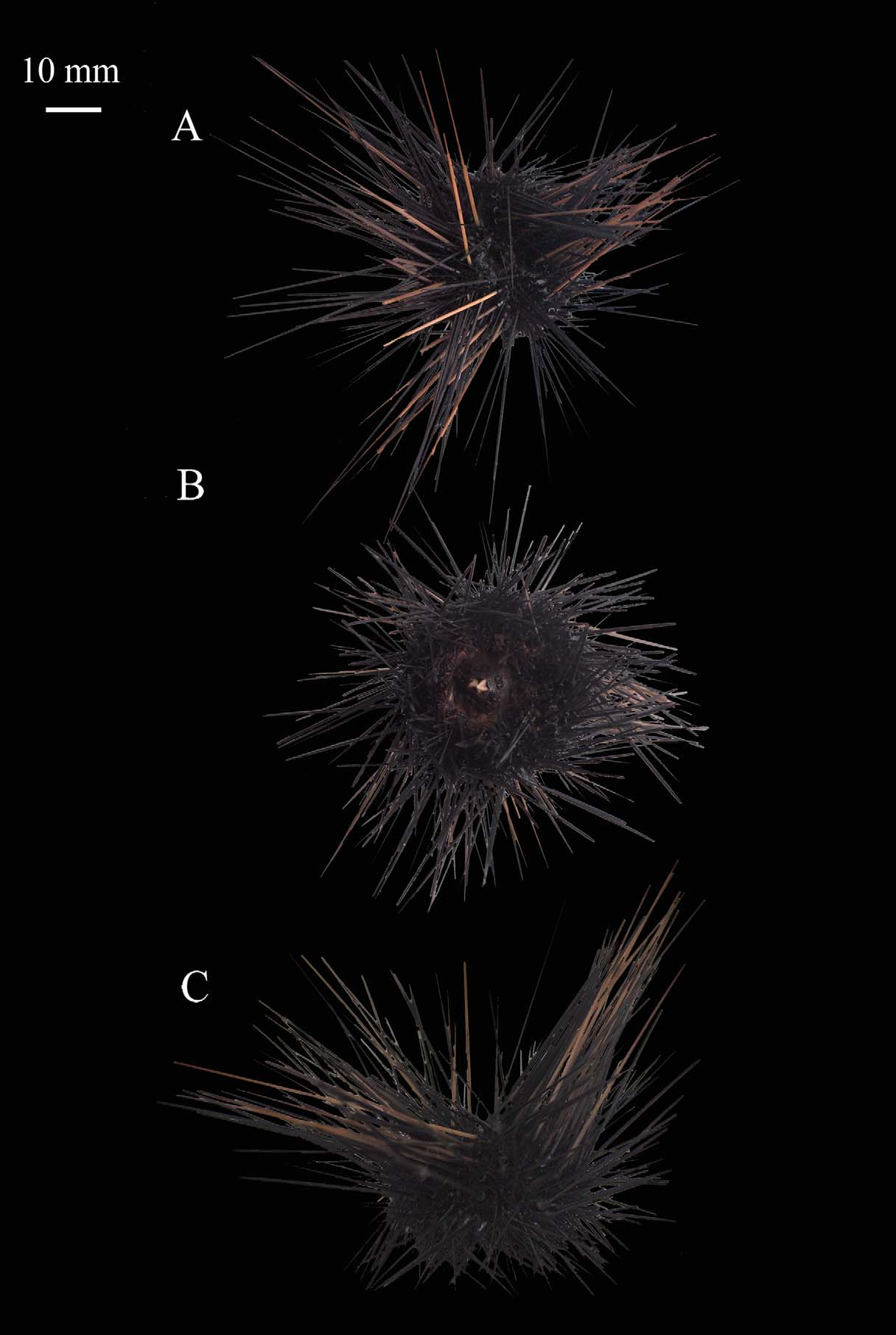
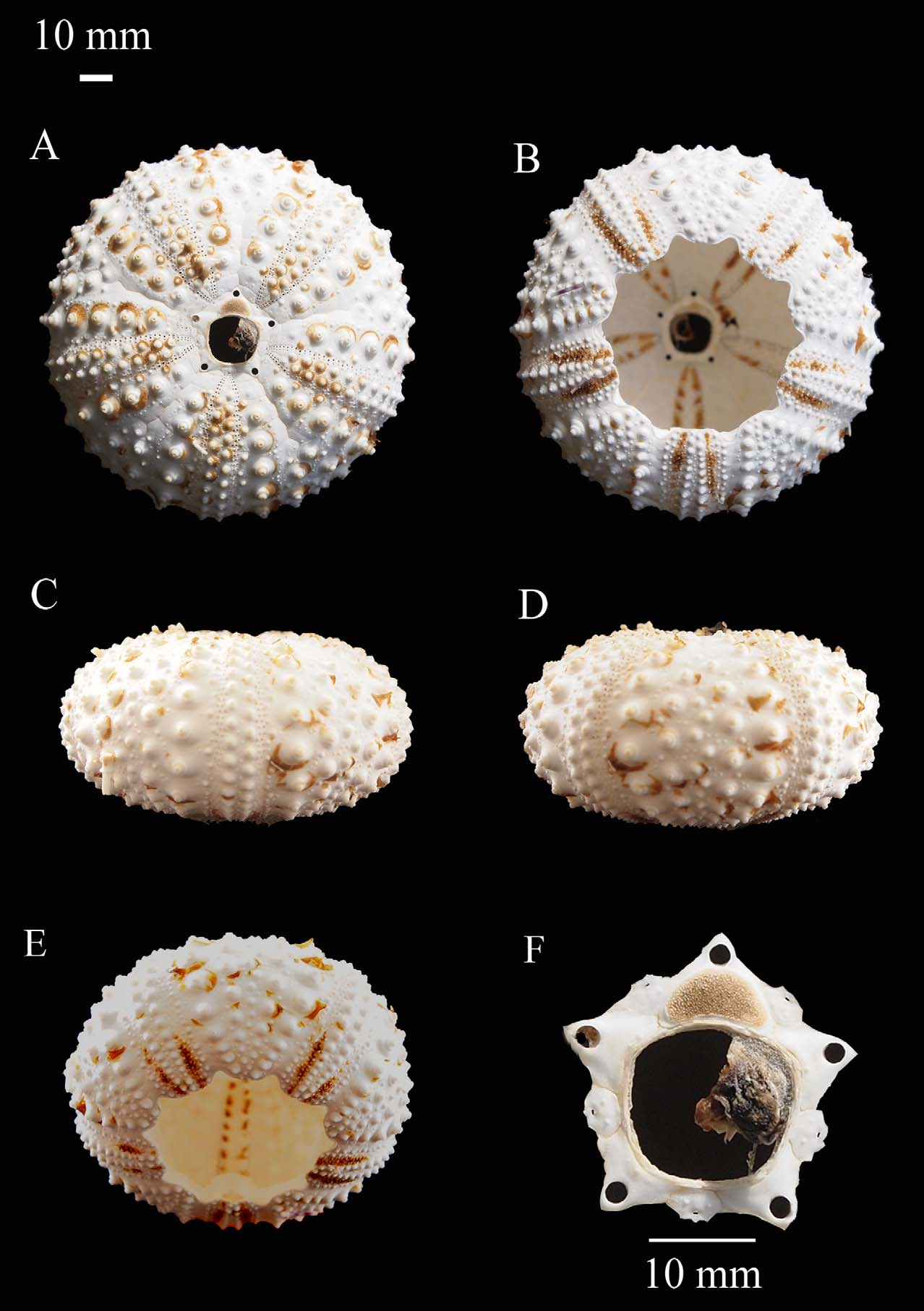
Neotype description. The test is hemispherical, with a horizontal diameter of 45.0 mm and a vertical diameter of 20.5 mm ( Fig 1 View FIGURE 1 ). The epithelium of the test is black ( Fig. 1 View FIGURE 1 A–C) with a red tinge. In living specimens, narrow blue lines of iridophores occur down either side of the naked median areas of the interambulacra and as a ring around the apical disc. The apical system is hemicyclic and measures 23.94 mm, 24% of the test’s horizontal diameter ( Fig. 2 View FIGURE 2 D & Table 1). The genital plates are wider than long, with one to three tubercles along their inner edge (Table 1 & Fig. 2 View FIGURE 2 F) and a genital pore that measures 45% the genital plate length. A faint arch-shaped depression occurs on the inner edge of each genital plate, forming the corners of the apical ring. Ocular plates are pentagonal in shape with two small tubercles in the centre of the plate. The periproctal cone is small and black, and does not have any platelets or markings on the skin ( Fig. 1 View FIGURE 1 A & Table 1). The ambulacra are slightly raised aborally; and measure 22% of the width of interambulacral measured at the ambitus (Table 1 & Fig. 2 View FIGURE 2 ). They have two rows of large crenulate and perforate primary tubercles ( Fig. 2 View FIGURE 2 ), with an offset inner series of small tubercles. The interambulacra measure 18 mm in width at the ambitus and contain four series of large primary tubercles, with an offset inner series of small tubercles. Each series contains 14 primary tubercles with areoles of moderate size ( Fig. 2 View FIGURE 2 & Table 1). The peristome measures 21.9 mm at the ambitus, 48.1% of the horizontal diameter. It is subcircular in shape and has five pairs of buccal tube feet. The peristomial membrane is black and is covered with a large number of triphyllous pedicellariae. Auricles are robust with high processes ( Fig. 2 View FIGURE 2 E).
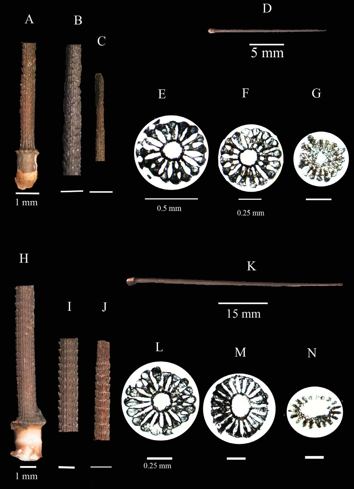
Ambulacral and interambulacral spines differ in their width and ornamentation. Ambulacral spines are black with a red tinge ( Fig. 3 View FIGURE 3 ), the longest measures 28.52 mm in length (63.34% of the test horizontal) on the neotype, 0.9 mm in width proximally and 0.5 mm distally ( Fig. 3 View FIGURE 3 E–G). These are verticillate with barbs distally ( Fig. 3 View FIGURE 3 A–C), and are typically composed of sixteen solid wedges.
Interambulacral spines are predominantly black with a red tinge, but a number of brown and white spines are present aborally ( Figs 1 View FIGURE 1 & 3 View FIGURE 3 ). They are long and slender, the longest measuring 47.37 mm in length on the neotype. The spines are verticillate, formed by 20 solid wedges, which radiate out from a hollow axial cavity. Spine width varies from 1.4 mm in diameter proximally to 0.9 mm distally, with numerous barbs present distally ( Figs 3 View FIGURE 3 H–J).
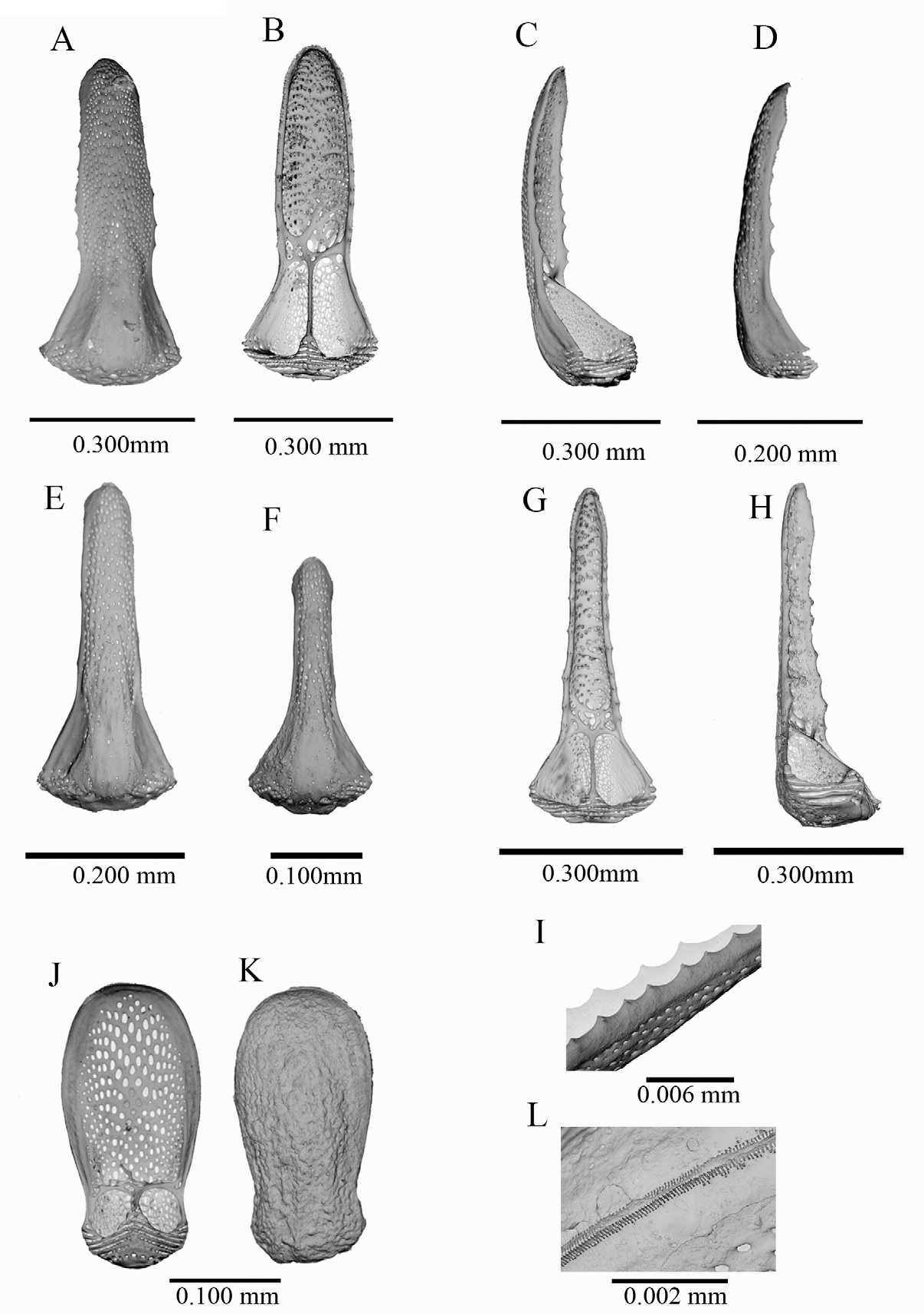
Only tridentate and triphyllous pedicellariae were found on the neotype, with no claviform ophicephalous pedicellariae observed ( Fig. 4 View FIGURE 4 &table 3). Tridentate pedicellariae occur as two forms, one with broad valves ( Fig. 4 View FIGURE 4 A–D) and a long broad neck on a short stalk, and a narrow form (comparing valves of equal length) with narrow valves, a short neck and a long stalk ( Fig. 4 View FIGURE 4 E–H). Both forms have moderately curved valves with serrations along edges of the blades of the valves ( Figs 4 View FIGURE 4 G–I). Both forms occur orally and aborally. However, the broad form is less abundant. Triphyllous pedicellariae are typical of the genus in having broad valves that are rounded distally, with numerous small peripheral teeth that occur in two rows. ( Figs 4 View FIGURE 4 J–L). The head of each pedicellaria is supported by a long muscular neck attached to a long stalk.
No known copyright restrictions apply. See Agosti, D., Egloff, W., 2009. Taxonomic information exchange and copyright: the Plazi approach. BMC Research Notes 2009, 2:53 for further explanation.
|
Kingdom |
|
|
Phylum |
|
|
Class |
|
|
Order |
|
|
Family |
|
|
Genus |
|
Kingdom |
|
|
Phylum |
|
|
Class |
|
|
Order |
|
|
Family |
|
|
Genus |