
Dujardinascaris madagascariensis ( Chabaud & Caballero, 1966 )
|
publication ID |
https://doi.org/ 10.11646/zootaxa.3893.2.6 |
|
publication LSID |
lsid:zoobank.org:pub:7559B95A-487B-464E-9EFB-1FCAF491C9AA |
|
DOI |
https://doi.org/10.5281/zenodo.5633287 |
|
persistent identifier |
https://treatment.plazi.org/id/847187CF-FFA1-FFA4-FF52-1B2CFEFC3997 |
|
treatment provided by |
Plazi |
|
scientific name |
Dujardinascaris madagascariensis ( Chabaud & Caballero, 1966 ) |
| status |
|
Dujardinascaris madagascariensis ( Chabaud & Caballero, 1966)
Figures 1 View FIGURE 1 , 2 View FIGURE 2 AB, 3, 5J
Description. Medium sized nematode. Males smaller than gravid females (average 59% of their length). Cuticle transversally striated. Fixed specimens usually slightly coiled. Lips shield-shaped, with interlocking processes ( Figs. 1 View FIGURE 1 B, 3B, C). Middle anterior edge of lips semicircular, with toothed oral plate, 21–23 teeth with slightly rounded tips ( Fig. 3 View FIGURE 3 C), highness and thickness of teeth 0.82–1.33 µm, 0.82–1.79 µm respectively; pulp of dorsal lip extended to the border of lip in four distinct lobes, and towards double papillae ( Fig. 3 View FIGURE 3 A). Pulps of subventral lips asymmetrical, main mass extending towards amphid and lateral papilla. Triangular interlabia well developed, reaching anteriorly to the middle of lip height ( Fig. 3 View FIGURE 3 B). Oesophagus slender, relatively long, terminating in minute oval ventriculus. Long anterior intestinal caecum present ( Fig. 1 View FIGURE 1 A). Nerve ring encircling oesophagus at approximately 13–20% of its length ( Fig. 1 View FIGURE 1 A). Excretory pore approximately at level of nerve ring ( Fig. 1 View FIGURE 1 A). Deirids not observed. Cervical alae absent.
Males (5 specimens): Length of body 10.2–13.1, maximum width 0.36–0.44 ( Fig. 2 View FIGURE 2 A). Ventriculus 0.099–0.113 long and 0.069–0.094 wide. Nerve ring and excretory pore approximately at the same distance from anterior extremity. Five pairs of preanal subventral papillae, last 2 pairs close together just anterior to cloaca, one papilla on upper cloacal lip, one pair of lateral paracloacal papillae, two pairs of subventral postanal papillae, and two pairs of subdorsal postanal papillae ( Figs. 1 View FIGURE 1 F, 3F, G). Lateral pair of phasmids in centre between dorsolateral pairs of postanal papillae ( Figs. 3 View FIGURE 3 G, J). Spicules equal or slightly subequal in length, very narrow, distal ends pointed ( Fig. 2 View FIGURE 2 B). Gubernaculum ( Fig. 5 View FIGURE 5 J) well sclerotised, its proximal end with dorsal projection in lateral view, longitudinally riffled straight tapering shaft and single sharp curved tip. Tail conical sharply pointed ( Fig. 3 View FIGURE 3 F, G). For detailed measurements see Table 2 View TABLE 2 .
Females (6 gravid specimens; measurements for one immature specimen is given in parentheses): Length of body 15.5–27.1, maximum width 0.63–1.46. Ventriculus 0.15–0.24 long and 0.099–0.173 wide ( Fig. 1 View FIGURE 1 A). Nerve ring and excretory pore approximately the same length from anterior extremity ( Fig. 1 View FIGURE 1 A). Vulva situated at first half of body, with the prominent papilla projecting outwards and forwards between two obtuse salient cuticular expansions ( Figs. 1 View FIGURE 1 C, 3D). Long vagina directed posteriorly. Eggs ranging from oval to almost spherical, thinwalled, with very fine reticulate surface; thickness of wall 0.85–1.73 µm. Content of eggs mostly uncleaved ( Fig. 1 View FIGURE 1 D) or cleaved into a maximum of 16 blastomeres. Tail slender, conical, with sharply pointed tip ( Fig. 3 View FIGURE 3 E); pair of phasmids outlets situated at 0.092–0.203 from tail tip. For detailed measurements see Table 3 View TABLE 3 .
Taxonomic summary.
Host: Crocodylus niloticus (Laurenti, 1768) (Crocodylidae) .
Site of infection: stomach.
Locality: Lake Turkana, north-western Kenya, vicinity of Todonyang village on the west margin of the Omo River delta (4°27'10"N, 35°56'30"E) (collected 23rd September 2009).
Intensity of infection: 29.
Deposition of voucher specimens: Helminthological Collection of the Institute of Parasitology, BC ASCR, in České Budějovice, Czech Republic (Cat. No. N-1024).
D. madagascariensis D. dujardini
Sprent (1977) Our data Our data Sprent (1977) Our data Our data Molecular analysis /DNA characterisation of D. madagascariensis— 18S and ITS2 region. The 637 bp nucleotide sequence of the 18S rDNA gene of D. madagascariensis was obtained. The gene has a 50.24% G+C content. The partial sequences of three species ( D. madagascariensis , D. waltoni , and M. heterotis ) were compared over an alignment length of 637 bp. Dujardinascaris madagascariensis had 99.5% identity with D. waltoni and 98.6% identity with M. heterotis . In the alignment of the sequences of D. madagascariensis and D. waltoni , three out of the 637 nucleotide positions were variable (three variable amino acid sites out of 212). The average nucleotide distance was 0.005 and amino acid distance was 0.015. Comparison of the 18S sequence of D. madagascariensis with that of M. heterotis revealed nine out of the 637 variable nucleotide sites (six variable amino acid sites out of 212). The values for average nucleotide and amino acid distances were 0.014 and 0.03. For D. madagascariensis , the length of the ITS2 sequence was 448 bp with G+C content of ITS2 51.12%. Comparison of ITS 2 sequence with that of D. waltoni was impossible due to lack of sequence data.
Phylogenetic relationships among selected representatives of Ascaridida ( Fig. 4 View FIGURE 4 ). Phylogenetic analyses based on partial 18S rDNA sequences produced four clades with high bootstrap support values (>50%). First two clades consist of five species of Heterocheilidae ; D. madagascariensis formed one clade with D. waltoni (92% bootstrap support), the second clade included Heterocheilus tunicatus Diesing, 1839 , M. heterotis and Kreftascaris sharpiloi Tkach, Kuzmin and Snyder, 2010 (71% bootstrap support). Pseudoterranova decipiens , Anisakis pegreffi and T. caballeroi represent third clade with 58% bootstrap support and A. lumbricoides and B. procyonis nest together in fourth clade with 82% bootstrap support. Tree topology strongly supports the separation of the heterocheilid ( Heterocheilidae ) clades, and groups nematode species from the hosts of the same class into highly supported clades.
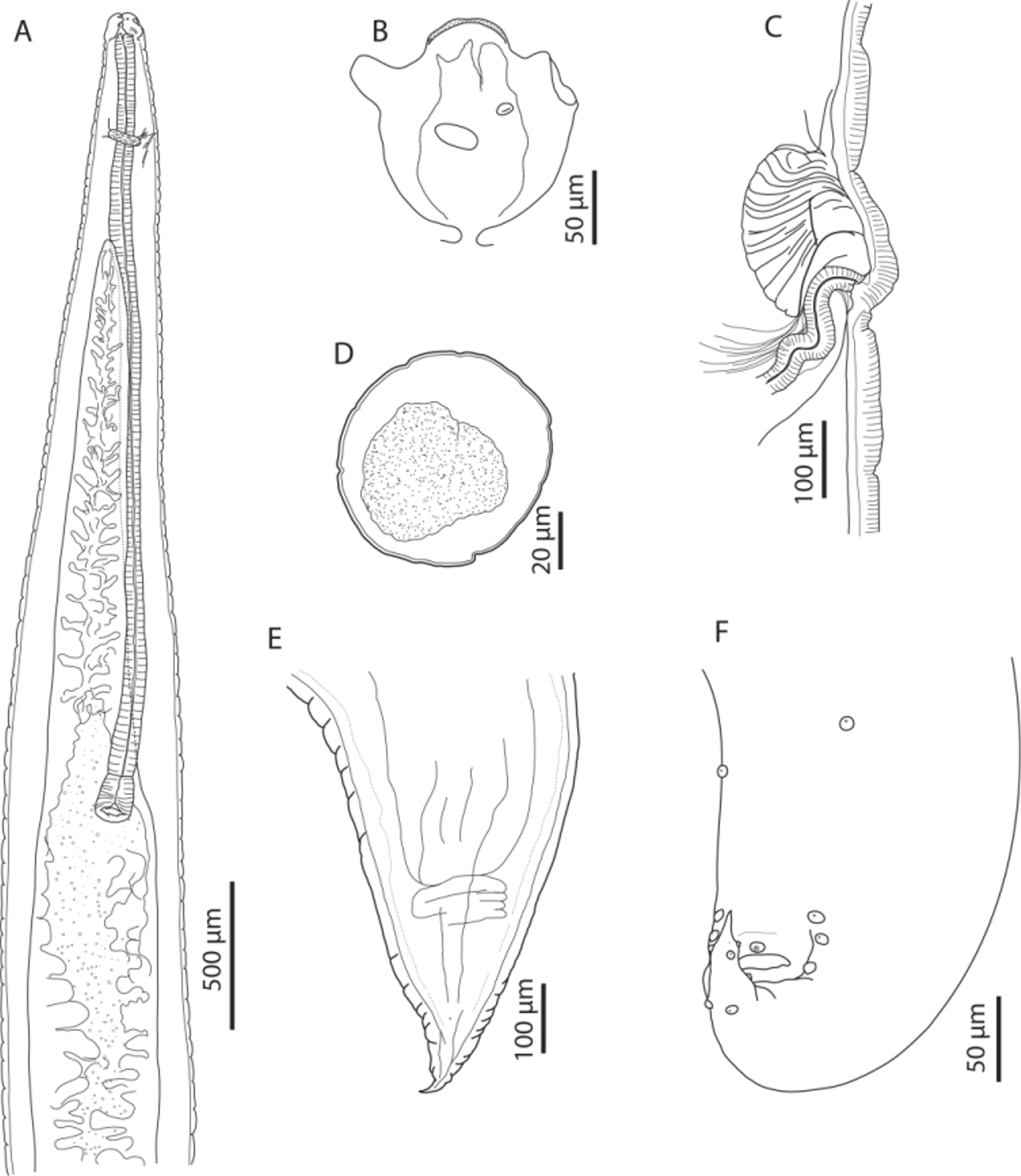
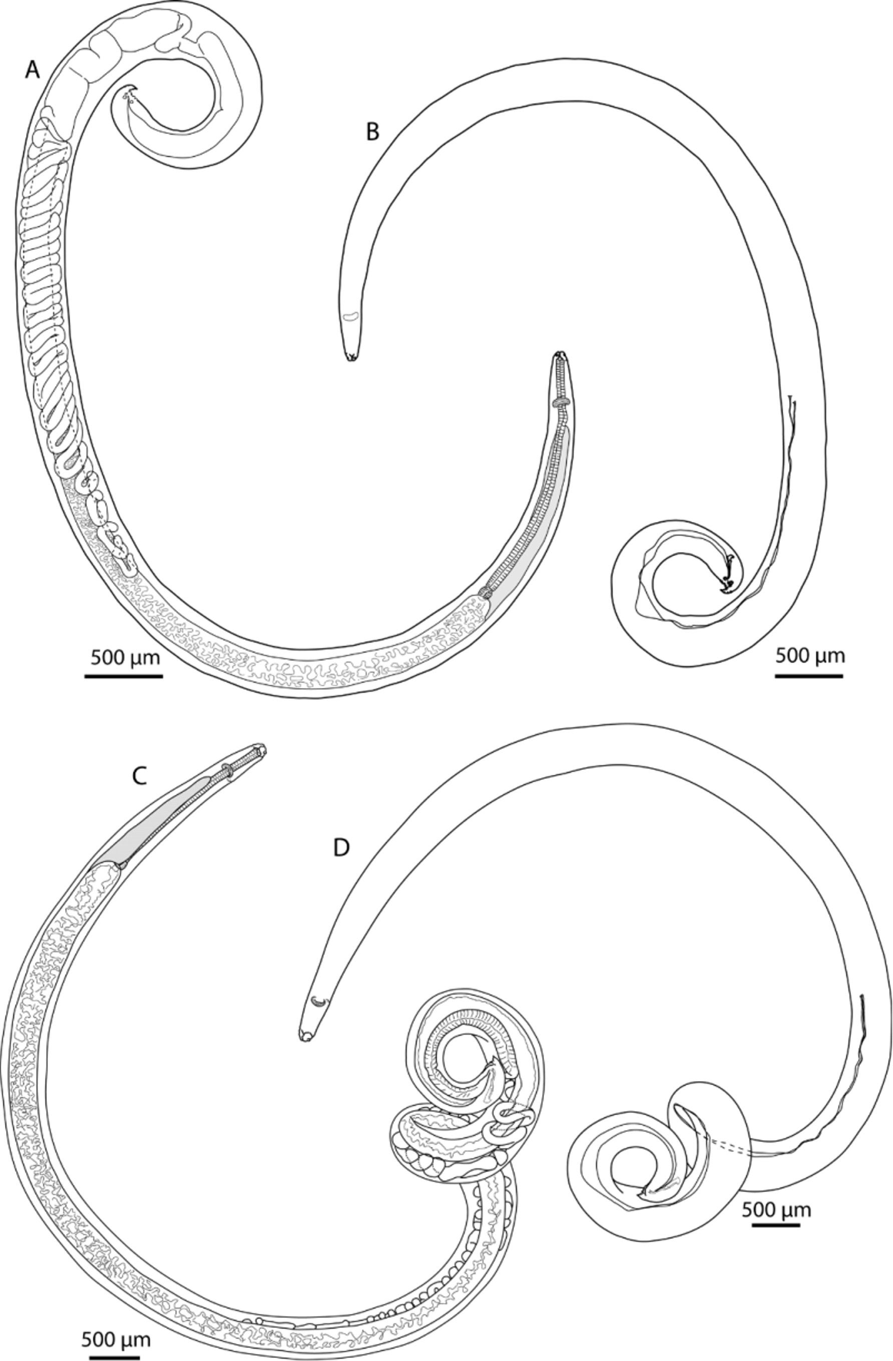
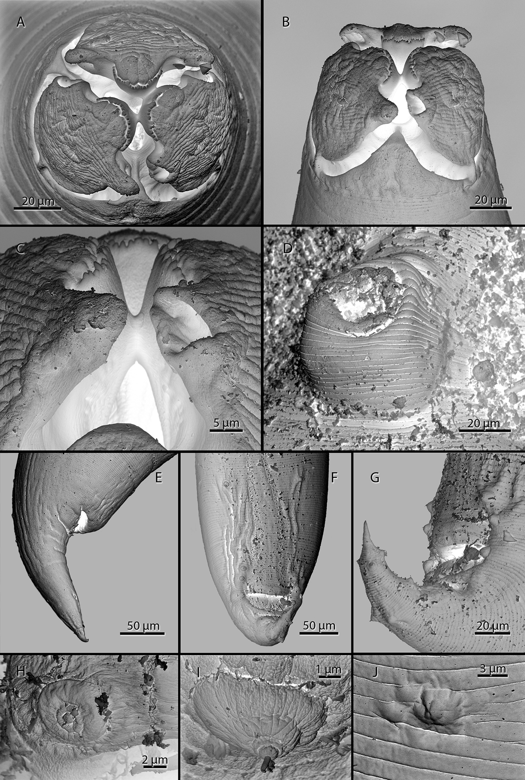

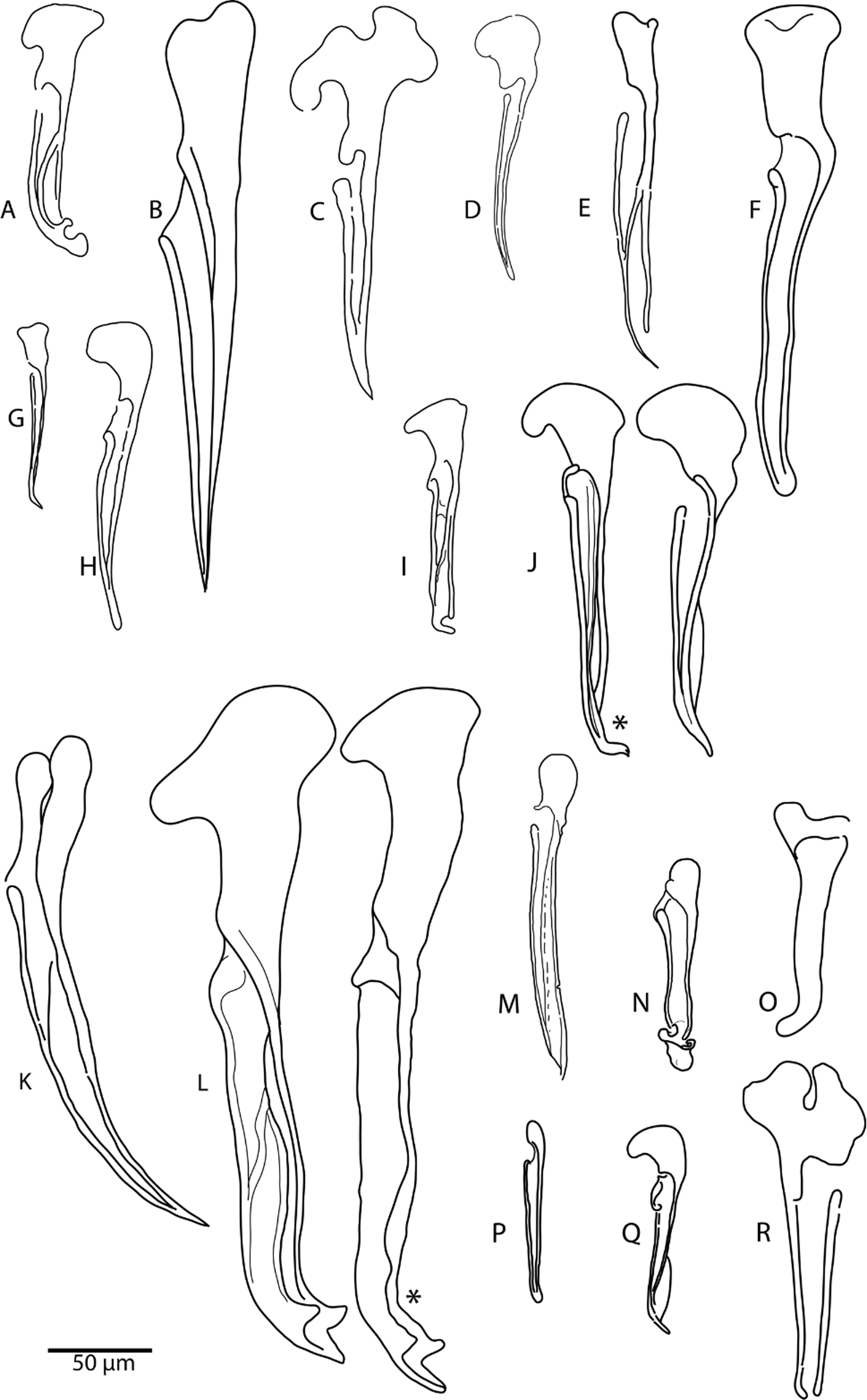
TABLE 2. Comparisons of the measurements of the males of D. madagascariensis and D. dujardini (dimensions in mm; mean values are given in parentheses).
| D. madagascariensis | D. dujardini | ||
|---|---|---|---|
| Sprent (1977) Our data | Sprent (1977) | Our data | |
| Number of specimen | 1 5 | 8 | 1 |
| Length | 8.8–12.3 10.2–13.1 (11.3) | 14.0–22.0 | 18.4 |
| Width (maximum) | --- 0.36–0.44 (0.41) | 0.48 | 0.55 |
| Width (at o/i junction) | 0.26 0.28–0.33 (0.31) | 0.25 | 0.38 |
| Subventral lip (length) | 0.070 0.060–0.084 (0.068) | 0.090 | 0.105 |
| Nerve ring (from anterior end) | 0.31 0.31–0.40 (0.350) | 0.52 | 0.40 |
| Excretory pore (from anterior end) | 0.33 0.35–0.48 (0.40) | 0.53 | 0.46 |
| Oesophagus (length) | 1.70 1.62–2.18 (1.91) | 2.70 | 1.97 |
| Oesophagus (%body length) | 18.68 % 16–19 % (17 %) | 15.07% | 11% |
| Caecum (length) | 1.30 1.2–1.56 (1.40) | 1.50 | 1.45 |
| Caecum (%body) | 14.3 % 12–14 % (12 %) | 8.7 % | 8 % |
| Caecum (%oesophagus) | 76.5 % 71–78 % (73 %) | 55.6 % | 73 % |
| Tail (length) | 0.150 0.070–0.148 (0.117) | 0.090 | 0.118 |
| Gubernaculum | 0.180 0.175–0.208 (0.189) | 0.330–0.340 | 0.339 |
| Tail (%gubernaculum) | 83 % 40–70 % (61 %) | 37.5 % | 35 % |
| Spicules | 5.90 3.60–5.23 (4.38) | 7.00–12.90 | 7.847 |
| Ejaculatory duct | 1.30 1.32–1.78 (1.57) | 1.40 | 2.27 |
| Ejaculatory duct (%body) | 14 % 12–16 % (14 %) | 8 % | 12 % |
TABLE 3. Comparison of the measurements of females of D. madagascariensis and D. dujardini (dimensions in mm, mean values are given in parentheses, abbreviations: o / i—oesophagus / intestine; L / W—length / width ratio).
| Immature | Gravid | Immature | Gravid | |
|---|---|---|---|---|
| Number of specimen | 1/not mentioned 1 | 6 | 1/not mentioned 2 | 1 |
| Length | 19.0–30.0 14.7 (28.0) | 15.5–27.1 (19.1) | 26.0–40.3 (26.0) 14.2–20.4 (17.3) | 23.4 |
| Width (at o/i junction) | 0.79 0.47 | 0.48–0.84 (0.59) | 0.49 0.48–0.54 (0.51) | 0.59 |
No known copyright restrictions apply. See Agosti, D., Egloff, W., 2009. Taxonomic information exchange and copyright: the Plazi approach. BMC Research Notes 2009, 2:53 for further explanation.