
Quercivir gounellei Lameere, 1912
|
publication ID |
https://doi.org/ 10.11646/zootaxa.4568.1.7 |
|
publication LSID |
lsid:zoobank.org:pub:0FB75C5C-3C04-4656-B083-3A7665CDE369 |
|
DOI |
https://doi.org/10.5281/zenodo.5940579 |
|
persistent identifier |
https://treatment.plazi.org/id/847787EA-7066-FFB6-FF04-FE4DFB73FCBC |
|
treatment provided by |
Plazi |
|
scientific name |
Quercivir gounellei Lameere, 1912 |
| status |
|
Quercivir gounellei Lameere, 1912 View in CoL
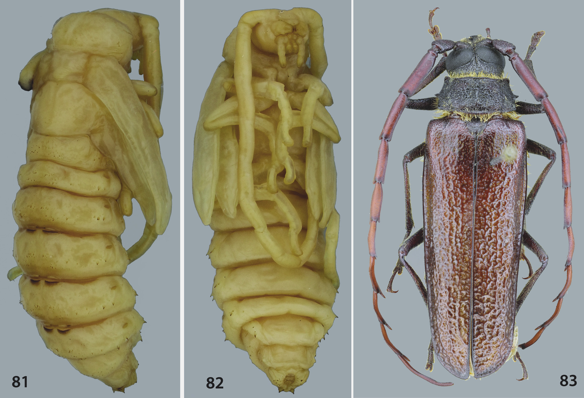
( Figs. 64–83 View FIGURES 64‒72 View FIGURES 73‒80 View FIGURES 81‒83 )
Description of larva based on larval exuvia. Epistomal margin ( Fig. 66 View FIGURES 64‒72 ) not projecting above anteclypeal base and with additional carina above epistoma; upper boundary slightly carinate and sinuous laterally, and lower boundary almost straight, slightly arched; three pairs of epistomal setae. Five stemmata on each side ( Fig. 71 View FIGURES 64‒72 ): three protuberant subcontiguous below antenna on elevated area, and two small, separated, below previous. Antennae ( Figs. 64, 65, 71 View FIGURES 64‒72 ) short, extensible with antennifer membranous, two elongate and one reduced antennomeres: basal antennomere with nine campaniform sensillae; median antennomere slightly truncate apically, with three campaniform sensillae near base, and at apex, one elliptical flat sensorium, two membranous small cupuliform appendices; distal antennomere reduced, rounded with four setae and one tiny membranous sensorium at apex. Clypeus ( Fig. 66 View FIGURES 64‒72 ) band-shaped, trapezoidal and membranous. Labrum ( Fig. 66 View FIGURES 64‒72 ) subelliptical, soft, convex, continuous ventrally; sclerotized on basal half; wide and long setae more concentrated on distal 2/3; each seta inserted on darker small patch. Epipharynx ( Fig. 67 View FIGURES 64‒72 ) densely covered with setae directed to middle; median region with several sensoria; one sclerite at each side. Mandibles ( Figs. 73–76 View FIGURES 73‒80 ) strongly sclerotized, symmetrical, wide; distal margin declivous, gouge-shaped; apical internal half with two inclined irregular keels; a wide and short dorsal subapical tooth; external margin with a transverse, semicircular depression parallel to distal margin, irregularly striate and rugose on distal half, and with sparse yellowish setae on basal half. Maxillolabial complex ( Figs. 69, 70 View FIGURES 64‒72 ) partially membranous; submentum, maxillary articulating areas and cardines fused, forming basal part. Basal part membranous with moderately long setae on maxillary articulating area. Distal maxilla: stipes with brown, inclined and irregular area near middle, bearing long setae, more concentrated near external margin; mala wide with rounded apex, bearing inclined sclerotized band near base and numerous long setae dorsally and ventrally. Palpifer membranous with sclerotized, narrow ventral basal band, and numerous long setae dorsally and ventrally; dorsally bearing a pointed well-developed lateroanterior membranous lobe (dorsolateral process of maxillary palpiger). Maxillary palpi trimerous; palpomeres partially sclerotized; basal palpomere transverse with three long dorsal setae near apex; median palpomere elongate with three ventral and dorsal setae; distal palpomere elongate with three ventral campaniform sensillae, and numerous peg-shaped sensillae at apex. Distal labium: mentum wider than long with lateral margins rounded; numerous long setae laterally; median region with barely sclerotized area with two setae and several small darker patches; one narrow irregular sclerite at each side of basal half. Prelabium with basal sclerotized irregular band projecting behind palpiger band; five campaniform sensillae between palpigers; palpiger with several long setae; numerous moderately long setae distributed on entire area, except basal region; setae more concentrated toward apex. Labial palpi dimerous and partially sclerotized; palpomeres elongate; palpomere basal with three small campaniform sensillae; palpomere distal with peg-shaped sensilla at apex. Hypopharynx ( Fig. 70 View FIGURES 64‒72 ) covered by stout setae and microspines near base; several campaniform sensillae at middle. Spiracles ( Fig. 72 View FIGURES 64‒72 ) elliptical. Leg ( Fig. 68 View FIGURES 64‒72 ) partially sclerotized, except coxa, membranous; 4- segmented: coxa short and wide with one seta and two campaniform sensillae; femur robust, wider than long, with numerous long and wide setae near distal middle; tibiotarsus elongate with numerous long and wide setae; pretarsus elongate, glabrous and tuberculate.
Abdomen glabrous with dorsal and ventral ampullae without spicules; segments I–VIII, each with a pair of elliptical spiracle, smaller than thoracic; first spiracle larger.
Description of pupa ( Figs. 77–82 View FIGURES 73‒80 View FIGURES 81‒83 ). Length: 38–40 mm. Coloration yellowish; pubescence whitish almost translucent, short and thin. Head longer than pronotum with short setae near base of antennal insertion. Mouthparts with short and thin setae. Pronotum wider than long, grooved longitudinally-medially, with lateral margins rounded. Mesonotum shorter than pro- or metanotum; scutellum slightly prominent. Metanotum grooved longitudinally-medially. Abdomen with tergites with band of microspines; microspines increasing in size toward apex; segments I-VI band-like: segments VII–VIII narrower and arched; segment IX reduced with two tiny urogomphi; segment X reduced, rounded and ventral; segments I–VI with a pair of functional spiracles; tergites IV–V, V–VI each with a pair of gin traps, each with posterior margin more strongly sclerotized. Pleural region of segments III–VII, each with a sclerotized and well developed microspine with one seta.
Material examined. Barzil. São Paulo: São José dos Lopes -Pq Est. Campos do Jordão, 16.X.1992, Exp. MZUSP 2 larvae reared until adult, 2 larval and 2 pupal exuviae, 2 pupae fixed (same-sex) ( MZSP8712 View Materials ).
Remarks. Based on the descriptions and on the examined material of Polyoza lacordairei and Quercivir gounellei , the larvae of Meroscelisini are characterized by having: lower boundary of epistoma almost straight, not projected over clypeus, except Tragosoma harrisii and Trichoderes pini , projected above clypeus in two distinct triangular lobes; upper boundary weakly carinate and slightly sinuous, except Microplophorus magellanicus , with a pair of paramedian dentate processes, Tragosoma harrisii with four flat teeth, and Trichoderes pini forming one triangular processes each side, similar to the lower boundary; antennae with two antennomeres; stemmata present in Microplophorus magellanicus , two pairs present in Trichoderes pini , 3–4 pairs in Tragosoma harrisii and five pairs (three contiguous) in Polyoza lacordairei and Quercivir gounellei ; legs short, 4-segmented, except, Microplophorus magellanicus 3-segmented; abdomen with retractile ambulatory ampullae dorsal and ventral; ampullae grooved and without asperites; anus trilobed.
Based on the description of Microplophorus magellanicus and the analyses of Polyoza lacordairei and Quercivir gounellei , the pupa of Meroscelisini is characterized by having: head almost totally covered by prothorax; pronotum wider than long, without lateral projection; gin traps absent in M. magellanicus , three pairs in Quercivir gounellei and four pairs in Polyoza lacordairei ; segment IX with about eight microspines at apex and lateral spines outside directed; in Q. gounellei apex of segment IX resembles a tiny urogomphus.


No known copyright restrictions apply. See Agosti, D., Egloff, W., 2009. Taxonomic information exchange and copyright: the Plazi approach. BMC Research Notes 2009, 2:53 for further explanation.
|
Kingdom |
|
|
Phylum |
|
|
Class |
|
|
Order |
|
|
Family |
|
|
Tribe |
Meroscelisini |
|
Genus |