
Libanopycnodus wenzi, Taverne & Capasso, 2018
|
publication ID |
https://doi.org/ 10.5852/ejt.2018.420 |
|
publication LSID |
lsid:zoobank.org:pub:F8E6FEEF-FC7A-4B40-AEEE-95DBC7BC61A4 |
|
DOI |
https://doi.org/10.5281/zenodo.5979271 |
|
persistent identifier |
https://treatment.plazi.org/id/2AD09A18-0416-4A26-AD08-E5307B901759 |
|
taxon LSID |
lsid:zoobank.org:act:2AD09A18-0416-4A26-AD08-E5307B901759 |
|
treatment provided by |
Plazi |
|
scientific name |
Libanopycnodus wenzi |
| status |
gen. et sp. nov. |
Libanopycnodus wenzi gen. et sp. nov.
urn:lsid:zoobank.org:act:2AD09A18-0416-4A26-AD08-E5307B901759
Figs 1–11 View Fig. 1 View Fig. 2 View Fig. 3 View Fig. 4 View Fig.5 View Fig. 6 View Fig. 7 View Fig. 8 View Fig.9 View Fig. 10 View Fig. 11
Diagnosis
Small, deep-bodied pycnodontid fish, with rounded dorsal and ventral profiles. Head triangular in shape, with a rectilinear frontal profile and a short postorbital region. Pointed snout, with a mouth gape ventrally inclined. Frontal short. Prefrontal very narrow. Dorsal region of parietal forming a marked angle with the ventral region. Parietal brush-like process present. No temporal fenestra. Dermosphenotic sutured to the skull roof. Large, deep, well-visible dilatator fossa surrounded by dermosphenotic and dermopterotic. Posterior region of the endocranium exposed behind the dermopterotic. Vomer with 10 rounded molariform teeth on the external dental row. Premaxilla bearing two incisiform teeth. Prearticular with three rows of molariform teeth. Ectopterygoid present. Large triangular first infraorbital. Preopercle much deeper than the exposed region of the hyomandibula-dermohyomandibula. Opercle small and coma-shaped. Notochord not completely surrounded by vertebral arches. Neural and haemal spines with an anterior wing-like component. Twenty-five neural spines before epichordal series. First eight neural spines autogenous. Twelve haemal spines before hypochordal series. Eleven pairs of ribs. Postcoelomic bone long, narrow, with an enlarged ventral extremity. Dorsal and anal fins strip-like. Dorsal fin supported by 42 pterygiophores. Anal fin supported by 34 pterygiophores. Caudal peduncle short. Five epichordals. Eleven hypochordals, with two moderately broadened. One urodermal. Caudal fin double emarginated, with 20 principal rays. A few scale bars on the abdominal region of the body. Small complete scales associated with the prepelvic ventral keel scutes. Bifid cloacal scale present. One postcloacal scale. Dorsal ridge with 11 scutes, of which the first two with a small spine. Ventral keel with 13 scutes, 11 prepelvic and two postcloacal with small spines.
Etymology
The specific epithet is in homage to Dr. Sylvie Wenz (Paris), who worked abundantly on the pycnodontiform fishes.
Material examined
Holotype
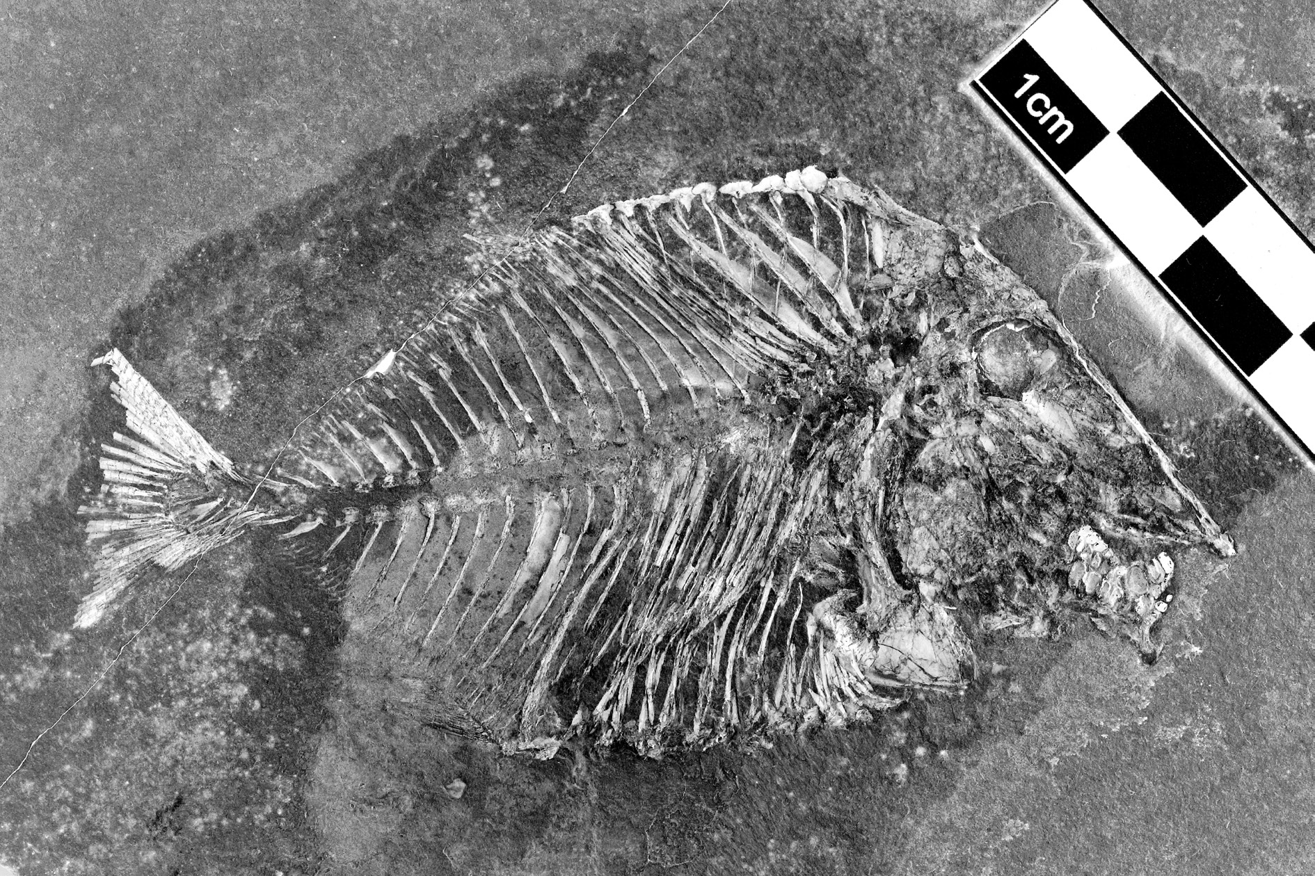
LEBANON: a complete specimen ( Figs 1–2 View Fig. 1 View Fig. 2 ), total length: 90 mm, standard length: 78 mm, marine Upper Cenomanian deposits of Ein Namoura ( CLC S-574 ).
General morphology and morphometric data
The fish is deep-bodied, the maximum body height being equal to a little more than the half of the standard length. The dorsal and the ventral profiles are rounded and devoid of marked apex.
The morphometric data are given in % of the standard length (78 mm):
Length of the head (opercle included) ……………………………………………………………34.0 %
Depth of the head (in the occipital region) ………………………………………………………44.7 %
Maximum depth of the body (just behind the head) ……………………………………………56.2 %
Prepelvic length …………………………………………………………………………………57.0 %
Predorsal length …………………………………………………………………………………68.1 %
Basal length of the dorsal fin ……………………………………………………………………35.7 %
Preanal length ……………………………………………………………………………………71.5 %
Basal length of the anal fin ………………………………………………………………………28.9 %
Depth of the caudal peduncle ………………………………………………………………………7.2 %
Osteology
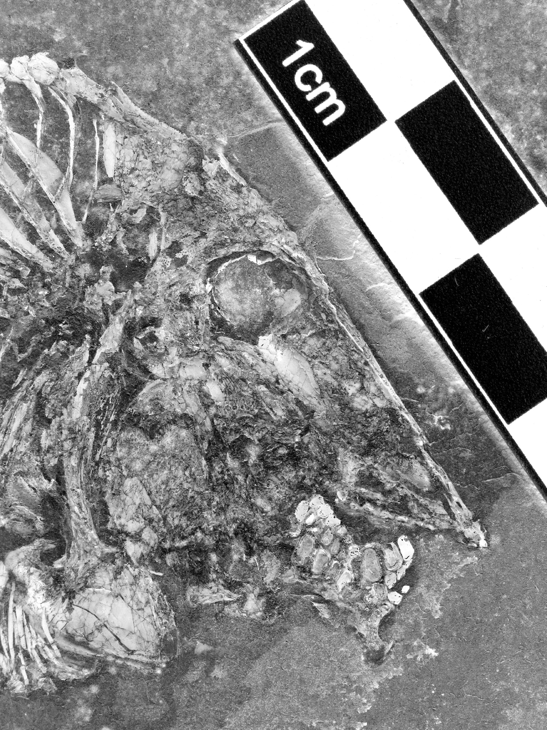
Skull ( Figs 3–6 View Fig. 3 View Fig. 4 View Fig.5 View Fig. 6 )
The head is large, triangular in shape, altogether long and deep, with an almost rectilinear frontal profile. The preorbital region is much longer than the postorbital one. The orbit is wide. The dermal bones of the skull are ornamented with small tubercles. The snout is pointed, with a mouth gape ventrally inclined.
The mesethmoid is elongate and broad. Its upper margin is covered posteriorly by a pair of very narrow prefrontals and anteriorly by the premaxillae. The orbitosphenoid is pressed against the posterior margin of the mesethmoid. The vomer, seen in profile, is long and deeper posteriorly than anteriorly. Only the lateral dental row, composed of 7 well-developed, rounded molariform teeth, is visible. There are traces of 3 other very small teeth located at the anterior extremity of the lower margin of the vomer.
The frontal is short and limited to the orbital region. The dermosupraoccipital is small, with a pointed dorso-posterior extremity. The parietal exhibits an unusual and strange shape. Its dorsal half forms a marked angle with its ventral part. This peculiar shape of the parietal is not due to an artefact of fossilisation. A short brush-like process (= branched peniculus) is attached to the parietal. The supratemporal is a rather broad scale that lies against the upper part of the parietal, just below the first dorsal ridge scute. The dermopterotic is deeper than long. The dermosphenotic is not a free bone. It is included in the lateral wall of the skull roof and is sutured with the frontal and the dermopterotic. The ventral margin of the dermosphenotic and the dermopterotic are located at the same level as the lower border of the orbit. The dermopterotic and the dermosphenotic surround a deep and wide dilatator fossa. The parasphenoid is long, broad, toothless and its trabercular region is obliquely inclined. The posterior region of the endocranium is well visible. A small part of the intercalar and the exoccipital are preserved behind the lower half of the parietal. The ventral margin of the exoccipital is pierced by a large foramen for the branches of the vagus nerve (X). The basioccipital, articulated with the first basiventral, is also present. The exoccipital and basioccipital are separated from the braincase main part as in Pseudopycnodus nardoensis ( Taverne 1997: fig. 2). A cartilaginous region probably linked the two cranial regions on the living fish. The autosphenotic and the sensory canals of the skull roof are not visible. A temporal fenestra is not present.
The quadratic arch includes wide metapterygoid and entopterygoid plus a small ectopterygoid. Only the anterior parts of the quadrate and the symplectic are preserved. Both bones articulate with the lower jaw. The articular head of the symplectic is very broad.
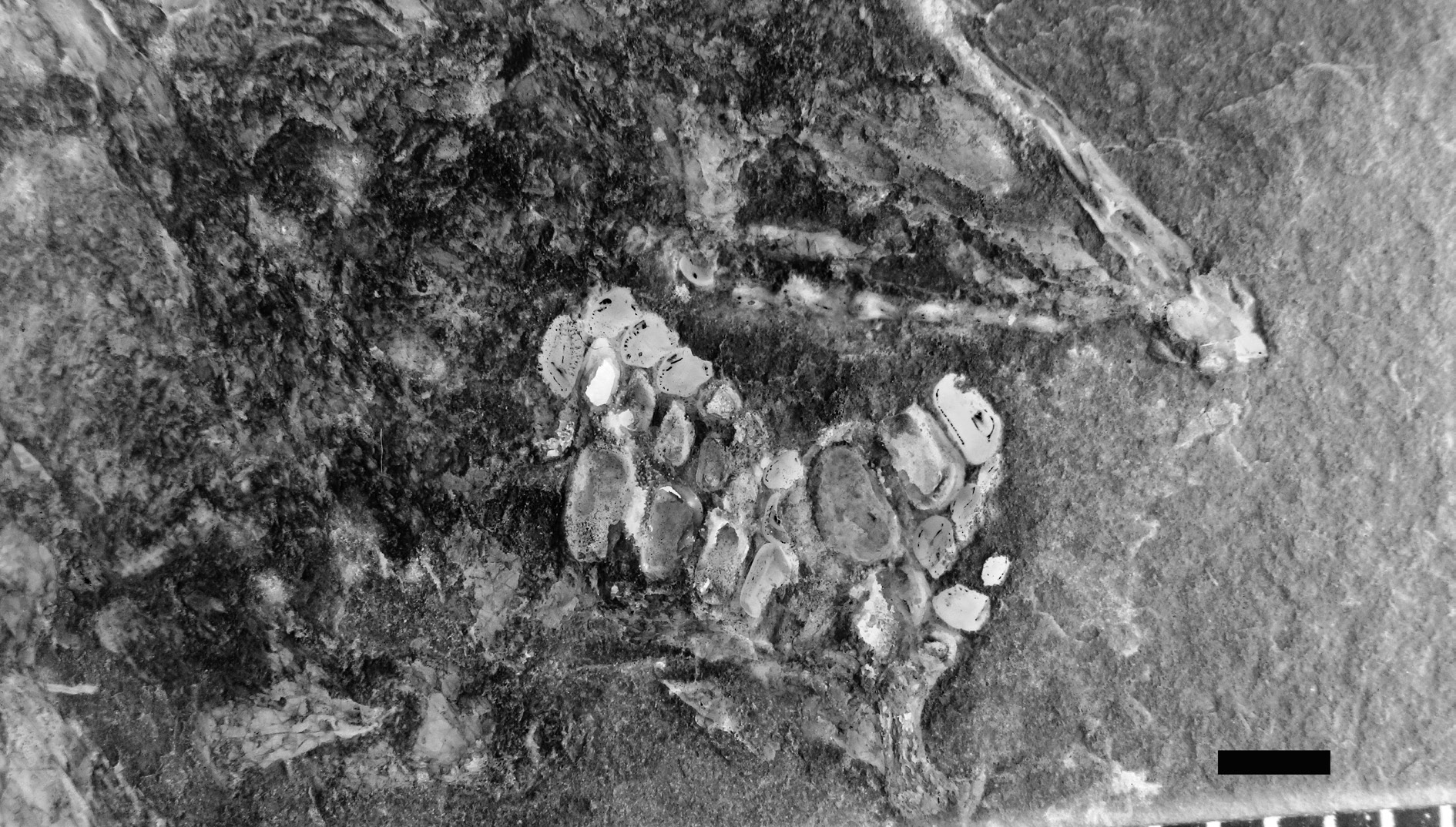
The premaxilla is long and narrow. It bears two incisiform teeth. The maxilla is lost. The posterior extremity of the dentary is preserved but not its anterior part with the teeth. The articular is small. The angular is not preserved. The prearticular is a large bone with a marked hook-like coronoid process, bearing three rows of molariform teeth. The dorsal row contains 6 teeth, the middle row 7 teeth and the ventral row at least 5 teeth. The teeth of the dorsal row are the smallest, with a slightly concave surface and their dorsal margin bears some little tubercles. The teeth of the lower row are the largest. They are deeper than long, with a slightly convex surface.
The orbital series contains four bones. The first infraorbital is a large triangular bone. The second and third infraorbitals are tubular bones. As already written, the dermosphenotic is sutured to the skull roof. A few fragments of the sclerotic bony ring are visible just under the frontal.
The exposed part of the hyomandibula-dermohyomandibula is much smaller than the preopercle to which it is sutured. The long ventral branch of the hyomandibula is covered by the preopercle but remains clearly visible. The upper margin of the hyomandibula bears a broad dorsal process that articulates in the dilatator fossa. The preopercle is deep and broad, with a rather narrow dorsal margin. The opercle is well developed, with an acuminate ventral tip and a broader upper part.
The anterior ceratohyal and two branchiostegal rays are visible below the preopercle.
Girdles ( Figs 3–4 View Fig. 3 View Fig. 4 )
A small posttemporal is preserved behind the skull, at the level of the suture between the parietal and the dermopterotic. The hypercleithrum (= supracleithrum) is a long and thin bone. The cleithrum is divided in a short but very broad ventral branch and an extremely long but narrow dorsal branch. No pterygiophore is preserved but fragments of short pectoral rays are visible.
The pelvic bones are not preserved but there are slight traces of the small pelvic fins under the cloacal region.
Axial skeleton ( Figs 1–2 View Fig. 1 View Fig. 2 , 7 View Fig. 7 )
Starting from the tail region, the axial skeleton progressively elevates to reach anteriorly the level of the orbit. The vertebrae are formed by the dorsal and ventral arcocentra. There are no ossified vertebral centra, as in all pycnodonts. The neural and haemal arches are small and there is no dorso-ventral contact between them. So, the notochord is not completely surrounded by bony elements. In the caudal region, one small pre- and one small postzygapophysis link each neural arch with the following one. There are 25 neural spines before the epichordal series and 12 haemal spines before the hypochordal pieces. These spines bear anterior sagittal wings, except the fifth to the eighth neural spines that are pressed the ones against the others. The first 8 neural spines are autogenous. There are at least 11 pairs of long ribs. These ribs do not reach the ventral margin of the body. The postcoelomic bone is long and very narrow, except in its ventral part that is greatly enlarged.
Dorsal and anal fins ( Figs 1–2 View Fig. 1 View Fig. 2 )
Both the dorsal and the anal fins are long and strip-like (type A 2 of Poyato-Ariza& Wenz2002: fig. 34). The origin of the dorsal fin is located far from the head. The dorsal fin is supported by 42 pterygiophores and the anal fin by 34 pterygiophores. Only a few fragments of the dorsal and anal fins are preserved. The precise number of these rays is not known.
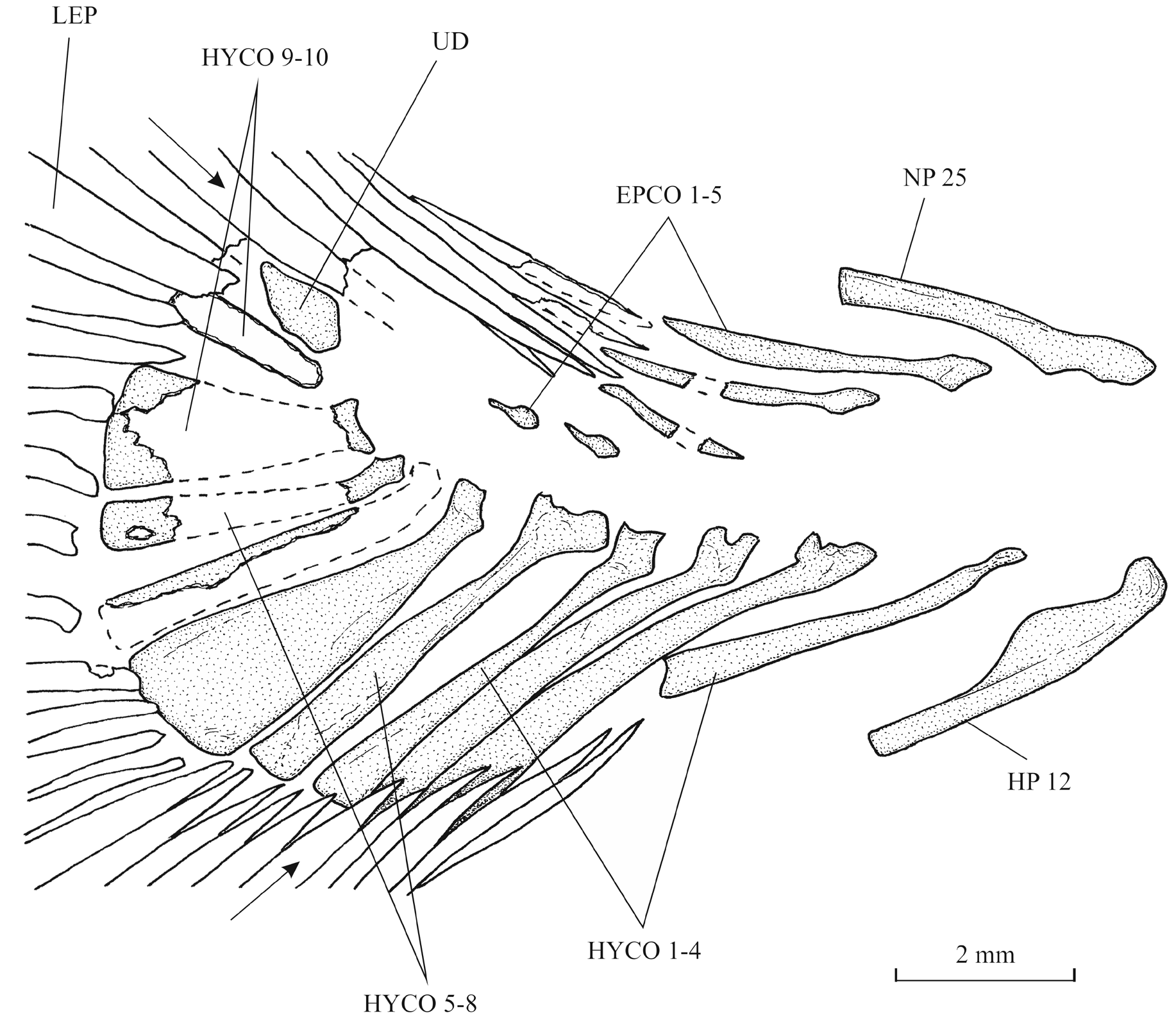
Caudal skeleton ( Figs 8–9 View Fig. 8 View Fig.9 )
The caudal peduncle is very short, the dorsal and anal fins ending near the tail. The caudal endoskeleton is composed of 5 epichordals, 11 hypochordals and 1 urodermal. The first three epichordals are thin and elongate. The last two elements of the series are extremely reduced. The hypochordals are longer and wider than the epichordals. The sixth and the ninth hypochordals are moderately broadened.
The caudal fin is double emarginated ( Poyato-Ariza& Wenz2002: fig. 36E). There are 20 principal caudal rays, five dorsal and five ventral procurrent rays.
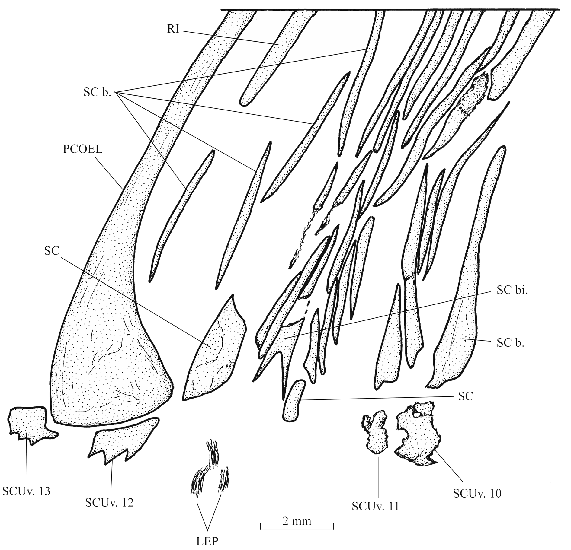
Squamation ( Figs 2 View Fig. 2 , 10–11 View Fig. 10 View Fig. 11 )
Flank scales are present only in the abdominal region of the body. Dorsally, there is a pair of elongate scale bars associated to each dorsal ridge scute, except the last one. The squamation is missing in the middle region of the body. Ventrally, the flank scale bars are shorter but more numerous. The most ventral scales are small and triangular in shape. They are associated with the ventral keel scutes.
A small scale, probably bearing the lateral line canal, is visible below the brush-like process of the parietal.
There is a small narrow bifid cloacal scale and one broader postcloacal scale. The bifid cloacal scale is surrounded by a series of small scale bars. A long and rather broad scale is present just before the cloacal region.
The dorsal ridge is formed by 11 scutes. The first dorsal scute is articulated with the dermosupraoccipital and is longer than the following scutes. The first two elements of the series bear a small spine.
The ventral keel contains 13 scutes of which 11 prepelvic and two postcloacal associated to the postcoelomic bone. The 11 prepelvic scutes are very badly preserved and their shape is not determinable. The first postcloacal scute exhibits three small spines.


No known copyright restrictions apply. See Agosti, D., Egloff, W., 2009. Taxonomic information exchange and copyright: the Plazi approach. BMC Research Notes 2009, 2:53 for further explanation.