
Sigmapycnodus giganteus, Taverne & Capasso, 2018
|
publication ID |
https://doi.org/ 10.5852/ejt.2018.420 |
|
publication LSID |
lsid:zoobank.org:pub:F8E6FEEF-FC7A-4B40-AEEE-95DBC7BC61A4 |
|
DOI |
https://doi.org/10.5281/zenodo.5979277 |
|
persistent identifier |
https://treatment.plazi.org/id/3ED26D58-2200-4F11-A9C2-FD6046E3ADA1 |
|
taxon LSID |
lsid:zoobank.org:act:3ED26D58-2200-4F11-A9C2-FD6046E3ADA1 |
|
treatment provided by |
Plazi |
|
scientific name |
Sigmapycnodus giganteus |
| status |
gen. et sp. nov. |
Sigmapycnodus giganteus gen. et sp. nov.
urn:lsid:zoobank.org:act:3ED26D58-2200-4F11-A9C2-FD6046E3ADA1
Figs 12–22 View Fig. 12 View Fig. 13 View Fig. 14 View Fig. 15 View Fig. 16 View Fig. 17 View Fig. 18 View Fig. 19 View Fig. 20 View Fig. 21 View Fig. 22
Diagnosis
Large pycnodontid fish, with a moderately deep body. Head deeper than long, with a sigmoid frontal profile and vertically oriented snout and mouth gape. Orbit small. Frontal short. Prefrontal long and broad. Parietal with a brush-like process. No temporal fenestra. Dermosphenotic sutured to the skull roof. Dermopterotic hypertrophied. Deep and extremely enlarged dilatator fossa surrounded by dermosphenotic and dermopterotic. Vomer bearing rounded globular-shaped teeth. Maxilla ovoid, with a notch in the posterior margin. Prearticular with two rows of molariform teeth. Ectopterygoid present. A series of six short tubular infraorbitals linking the orbital region to the snout. Preopercle as deep as the exposed region of the hyomandibula-dermohyomandibula. Small coma-shaped opercle. Notochord completely surrounded by vertebral arches. Arcocentra in hypercomplex contact in the caudal region. Neural and haemal spines with a wing-like component. Forty-one neural spines anterior to the epichordal series. Seventeen haemal spines before the hypochordal series. Eleven or 12 pairs of ribs. Postcoelomic bone long and thin. Origin of dorsal fin anterior to origin of anal fin. Caudal peduncle long. Six epichordals. Eleven hypochordals, of which some moderately broadened. One urodermal. Scale bars and a few ventral complete scales in the abdominal region of the body.
Etymology
From the Latin ‘ giganteus , a, um ’, gigantic. The specific name refers to the huge shape of the new fossil fish.
Material examined
Holotype
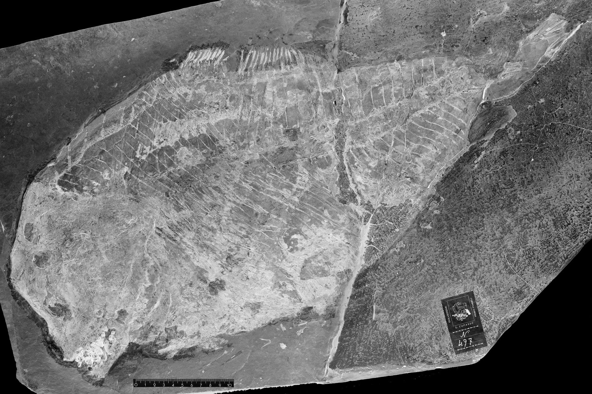
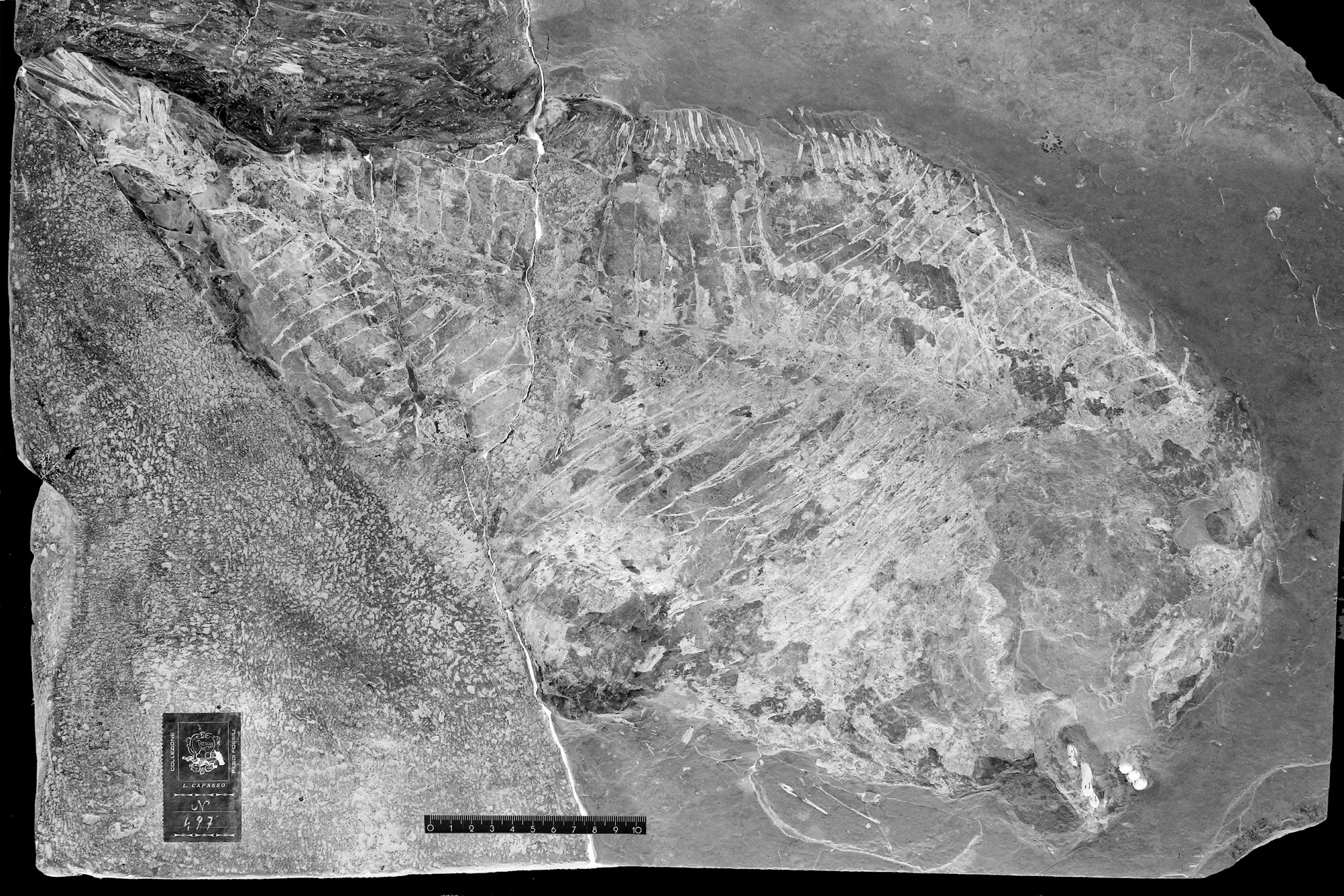
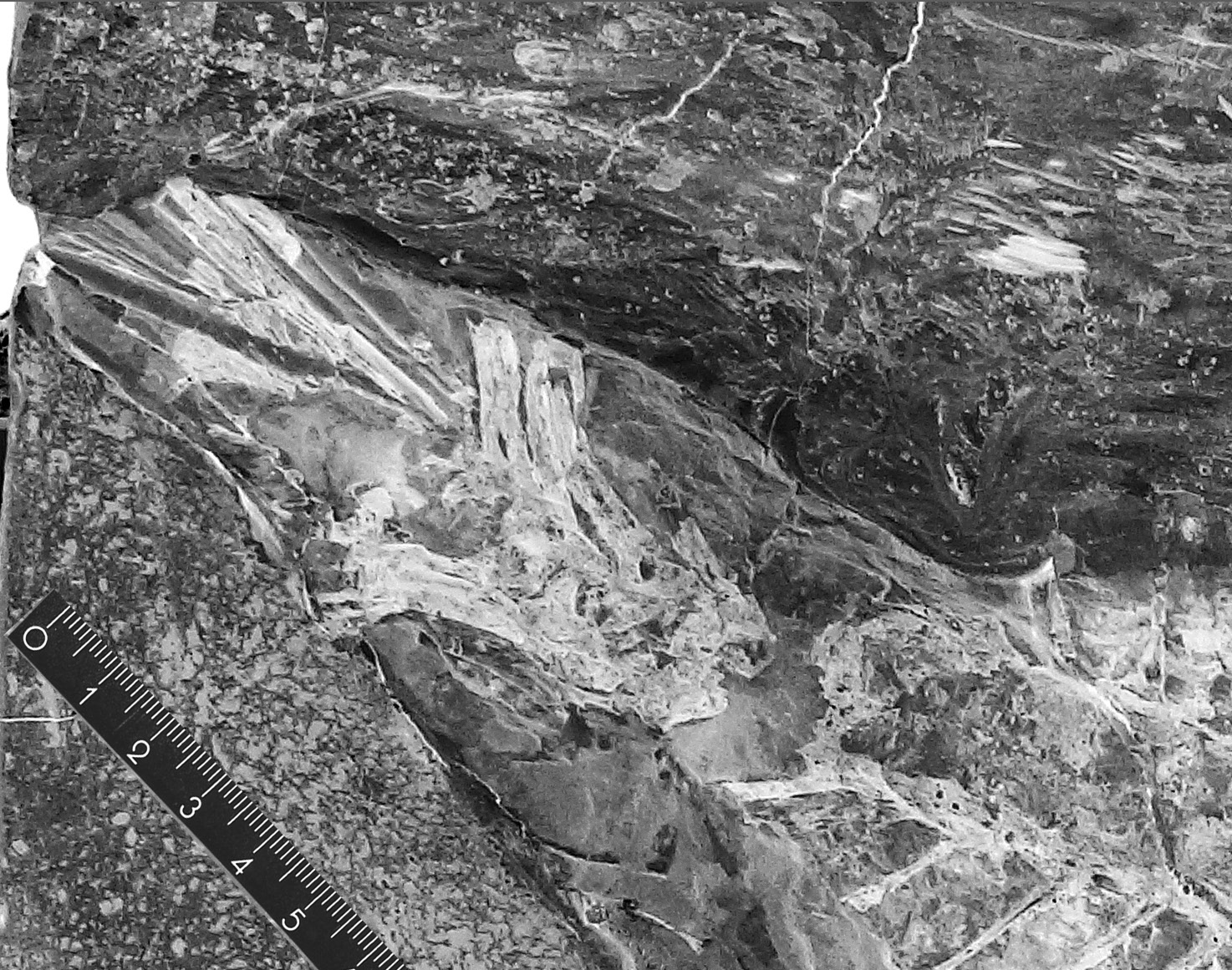
LEBANON: part and counterpart of a nearly complete specimen ( Figs 12–13 View Fig. 12 View Fig. 13 ), total length: 66 cm (the posterior part of the caudal fin is missing), standard length: 59cm, marine Upper Cenomanian deposits of Haqel ( CLC S-497a, b ).
General morphology and morphometric data ( Fig. 14 View Fig. 14 )
Sigmapycnodus giganteus gen. et sp. nov. is a large pycnodontiform fish. Its total length is assumed to be ca 70cm with the complete caudal fin. The body is not very high for a pycnodont, its depth being only equal to the half of the standard length.
The morphometric data are given in % of the standard length (59 cm):
Length of the head (opercle included) ……………………………………………………………24.5 %
Depth of the head (in the occipital region) ………………………………………………………38.1 %
Maximum depth of the body (just before the dorsal fin) …………………………………………49.8 %
Prepelvic length………………………………………………………………pelvic girdle not preserved
Predorsal length……………………………………………………………………………………47.2 %
Basal length of the dorsal fin………………………………………………………………fin incomplete
Preanal length ………………………………………………………………………………………58.5 %
Basal length of the anal fin …………………………………………………………………fin incomplete
Depth of the caudal peduncle………………………………………………………not entirely preserved
Osteology
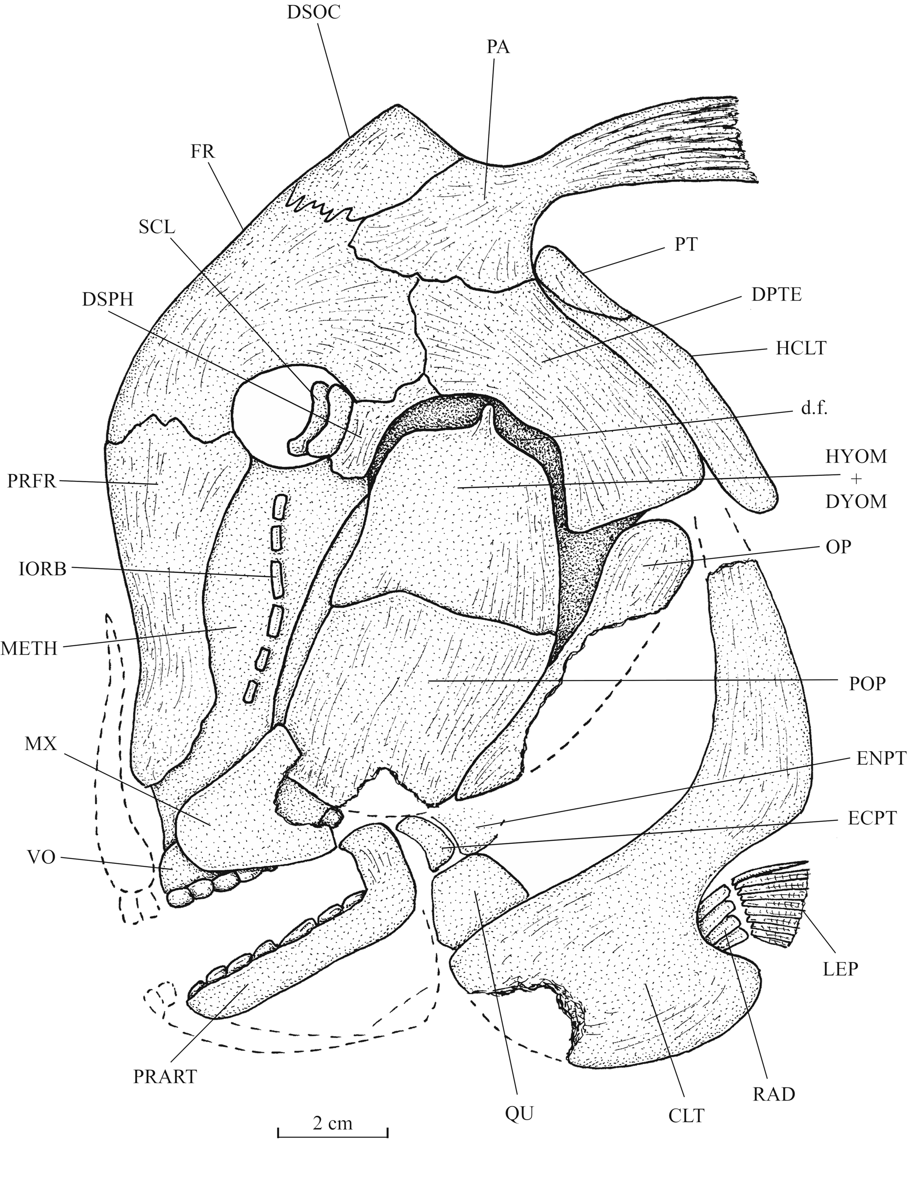
Skull ( Figs 15–19 View Fig. 15 View Fig. 16 View Fig. 17 View Fig. 18 View Fig. 19 )
The head is crushed and badly preserved. However, when combining the data given by the two slabs of the holotype, it is possible to reconstruct almost completely the skull.
The snout is short and vertically oriented. The frontal profile is sigmoid in shape, with a convex external margin at the level of the suture between the prefrontal and the frontal and a concave external margin lower on the prefrontal. The very small orbit is surrounded by the mesethmoid, the prefrontal, the frontal and the dermosphenotic. No endochondral bone of the braincase is visible, except the mesethmoid. The dermal bones of the skull bear some thin striae but this ornamentation is weakly developed.
The wide and deep mesethmoid is partly hidden by the long and broad prefrontal. The vomer is a large bone. Ten large molariform, rounded and globular-shaped teeth are visible. The first five are ranged in two rows and the posterior five in one row.
The frontal is short but rather broad. The dermosphenotic and the parietal are quite smaller. The parietal bears a long posterior brush-like process (= parietal peniculus) that is preserved as an imprint. The dermopterotic is hypertrophied, broad and high. There is no temporal fenestra. Only a small part of the parasphenoid is visible. The dermosphenotic is sutured to the dermopterotic and the frontal, becoming a lateral part of the dermal skull roof. The ventral margin of the dermosphenotic and of the dermopterotic is located at the level of the ventral border of the orbit. A deep and extremely enlarged dilatator fossa is present between the dermosphenotic and the dermopterotic. The autosphenotic and the sensory canals of the skull roof are not visible.
The quadrate, a part of the entopterygoid and a very small ectopterygoid are preserved on the slab b ( Fig. 13 View Fig. 13 ) of the holotype, but not the symplectic.
The maxilla is a large plate-like bone, broader posteriorly than anteriorly. There is a deep and broad notch in its posterior margin. The prearticular is long but rather narrow, with a strongly developed coronoid process. It bears at least two rows of molariform teeth still larger than those on the vomer. The dorsal row contains six teeth and the ventral row three teeth, with the last one longer than the others. These teeth are ovoid in shape with a more or less flat surface. The premaxilla, the dentary, the angular and the articular are not preserved.
A series of six small tubular infraorbitals, vertically oriented, is present along the mesethmoid. They connect the infraorbital and the rostral sensory canals. Paired sclerotic bones are visible in the orbit, close to the dermosphenotic.
The hyomandibula-dermohyomandibula and the preopercle are sutured together. The exposed part of the hyomandibula-dermohyomandibula has the same depth as the preopercle. As seen on slab a ( Fig. 12 View Fig. 12 ) of the holotype, the dorsal margin of the hyomandibula bears a small pointed process that penetrates the dilatator fossa.
A large hypohyal, a short anterior ceratohyal, a very small fragment of the posterior ceratohyal, two broad branchiostegal rays and a possible urohyal are preserved on slab b ( Fig. 13 View Fig. 13 ) of the holotype.
Girdles ( Figs 15–17 View Fig. 15 View Fig. 16 View Fig. 17 )
The short posttemporal and the long hypercleithrum (= supracleithrum) are pressed against the posterior margin of the dermopterotic. The cleithrum has a short but very broad ventral branch and a much narrower dorsal branch. There is a sinus in the posterior margin of the bone between the two branches to house the short pectoral fin. At least four pterygiophores are present. The number of pectoral rays is not determinable.
The pelvic girdle is lost, due to the bad preservation.
Axial skeleton ( Figs 12–14 View Fig. 12 View Fig. 13 View Fig. 14 , 20 View Fig. 20 )
The skeletal axis is badly preserved. In the rare places where the neural and haemal archocentra are more or less complete, these arches surround totally the notochord. In the caudal region of the body, the arcocentra are in hypercomplex contact, involving three or for pre- and postzygapophyses for each centrum. There are 41 neural spines before the epichordal series. Only 39 neural spines are visible on slab a ( Fig. 12 View Fig. 12 ), but two more posterior spines are present on slab b ( Fig. 13 View Fig. 13 ). It is not possible to see if the first neural spines were autogenous or not. There are at least 17haemal spines anterior to the hypochordal elementss but one or two spines seemingly are missing. There are 11 or 12 pairs of ribs. The postcoelomic bone is thin but very long.
Dorsal and anal fins
The dorsal fin origin is far posterior to the head. The fin is incomplete. The first 29 dorsal pterygiophores are preserved on slab a ( Fig.12 View Fig. 12 ). The basis of the first 11 rays is visible on slab b ( Fig.13 View Fig. 13 ).
The anal fin inserts at a posterior level than the dorsal one. Small fragments of a few anterior anal pterygiophores are present on slab b ( Fig. 13 View Fig. 13 ).
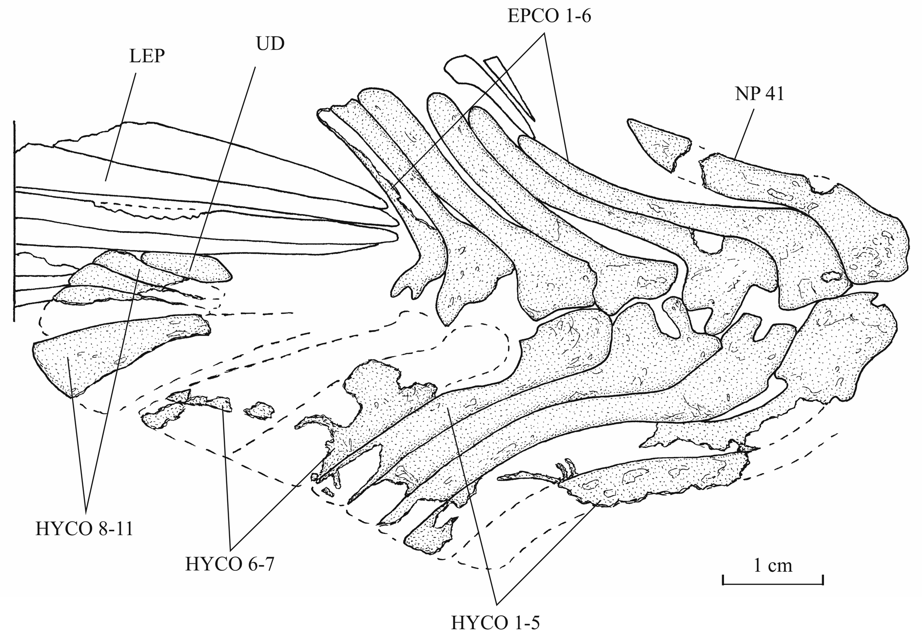
Caudal skeleton ( Figs 21–22 View Fig. 21 View Fig. 22 )
The caudal peduncle is well marked but its dorsal and ventral margins are not visible and its depth is not determinable. The caudal skeleton is much better preserved on slab b ( Fig. 13 View Fig. 13 ) than on slab a ( Fig. 12 View Fig. 12 ). There are six epichordals, 11 hypochordals and one urodermal. The epichordals are long and the first six hypochordals are comparably longer. The sixth and probably the seventh hypochordals are broadened but there is no real hypertrophy.
Only parts of a few caudal rays are preserved. The size and shape of the caudal fin are unknown.
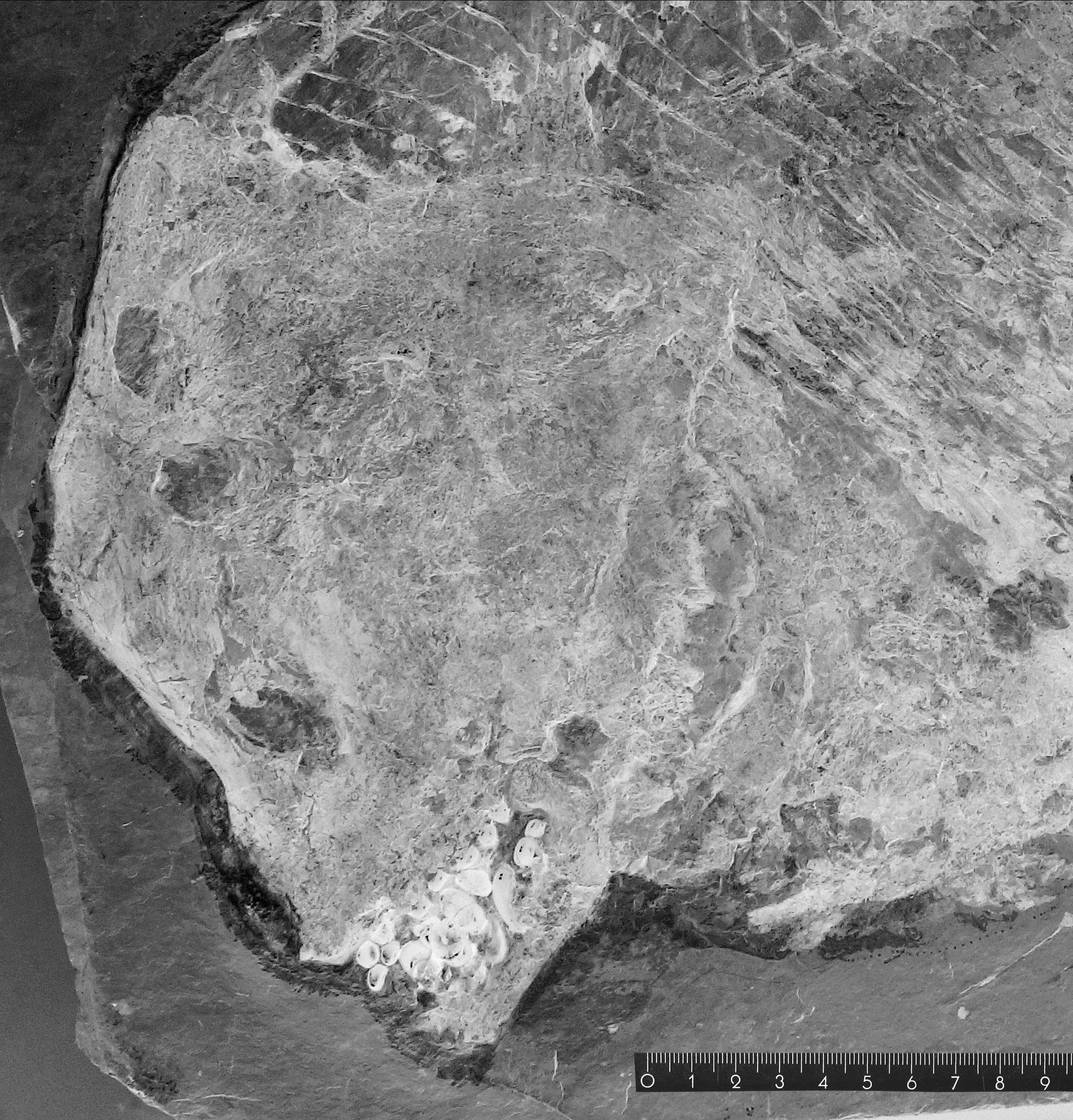
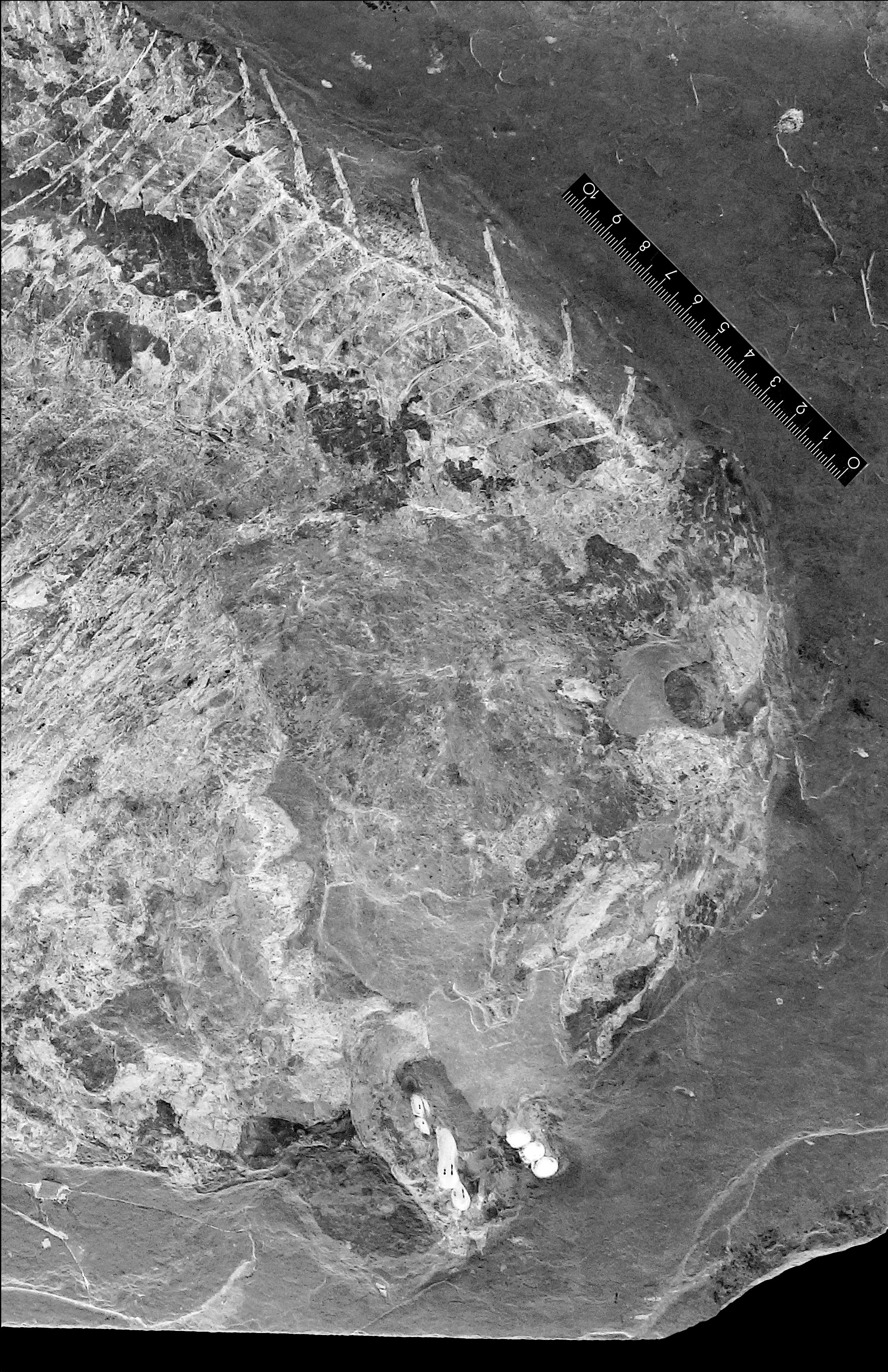
Squamation
The scales cover only the abdominal region of the body. Most of them are reduced to scale bars but ventrally a few complete scales are present.
Dorsal ridge scutes and the scales of the cloacal region are not preserved.
Some spiny scutes of the ventral keel are visible but the total number of these elements is not determinable.


No known copyright restrictions apply. See Agosti, D., Egloff, W., 2009. Taxonomic information exchange and copyright: the Plazi approach. BMC Research Notes 2009, 2:53 for further explanation.