
Pseudoscopelus parini Prokofiev & Kukuev 2006
|
publication ID |
https://doi.org/ 10.11646/zootaxa.2710.1.1 |
|
persistent identifier |
https://treatment.plazi.org/id/852E9C20-FFC0-FFD8-FF3C-FE9248646568 |
|
treatment provided by |
Felipe |
|
scientific name |
Pseudoscopelus parini Prokofiev & Kukuev 2006 |
| status |
|
Pseudoscopelus parini Prokofiev & Kukuev 2006 View in CoL
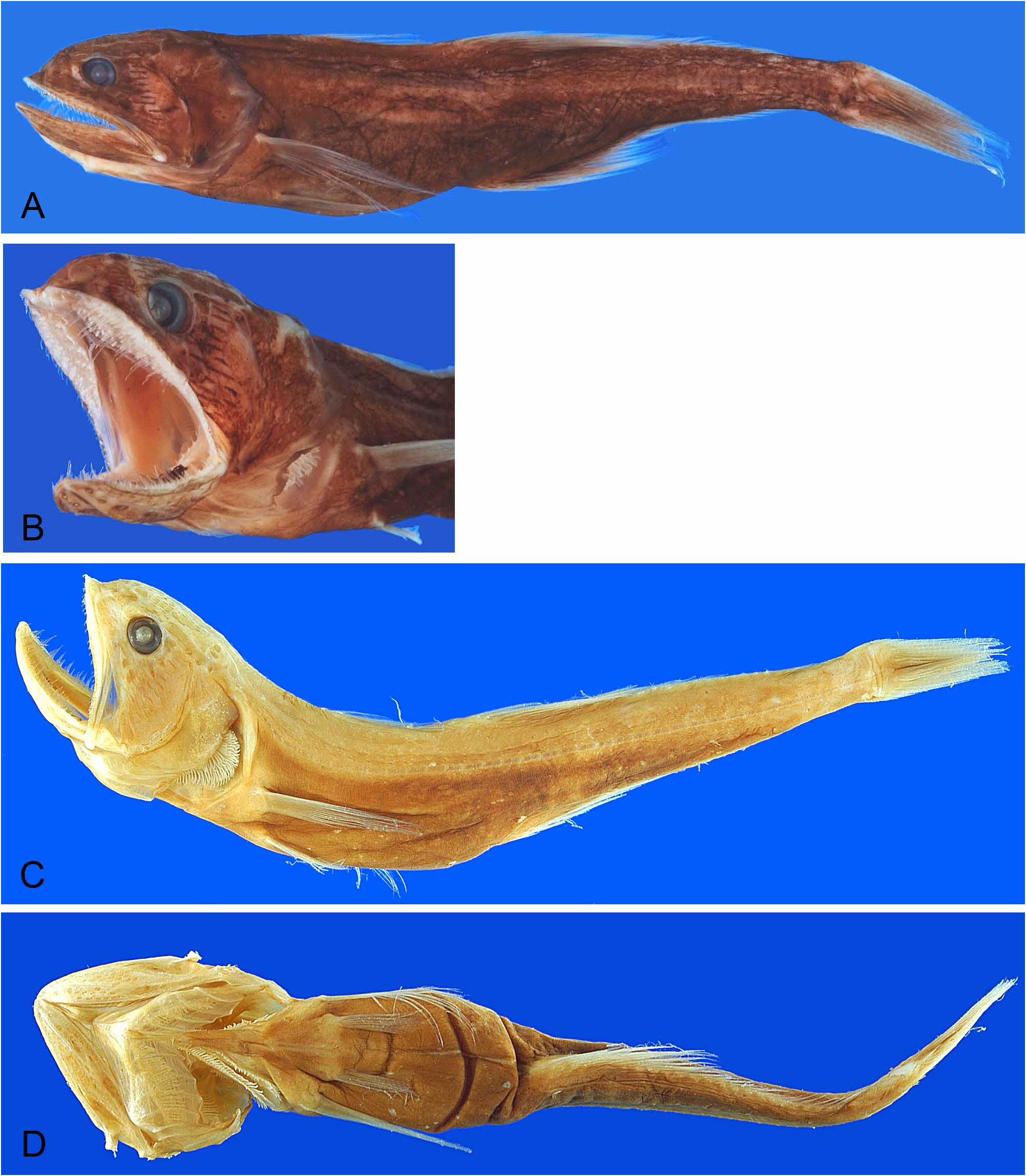
Figures 22 C–D View FIGURE 22 , 23 A View FIGURE 23 , 28 P; Table 8.
Pseudoscopelus parini Prokofiev and Kukuev 2006b: 481–484 View in CoL [type locality: western North Pacific, Marcus-Nekker Ridge, 24°07’ N, 150º00’ W, holotype ZMMU 21250, 168.0 mm]; Prokofiev and Kukuev 2008: 76–81, figures 15, 42, 68, 125–128 [species account].
Pseudoscopelus clarkei Lavenberg 1974: 211–222 , figures 45–46 [Pacific Ocean; name not available in accordance to the ICZN 1999].
Pseudoscopelus sp. Prokofiev and Kukuev 2008: 135-136 [species diagnosis].
Pseudoscopelus vityazi Prokofiev and Kukuev 2008: 158–162 View in CoL , figures 101, 197–200 [type locality: Indian Ocean, 15°07’ N, 150º00’ W, 3400–3473 m, holotype ZM MGU nr. 21254, 46.0 mm]. NEW SYNONYM.
Diagnosis. A species of the Pseudoscopelus aphos species group, which can be distinguished from P. aphos by the presence of whitish lines in the ventral part of body (vs. photophores completely absent in P. aphos , and small discrete circular photophores present on body and head in other species).
Description. Moderate-sized species of Pseudoscopelus , largest specimen examined 82.9 mm SL. Morphometric data summarized in Table 8. General body shape as described for genus with diagnostic characteristics of species and species group.
First dorsal-fin rays vi (1), vii (1), ix (5); second dorsal-fin rays ii+23 (3), ii+24 (4); anal-fin rays iii+21 (3), iii+22 (3), iii+24 (1); pectoral-fin rays 13 (3), 14 (3), 16 (1); pelvic-fin rays I+5 (7); caudal-fin rays i+7+8+i (7). Branchiostegal rays 7 (7). Pre-caudal vertebrae 17 (1), 18 (5), 19 (1); total vertebrae 36 (7).
Lateral line complete; lateral-line pores 77 (4), 78 (3). Pores in temporal canal 2 (7); supratemporal canal 3 (7); otic canal 2 (7); supraorbital canal 5 (3), 6 (4); supranasal pore 3 (1), 4 (6); epiphyseal branch 3 (7); infraorbital canal 12 (7); preopercular canal 5 (7); mandibular canal 6 (7); fifth pore of mandibular canal 1 (2), 2 (5).
Dentition. Enlarged teeth on premaxilla, dentary and palatine. Teeth arrangement as illustrated for Pseudoscopelus scriptus . Premaxilla moderately wide, widest point on body 15–20 % in premaxillary length.
Premaxillary teeth on head, neck, body and caudal process. Lateral series in single longitudinal row, along lateral edge of premaxillary head, neck, body and caudal process; teeth conical, slightly curved. Canine and fang on ventral shelf of premaxillary head. Middle and mesial series of premaxilla on ventral shelf of body. Middle series in two, irregular, longitudinal rows; teeth needle-like, straight, gradually increasing in size from lateral to medial. Mesial series in transverse rows, each row with three to four teeth; teeth needle-like, slightly curved, gradually increasing in size from medial to mesial.
Dentary teeth in lateral and mesial series. Lateral series along lateral shelf of dentary, in single, longitudinal row, extending from symphysis to posterior tip; teeth conical, slightly curved. Mesial series in transverse rows of one to four teeth; teeth straight, needle-like, gradually increasing in size from lateral to mesial. Palatine teeth 3 (1), 4 (2), 5 (3).
Teeth on infrapharyngobranchials and fifth ceratobranchial, conical, curved. Teeth on second basibranchial 15 (2), 19 (2), 21 (2), 25 (1), conical, in two rows. Teeth absent on basihyal and other basibranchials. Gill rakers on first epibranchial 0 (7); first ceratobranchial 8 (1), 11 (1), 13 (2), 14 (1), 15 (1), 16 (1); first hypobranchial 5 (2), 9 (3), 13 (1), 18 (1). Gill rakers absent on other elements.
Color. Most specimens examined faded to light brown or bleached white; description based on LACM 44392-1 and 56622-1. Body dark brown, except for triangular area on epiphyseal branch. Pectoral, pelvic, first and second dorsal fins, and anal fin slightly pigmented at their bases; caudal fin pigmented. Internal part of mouth and gill dark including skin on toothed area of premaxilla and dentary, over basihyal, roof, floor and lateral walls of mouth, internal part of opercle, membrane between dentaries and premaxillae; basibranchials, gill arches and filaments pale.
Luminescent organs. Photophores on head and body absent. White longitudinal lines present in place of if, prvf, ptvf, pf, paf, saf, and vf photophore groups.
Distribution. In the Pacific Plate and West Pacific; from 19° N to 19° S, 150° E to 150° W ( Fig. 23 A View FIGURE 23 ).
Prokofiev (2008) recorded the species from the western Indian as the type locality for P.vityazi . Bathymetric distribution. Meso- to bathypelagic, from 310 to 1250 m (mean 802 m). The status of Pseudoscopelus vityazi . There is a lot of misleading information concerning the identity of
Pseudoscopelus parini . Prokofiev and Kukuev (2006b) based the description of P. parini on discoveries of
Nikolai V. Parin ( Parin et al. 1977; Parin 1978). A single specimen collected in 1977 was used in the description and their subsequent work ( Prokofiev and Kukuev 2008); its condition of fixation and preservation is as poor as the holotype of P. aphos (compare Prokofiev and Kukuev 2005, figure 6 A to Prokofiev and Kukuev 2006b, figure 1 A), and a re-fixation in 75% alcohol [sic] was proposed by Prokofiev and Kukuev (2008: 80). Moreover, the original description includes a number of errors, which are discussed below.
The first mention of P. parini made by Prokofiev and Kukuev (2005: 727) was during the description of Pseudoscopelus aphos , “in contrast to P. aphotos [sic] in the Pacific species, in place of photophores there are bands of light (probably luminescent tissue) and fewer rays in P [pectoral fin] (13 vs. 15)”. During the description of P. parini, Prokofiev and Kukuev (2006b) identified and described the whitish bands in place of photophores in the species. In the introduction (p. 481) they stated that “the existence of Pseudoscopelus that lack [sic] normally developed photophores, arranged in series, but having bands of presumably luminescent tissue have been documented in the literature (…). We obtained an adult specimen, obviously belonging to the same species”. Along the description (p. 483) the photophores were described as “a ring of blackish band tissue around the anus (…) base of pelvic fin (in place of group vaf photophores) (…) [and] the upper half of the base of A [anal fin, saf]. (…) There is a well-developed band of light tissue (…) in front of the base of A [if, prf, ptvf?], and along the lower margin of the pectoral fin (more at its base) [pf and paf]”. During the discussion, they added the presence of such structure “in the place of the prcf series, and (…) at the anal and paired fins”.
In Prokofiev and Kukuev (2006c: S21) Pseudoscopelus parini was regarded as “perhaps” the equivalent of Lavenberg’s (1974) “ P. clarkei ”, due to the whitish tissues in place of the photophores. Prokofiev and Kukuev (2008: 76) reiterated their previous conclusions, diagnosing P. parini by “no photophores or other kinds [sic] of luminous tissues (perhaps except a weak modified skin cover in some regions on the ventral region of the body)” and (p. 79) “luminescent tissues are completely absent, perhaps except for a few patches of modified skin tissue occurring at the bases of P [pectoral fin] and V [pelvic fin], on the belly in front of A [anal fin] and in place of the prcf series”.
Another serious problem from Prokofiev and Kukuev (2006b) is the erroneous counts second dorsal-fin rays (I, 25) and anal-fin rays (II, 24) [no differenciation was made between a soft-ray and a spine thorough the description]. The counts were corrected in Prokofiev and Kukuev (2008) with second dorsal-fin rays 23 (total elements) and 23 (total elements), but this time no discrimitation between unbranched and branched rays was made. Moreover, faded color of the holotype, made the authors to believe that the specimen was yellowish-tan to dark brown, with lighter head and yellowish mouth. Such coloration is typical for specimens kept in ethanol for long time, especially when they are in such poor condition of preservation. Upon request, Prokofiev (pers. comm., VIII–2006), sent me a few pictures of ventral parts of the holotype of P. parini to illustrate the whitish bands of tissue.
Surprisingly, Prokofiev and Kukuev (2008: 135–136) introduced Pseudoscopelus sp. based on Lavenberg (1974) diagnosis to P. clarkei , but listed “none” in the material examined. Latter, in the addendum, Prokofiev and Kukuev (2008: 158–162) described P. vityazi listing the “photophores absent but replaced by trough-like bands of luminous tissue” as diagnostics; i.e., the same characteristic used for P. parini . Once again, it was used a single, poorly preserved specimen in the description, and the illustrations provided are of low quality. On a species key, (p. 137), P. aphos was distinguished from P. parini by the dark-brownish versus yellowishwhite [sic] orobranchial cavity. Those characteristics, however, were erroneously evaluated for both species (see further discussion under P. aphos ).
With so many mistakes and such confusing diagnosis it is hard to rely on any data presented for Pseudoscopelus parini and P. viatzy by Prokofiev and Kukuev (2006b, 2008). It is clear for me that Pseudoscopelus parini and P.vityazi are the same species which was described twice using the same diagnostic characteristics. Therefore, P. vityazi is herein formerly being placed in synonymy with P. parini .

No known copyright restrictions apply. See Agosti, D., Egloff, W., 2009. Taxonomic information exchange and copyright: the Plazi approach. BMC Research Notes 2009, 2:53 for further explanation.
|
Kingdom |
|
|
Phylum |
|
|
Class |
|
|
Order |
|
|
Family |
|
|
Genus |
Pseudoscopelus parini Prokofiev & Kukuev 2006
| Melo, Marcelo R. S. 2019 |
Pseudoscopelus sp.
| Prokofiev, A. M. & Kukuev, E. I. 2008: 135 |
Pseudoscopelus vityazi
| Prokofiev, A. M. & Kukuev, E. I. 2008: 162 |
Pseudoscopelus parini Prokofiev and Kukuev 2006b: 481–484
| Prokofiev, A. M. & Kukuev, E. I. 2008: 76 |
| Prokofiev, A. M. & Kukuev, E. I. 2006: 484 |
Pseudoscopelus clarkei
| Lavenberg, R. J. 1974: 222 |