
Pseudoscopelus obtusifrons ( Fowler 1934 )
|
publication ID |
https://doi.org/ 10.11646/zootaxa.2710.1.1 |
|
DOI |
https://doi.org/10.5281/zenodo.5459817 |
|
persistent identifier |
https://treatment.plazi.org/id/852E9C20-FFEB-FFF3-FF3C-FB8F4FFB6540 |
|
treatment provided by |
Felipe |
|
scientific name |
Pseudoscopelus obtusifrons ( Fowler 1934 ) |
| status |
|
Pseudoscopelus obtusifrons ( Fowler 1934) View in CoL
Figures 5 B View FIGURE 5 , 7 B View FIGURE 7 , 8 B View FIGURE 8 , 10 B View FIGURE 10 , 11 A–E View FIGURE 11 , 28 D; Table 4.
Myersiscus obtusifrons Fowler 1934: 362 View in CoL , figures 112–3 [type locality: western Central Pacific GoogleMaps , Sulawesi, Indonesia, 3º32’ S, 120º31’ E].
Pseudoscopelus obtusifrons de Beaufort in de Beaufort and Chapman 1951: 6–7 View in CoL [western Central Pacific]; Lavenberg 1974: 161–169, figures 14, 37–38, 40 [Indo-Pacific and Gulf of Mexico]; Moore et al. 2003: 227 [western North Atlantic]; Prokofiev and Kukuev 2006a: 225–228, figure 4 [western North Pacific, off Japan]; 2006c [key to species]; 2008: 89–102, figures 6, 7, 17, 43, 53, 59, 79–81, 84, 136–153 [redescription].
Pseudoscopelus obtusirostris Krefft 1971: 165 [misspelled].
Pseudoscopelus pauciporus Lavenberg 1974: 171–182 , figures 39–40 [ Indo-Pacific , Gulf of Mexico; name not available in accordance to the ICZN 1999 ].
Diagnosis. A species of the Pseudoscopelus scriptus species group, which can be distinguished from its congeners by two unique characteristics: spf present (vs. spf absent); and anterior teeth of lateral series in dentary and premaxillary hook-like curved, inserting on the lateral edge of bone and positioned outside of mouth so that teeth are visible in ventral and dorsal views (vs. lateral series of dentary and premaxillary teeth straight, or if curved, not hook-like, and inserted at oral edge of bone, not allowing them to be seen in dorsal or ventral views).
Description. Middle-sized species, largest specimen examined 144.0 mm SL. Morphometric data summarized in Table 4. General body shape as described for genus with diagnostic characteristics of species and species group.
First dorsal-fin rays ii (1), vii (2*), viii (9), ix (4); second dorsal-fin rays ii+17 (1), ii+18 (1), ii+19 (1), ii+20 (2), ii+21 (6), ii+22 (2), iii+19 (2*), xx (1); anal-fin rays ii+17 (1), ii+18 (2), ii+20 (1), iii+17 (2), iii+18 (1*), iii+19 (2), iii+20 (6), iv+18 (1); pectoral-fin rays 12 (6), 13(8*), 15 (2); pelvic-fin rays I+4 (1), I+5 (15*); caudal-fin rays i+7+8+i (15), i+7+7+i (1*). Branchiostegal rays 7 (16*). Pre-caudal vertebrae 16 (4), 17 (6*), 18 (2); total vertebrae 35 (1), 36 (11*), 37 (1).
Lateral line complete; lateral-line pores 52 (1), 57 (1), 60 (1), 62 (1), 63 (1), 65 (1), 67 (1), 69 (2), 70 (3*), 73 (2), 74 (2). Pores in temporal canal 2 (16*); supratemporal canal 3 (16*); otic canal 2 (16*); supraorbital canal 5 (16*); supranasal pore 1 (16*); epiphyseal branch 2 (14*), 3 (2); infraorbital canal 11 (14*), 12 (2); preopercular canal 5 (16*); mandibular canal 6 (16*); fifth pore of mandibular canal 1 (15*), 2 (16).
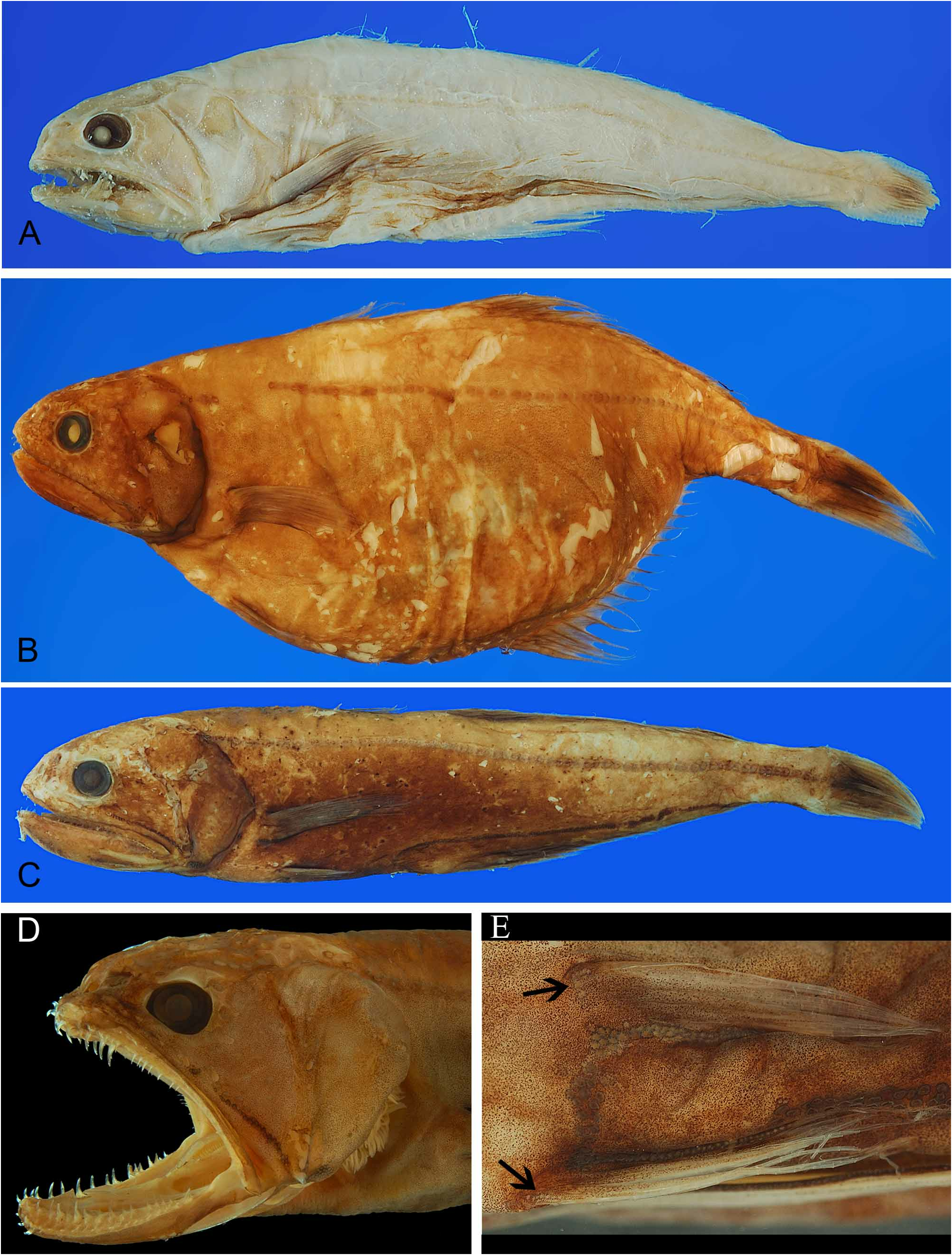
Dentition. Enlarged teeth on premaxilla, dentary and palatine. Premaxilla and dentary illustrated in Figure 5 B View FIGURE 5 . Premaxilla moderately wide, widest point of premaxillary body 10–14% in premaxillary length.
Premaxillary teeth on head, neck, body and caudal process. Lateral series in single longitudinal row, along lateral edge of premaxillary head, neck, body and caudal process; teeth on head, five to six, hook-like, attached to external edge of premaxilla, flared outwards; posterior teeth conical, curved caudally. Canine hook-like on antero-lateral shelf of dentary, flared outwards. Fang on ventral shelf of premaxillary head. Middle and mesial series on ventral shelf of premaxillary body. Middle series in single, longitudinal row; teeth conical, curved. Mesial series in single, longitudinal row; teeth conical, curved.
Dentary teeth in lateral and mesial series. Lateral series along lateral shelf of dentary, in single, longitudinal row, extending from symphysis to posterior tip; anteriormost four to six teeth hook-like, attached to external edge of bone, flared outwards; posterior teeth conical, slightly curved. Mesial series in single, longitudinal row, teeth curved, type 4. Palatine teeth in single, longitudinal row; teeth conical, slightly curved. Palatine teeth 3(2), 4 (1), 5 (4*), 6 (3), 7 (3), 8 (3); in single, longitudinal row.
Teeth on infrapharyngobranchials and fifth ceratobranchial, conical, curved. Teeth on second basibranchial 3 (2), 4 (1), 5 (3*), 6 (3), 7 (2), 8 (3), conical, in V -shaped or single, irregular row. Teeth absent on basihyal and other basibranchials. Gill rakers on first epibranchial 0 (14*); first ceratobranchial 3 (1), 5 (1), 6 (2*), 7 (2), 8 (3), 10 (1), 11 (1), 12 (1), 15 (2); first hypobranchial 3 (3), 4 (1), 5 (6), 6 (2), 8 (1), 9 (1). Gill rakers absent on other elements.
Luminescent organs. Luminescent organs present as discrete photophores on head and body ( Fig. 10 B View FIGURE 10 ). Photophores on head: apf, dnf, inof 1–2, lpf, opf, and pof absent; mxf elongated, in single row, parallel to maxilla, from level of posterior margin of eye to angle between preopercle and dentary; vnf in triangular patch, or straight line of few photophores; ppf in small patch on ventral edge of interopercle; amf in one to three rows, medial to mandibular canal, from first pore to halfway between third and fourth pores; pmf in one to three rows, lateral to mandibular canal, from halfway between fourth and fifth pores to halfway between fifth and sixth pores.
Photophores on body: scf and svf absent; rtf on single specimen (USNM 268492, Fig. 13 C View FIGURE 13 ); spf present in at least in one side in all specimens larger than 100.0 mm, from two to 16 small photophores; pf in single row along ventral-most pectoral-fin ray; paf in row of up to three photophores at pectoral-fin axil; vf in single row, along mesial pelvic-fin ray; lvf at base of first pectoral fin; vaf continuous with vf and trf, extending over base of pelvic rays 4–5, and first pelvic-fin ray; if and prvf continuous, in irregular two rows, from isthmus to anterior part of pelvic girdle; ptvf in two rows, from posterior half of pelvic fin to close to anus; trf laterally in single row, with medial circular group of photophores; saf in two rows, with smaller photophores closely spaced and ventral, and larger photophores widely spaced and dorsal, heart-shaped, extending to level of or slightly anterior to anus, and connected posteriorly; prcf in posterior half of peduncle, elongated, three-pronged, medial prong extending over anteriormost lower procurrent rays.
Color. Most specimens examined faded brown; holotype bleached white. Color description based on BMNH 2003.6.10.7. Body uniformly black, except for triangular area on epiphyseal branch. Pectoral, pelvic and first dorsal fins mostly hyaline, with few melanophores over fin-rays; second dorsal, anal and caudal fins with melanophores distributed over rays, more concentrated at fins base. Internal area of mouth and gill arches pale.
Distribution. In the western Atlantic, from the United States to Suriname; in the eastern Atlantic, from Canary Islands to off Liberia in the East; from 32º N to 20º S, 93º W to 14º W. A single record in the Indian Ocean, from 0º21’ S, 65º05’ E. In the Pacific Ocean, from Hawaii to east of Northfolk Islands, from 24º N to 26º S 120º E to 153º W ( Fig. 7 B View FIGURE 7 ).
Bathymetric distribution. Meso- to bathypelagic, from 124 to 2250 m (mean 718 m).
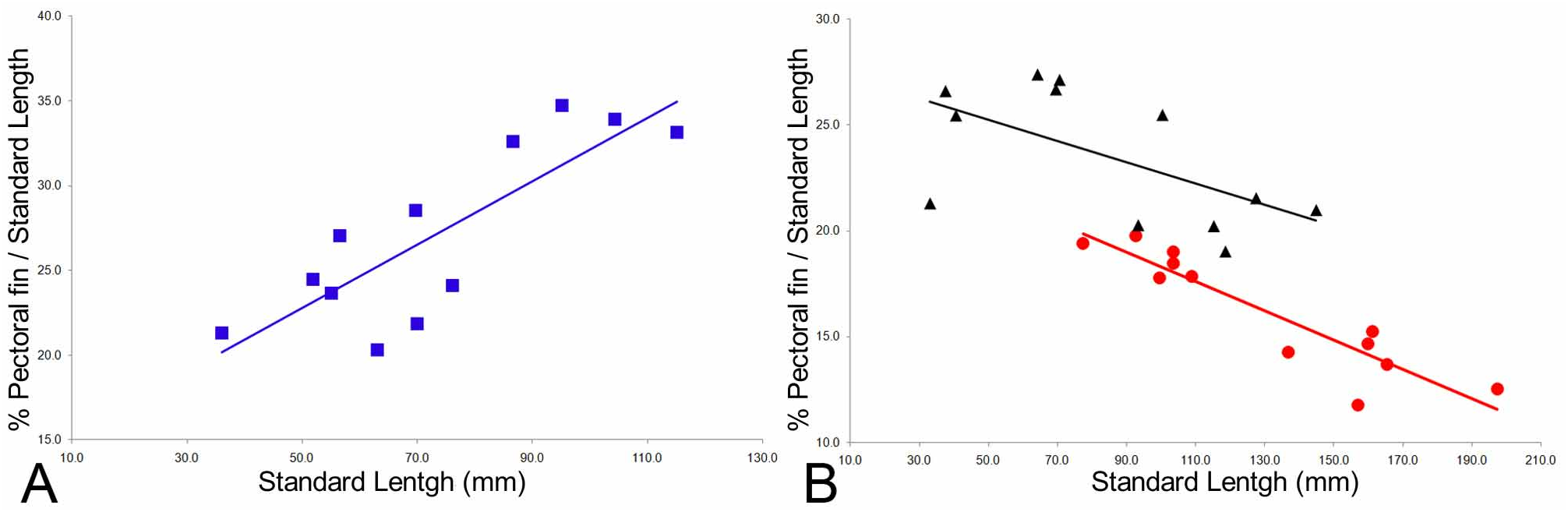
Ontogenetic changes. Pseudoscopelus obtusifrons has a negative, statistically significant allometric growth of the pectoral fin (p=0.042). The variation is illustrated in Figure 8 B View FIGURE 8 , and this explains the large standard deviation variation found for this characteristic (SD=3.2). Juveniles of at least 20.0 mm (e.g., USNM 200535) could already be identified because of their distinguishable hooked teeth.
Remarks. Lavenberg (1974) considered Pseudoscopelus obtusifrons as valid, but proposed the existence of a closely related species distinguished by fewer lateral-line pores: P. pauciporus . According to his conclusions, the distribution of the two species overlaps. Among the specimens of P. obtusifrons the lateral-line pores varies from 52 to 74; in two specimens (LACM 31528 and HUMZ 310746) the number of pores varies even from one side to the other. The number of pores in the lateral line forms a gradient without a clear break of means between populations. Lavenberg (1974) only listed two specimens for P. obtusifrons and five for P. pauciporus (vs. 24 specimens for this contribution), a little material for determining such gradient. The specimens listed by Lavenberg (1974) were examined and x-rayed, but no further evidences that P. obtusifrons should be divided into two species were found.


No known copyright restrictions apply. See Agosti, D., Egloff, W., 2009. Taxonomic information exchange and copyright: the Plazi approach. BMC Research Notes 2009, 2:53 for further explanation.
|
Kingdom |
|
|
Phylum |
|
|
Class |
|
|
Order |
|
|
Family |
|
|
Genus |
Pseudoscopelus obtusifrons ( Fowler 1934 )
| Melo, Marcelo R. S. 2019 |
Pseudoscopelus pauciporus
| Lavenberg, R. J. 1974: 182 |
Pseudoscopelus obtusirostris
| Krefft, G. 1971: 165 |
Pseudoscopelus obtusifrons de Beaufort in de Beaufort and Chapman 1951: 6–7
| Prokofiev, A. M. & Kukuev, E. I. 2006: 225 |
| Moore, J. A. & Hartel, K. E. & Craddock, J. E. & Galbraith, J. K. 2003: 227 |
| Lavenberg, R. J. 1974: 161 |
| de Beaufort, L. F. & Chapman, W. M. 1951: 7 |
Myersiscus obtusifrons
| Fowler, H. W. 1934: 362 |