
Microporella pauciperforata Chowdhury & Di Martino, 2024
|
publication ID |
https://doi.org/ 10.5852/ejt.2024.932.2509 |
|
publication LSID |
lsid:zoobank.org:pub:231BF669-4E64-4EAD-8305-4AEA0481D807 |
|
DOI |
https://doi.org/10.5281/zenodo.11030378 |
|
persistent identifier |
https://treatment.plazi.org/id/8561E974-BC32-FFBE-78D9-CD03FD04E1B3 |
|
treatment provided by |
Plazi |
|
scientific name |
Microporella pauciperforata Chowdhury & Di Martino |
| status |
sp. nov. |
Microporella pauciperforata Chowdhury & Di Martino sp. nov.
urn:lsid:zoobank.org:act:B17B841F-E7A3-4548-AFA7-A2B04E5A8674
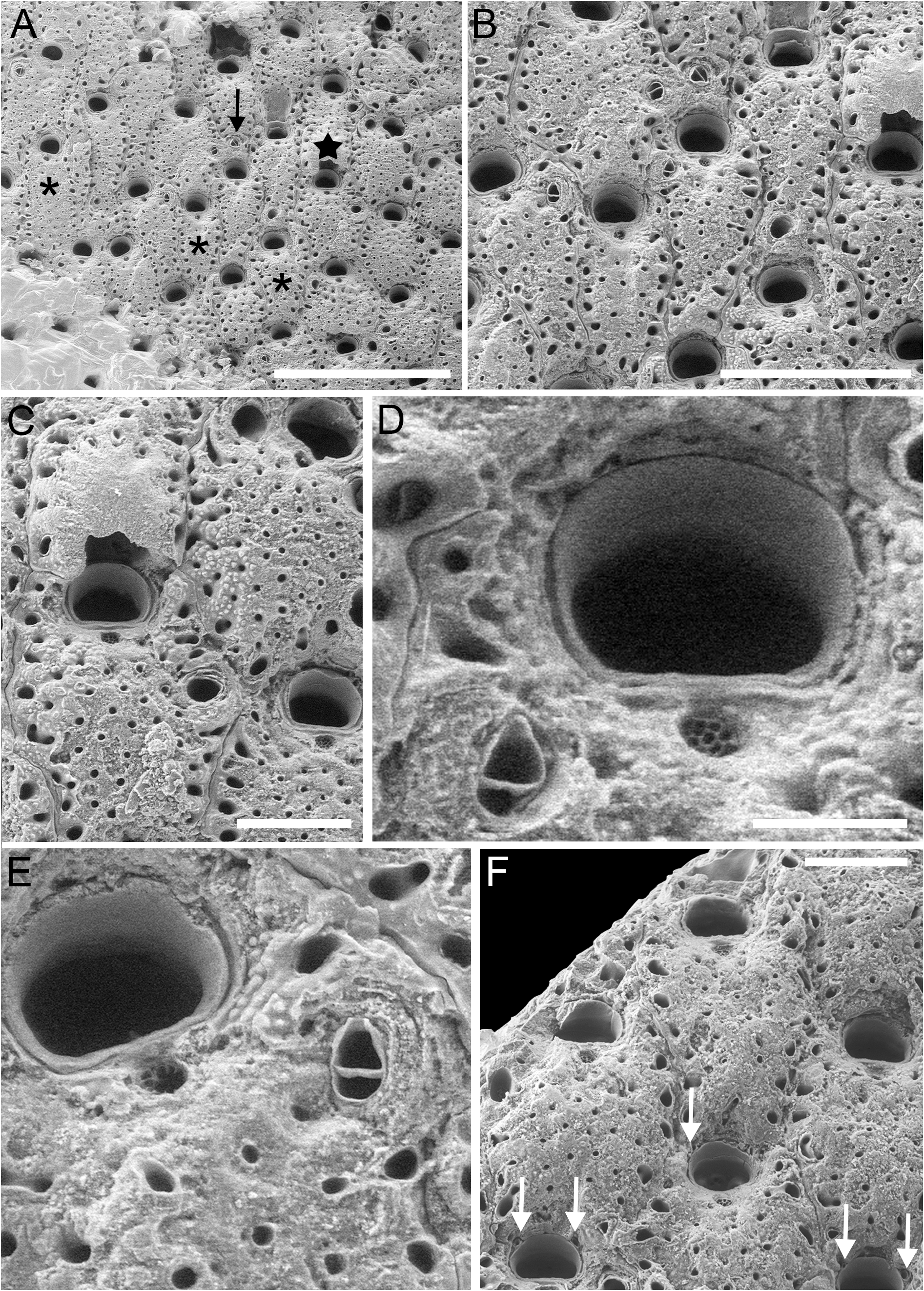
Fig. 3 View Fig , Table 2 View Table 2
Diagnosis
Encrusting Microporella ; zooids with single, small, distally directed adventitious avicularium, proximolateral to orifice, or lacking avicularium; some zooids with avicularium in proximolateral corner of zooid; primary orifice with smooth proximal margin and rounded corners lacking condyles; 2–4 ephemeral oral spines; frontal shield with relatively few pseudopores and distinct, elliptical marginal areolae; ascopore cribrate, close to the proximal margin of the orifice; ovicells rounded-quadrate, imperforate except for marginal pores.
Etymology
Latin ‘ paucus ’, few, plus ‘ perforatus ’, pierced, alluding to the limited number of pseudopores in the frontal shield of this species.
Type material
Holotype USA • colony of 40 zooids, one ovicellate, on rock; California, Trinidad Head North ; 41°3′25.1928″ N, 124°9′4.1826″ W; 7 Feb. 2020; I.A. Chowdhury and H. Lee leg.; SBMNH 704788 About SBMNH . GoogleMaps
Description
Colony encrusting, multiserial, unilaminar, forming a subcircular patch; pore chamber windows not observed.
Autozooids ( Fig. 3A–B View Fig ) generally elongate and hexagonal but sometimes irregularly shaped, delineated by narrow grooves and suture lines; ZL = 361–752 µm (523±99 µm, N = 22), ZW = 223–446 µm (323±59 µm, N = 22) µm, mean ZL/ZW = 1.62. Frontal shield ( Fig. 3B–C View Fig ) flat to slightly convex centrally, coarsely granular, with 13–36 circular pseudopores (D = 8–15 µm) sparsely scattered proximal to ascopore; 5–8 circular to elliptical, marginal areolae per side, clearly distinguishable from pseudopores because much larger (D = 20–55 µm).
Primary orifice transversely D-shaped, wider than long; OL = 64–109 µm (86±12 µm, N = 22), OW = 106–149 µm (120±10 µm, N = 22), mean OL/OW = 0.72, mean ZL/OL = 6.08; hinge-line straight, smooth, lacking denticles; proximolateral corners rounded without condyles. Two to four articulated spines visible in some zooids, often hidden by secondary calcification spreading from the distal zooid ( Fig. 3F View Fig ).
Ascopore depressed relative to adjacent frontal shield, transversely elliptical to subcircular, D = 22– 35 µm, close to orifice, roughly no more than 0.5–1.5 ascopore widths from proximal margin; ascopore in some zooids outlined by a rim of gymnocystal calcification that also encircles orifice; ascopore opening cribrate ( Fig. 3D View Fig ).
Avicularium usually single but often absent, located laterally on either side, in some zooids at zooidal mid-length, in some others at orifice level ( Fig. 3E View Fig ); AvL = 40–78 µm (52±11 µm, N = 11), AvW = 27– 57 µm (40±8 µm, N = 11) µm, mean AvL/AvW = 1.3; crossbar complete; triangular rostrum, usually directed distally but sometimes laterally, slightly depressed relative to adjacent frontal shield; in some zooids, slightly larger avicularium was observed in one of the proximolateral corners, directed proximally or proximolaterally inwards ( Fig. 3B View Fig ). Mandible not observed.
Ovicell prominent, globose, roughly as long as wide, with a rounded-quadrate appearance because narrower than frontal shield of underlying zooid, OvL = 272 µm, OvW = 282 µm (N = 1), OvL/ OvW = 0.96; continuous with frontal shield of the distal zooid, obscuring distal margin of orifice ( Fig. 3C View Fig ); calcification finely granular, smoother than the frontal shield, imperforate except for peripheral row of elliptical marginal areolae (12–36 µm, N = 10); peripheral interareolar ridges around margin.
Ancestrula not observed.
Remarks
The main features distinguishing M. pauciperforata Chowdhury & Di Martino sp. nov. from other congeners of the NE Pacific are the small, distally directed adventitious avicularia, the relatively flat frontal shield, and subordinately the rounded-quadrate ovicell, although we observed only one. In NW Pacific species of Microporella with a cribrate ascopore, the avicularium is usually larger [e.g., AvL 92–137 µm (112±14 µm, N = 20), AvW 39–65 µm (57±7 µm, N = 20) in an unregistered specimen of Microporella cribrosa from Korea measured from SEM image pdt4289 made available by Dr P.D. Taylor; AvL 71–91 µm (83±8 µm, N = 5), AvW 35–44 µm (40±4 µm, N = 5) in M. neocribroides measured from Dick et al. 2005: fig. 19] and directed distolaterally, while the ovicells are globular (e.g., M. neocribroides Dick & Ross, 1988 ) and pseudoporous (e.g., M. cribrosa Osburn, 1952 ) ( Osburn 1952; Dick & Ross 1988). In addition to the cribrate ascopore, M. pauciperforata resembles both M. cribrosa and M. neocribroides in having the ascopore close to the proximal margin of the orifice and the latter species in the limited number of pseudopores on the frontal shield. However, M. pauciperforata also has very distinct elliptical marginal areolae all along the lateral margins that are lacking in M. neocribroides .
The presence of avicularia located proximolaterally is an unusual feature for Microporella ; this might be induced by the presence of predators and the necessity to repair some boreholes as seen in colonies of M. hyadesi (Jullien, 1888) (see Di Martino et al. 2020: fig. 6c).
The signs of abrasion on the frontal shield seen in some zooids ( Fig. 3B–C View Fig ) might be due to the high wave action typical of the intertidal environment in which the colony was found. Similar signs of abrasion and/or dissolution were observed in fossil species of Microporella from the Florida Tamiami Formation ( Di Martino et al. 2019). In that case, the cause was unknown.
Distribution and ecology
Microporella pauciperforata Chowdhury & Di Martino sp. nov. is only known from a single specimen from Trinidad Head North in California, USA. The single colony was found encrusting a boulder in a highly exposed intertidal boulder field.
No known copyright restrictions apply. See Agosti, D., Egloff, W., 2009. Taxonomic information exchange and copyright: the Plazi approach. BMC Research Notes 2009, 2:53 for further explanation.
|
Kingdom |
|
|
Phylum |
|
|
Class |
|
|
Order |
|
|
SuperFamily |
Schizoporelloidea |
|
Family |
|
|
Genus |