
Microporella similis Chowdhury & Di Martino, 2024
|
publication ID |
https://doi.org/ 10.5852/ejt.2024.932.2509 |
|
publication LSID |
lsid:zoobank.org:pub:231BF669-4E64-4EAD-8305-4AEA0481D807 |
|
DOI |
https://doi.org/10.5281/zenodo.11035531 |
|
persistent identifier |
https://treatment.plazi.org/id/8561E974-BC35-FFA7-7894-CFDCFEF3E504 |
|
treatment provided by |
Plazi |
|
scientific name |
Microporella similis Chowdhury & Di Martino |
| status |
sp. nov. |
Microporella similis Chowdhury & Di Martino sp. nov.
urn:lsid:zoobank.org:act:FD102F85-A00A-44F7-BD69-744CF802D773
Fig. 6 View Fig , Table 2 View Table 2
Microporella cribrosa View in CoL – Soule et al. 1995: 144, pl. 50 figs a–c.
Diagnosis
Encrusting Microporella with interzooidal communications through multiporous septula; zooids with paired, rarely single, distolaterally directed adventitious avicularia, with crossbar at same level as ascopore; primary orifice with smooth, straight proximal margin, and small condyles at corners; 5–7 robust oral spines, two or none retained in ovicellate zooids; frontal shield tubercular, smooth, with reticulate pseudopores and distinct, elliptical marginal areolae; ascopore delicately cribrate with thin distal projection and eight-shaped/trifoliate pores, close to orifice, with proximal, smooth umbo, becoming squared and curved inwards; ovicells prominent, large, with numerous, well defined radial ribs, few pseudopores in grooves between ribs, and central to proximal smooth, imperforate, umbonate area.
Etymology
Latin ‘ similis ’, similar, alluding to the resemblance of this species to both Microporella cribrosa and M. rota Chowdhury & Di Martino sp. nov.
Type material
Holotype USA • colony of ca 100 zooids, several ovicellate, detached from the substrate; California, Los Angeles County, Velero 1232–41, 5 miles from San Pedro Breakwater ; depth 35 m; 33°38′15″ N, 118°12′15.00012″ W; 15 Feb. 1941; T.B. Scanland leg.; SBMNH 704271 About SBMNH . GoogleMaps
Paratypes USA • 1 colony of 30 zooids, several ovicellate, detached from the substrate; California, Orange County, Laguna Beach ; 33°32′31.2″ N, 117°47′16.8″ W; Allan Hancock Foundation leg.; SBMNH 702583 About SBMNH GoogleMaps • 1 colony of 40 zooids, none ovicellate, on echinoid spine; California, Los Angeles County, Channel Islands, Santa Catalina Island ; 33°27′00″ N, 118°25′0.00012″ W; 27 Aug. 1972; Allan Hancock Foundation leg.; SBMNH 703675 About SBMNH (slide 2) GoogleMaps .
MEXICO • 1 colony of 70 zooids, several ovicellate, detached from the substrate; Mexico, Bahia California (Norte), Bahia Todos Santos ; 31°47′59.997254″ N, 116°41′59.989014″ W; depth 25–30 m; 2 Jul. 1938; J.Q. Burch leg.; SBMNH 668408 About SBMNH GoogleMaps .
Additional material
USA • 1 colony of 40 zooids, several ovicellate, detached from the substrate; California, Marin County, Dillon Beach ; 38°15′00″ N, 122°56′59.989014″ W; R.J. Menzies leg.; SBMNH 668407 About SBMNH GoogleMaps • 1 colony of ca 100 zooids, several ovicellate, detached from the substrate; California, Monterey County, Lighthouse Point ; 36°37′59.97864″ N, 121°56′22.92″ W; intertidal; Nov. 1940; Allan Hancock Foundation leg.; SBMNH 673403 About SBMNH GoogleMaps • 1 colony of 60 zooids, several ovicellate, detached from the substrate; California, Ventura County, Channel Island, San Nicolas Island ; 33°14′57.99984″ N, 119°29′57.99984″ W; 26 Aug. 1976; Allan Hancock Foundation leg.; SBMNH 695087 About SBMNH GoogleMaps .
Description
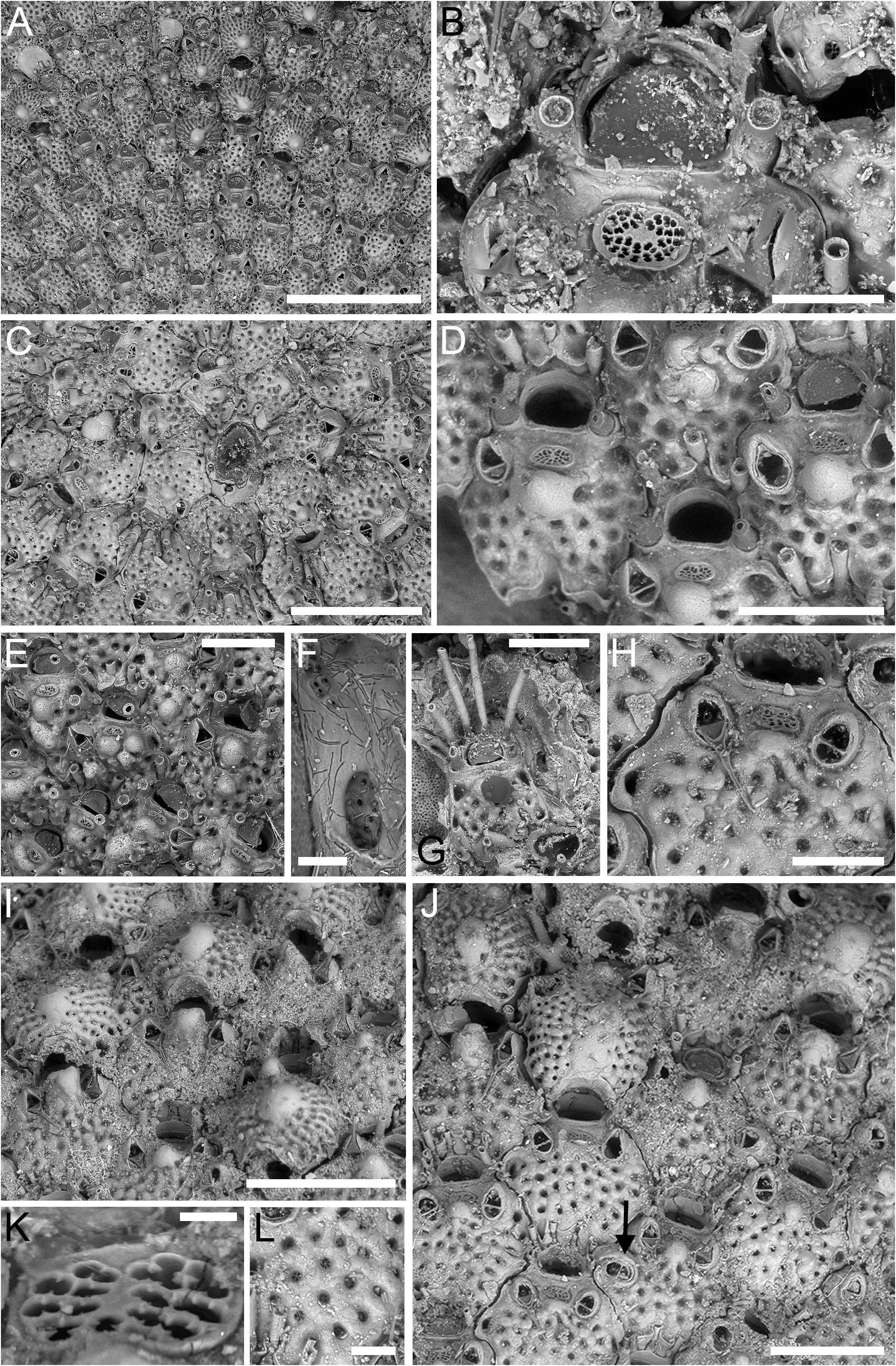
Colony encrusting, multiserial, unilaminar; interzooidal communications through multiporous septula ( Fig. 6F View Fig ).
Autozooids hexagonal, ZL = 398–563 µm (475±37 µm, N = 20), ZW = 259–467 µm (378±51 µm, N = 20), mean L/W = 1.26, boundaries marked by deep grooves ( Fig. 6A View Fig ). Frontal shield flat to slightly convex, tubercular, smooth, with elliptical marginal areolae (30–60 µm long) at zooidal lateral corners usually distinguishable from much smaller, circular pseudopores; pseudopores reticulate ( Fig. 6L View Fig ), D = 10–25 µm, numbering 20–30, evenly distributed on the frontal shield proximal to ascopore.
Primary orifice transversely D-shaped; OL = 75–97 µm (83±7 µm, N = 9), OW = 114–160 µm (138±14 µm, N = 15), mean OL/OW = 0.60, mean ZL/OL = 5.70; hinge-line straight, smooth, with small triangular condyles at corners ( Fig. 6D View Fig ). Five to seven robust, long (190–260 µm), articulated, oral spines visible in non-ovicellate zooids ( Fig. 6A, C, E, G View Fig ); two spines or none visible in ovicellate zooids ( Fig. 6A, I–J View Fig ); proximalmost pair of spines thicker, placed almost at level with the proximal margin of orifice ( Fig. 6B, D View Fig ); spines indenting proximal margin of distal zooid and becoming embedded until completely obliterated by frontal shield calcification ( Fig. 6D, J View Fig ).
Ascopore ( Fig. 6B, K View Fig ) moderately depressed or at level with adjacent frontal shield, transversely elliptical, D = 27–81 µm, less than one ascopore width from orifice; ascopore opening delicately cribrate with thin distal projection expanding proximally; pores of ascopore plate eight-shaped or trifoliate; smooth, low, knobby, rounded umbo developing proximally to ascopore, sometimes becoming quadrangular and curved inwards ( Fig. 6I View Fig ), hiding ascopore. In some zooids multiple umbones forming on frontal shield with age ( Fig. 6E View Fig ).
Avicularia more often paired ( Fig. 6A, D, H View Fig ), sometimes single ( Fig. 6D–E View Fig ); in two zooids third avicularium was observed at zooidal mid-length, budded from one of lateral areolae ( Fig. 6J View Fig , see arrow); relatively large, AvL = 77–115 µm (95±17 µm, N = 20), AvW = 56–80 µm (69±6 µm, N = 20), mean AvL/AvW = 1.38; located laterally in zooidal distal half, on either side, with thin complete crossbar usually at same level as ascopore; rostrum acutely triangular, narrowly channelled distally, directed distolaterally, distally raised. Mandible long (120–150 µm), with setose end, and pair of basal hooks ( Fig. 6H View Fig ).
Ovicell globular, prominent ( Fig. 6A, I–J View Fig ), large, OvL = 286–335 µm (306±18 µm, N = 6), OvW = 338– 430 µm (384±37 µm, N = 6), mean OvL/OvW = 0.80; occupying frontal shield of next distal zooid up to the proximal margin of avicularia; calcification smooth, radially ribbed, with about 20 well defined ribs, and five circular pseudopores, D = 5–20 µm, arranged in radial rows between ribs, extending from ovicell periphery but not reaching centre, which is occupied by more or less raised, smooth and imperforate umbo; in some ovicells umbo more proximally placed ( Fig. 6J View Fig ).
Ancestrula tatiform ( Fig. 6C View Fig ), elliptical, 370 µm long by 260 µm wide, with 10 circumopesial spines, arranged seven distolaterally closely spaced and three proximally widely spaced, indenting opesia outline which appears undulate, opesia 260 µm long by 185 µm wide, surrounded by seven zooids; first two zooids budded distolaterally, lacking avicularia.
Remarks
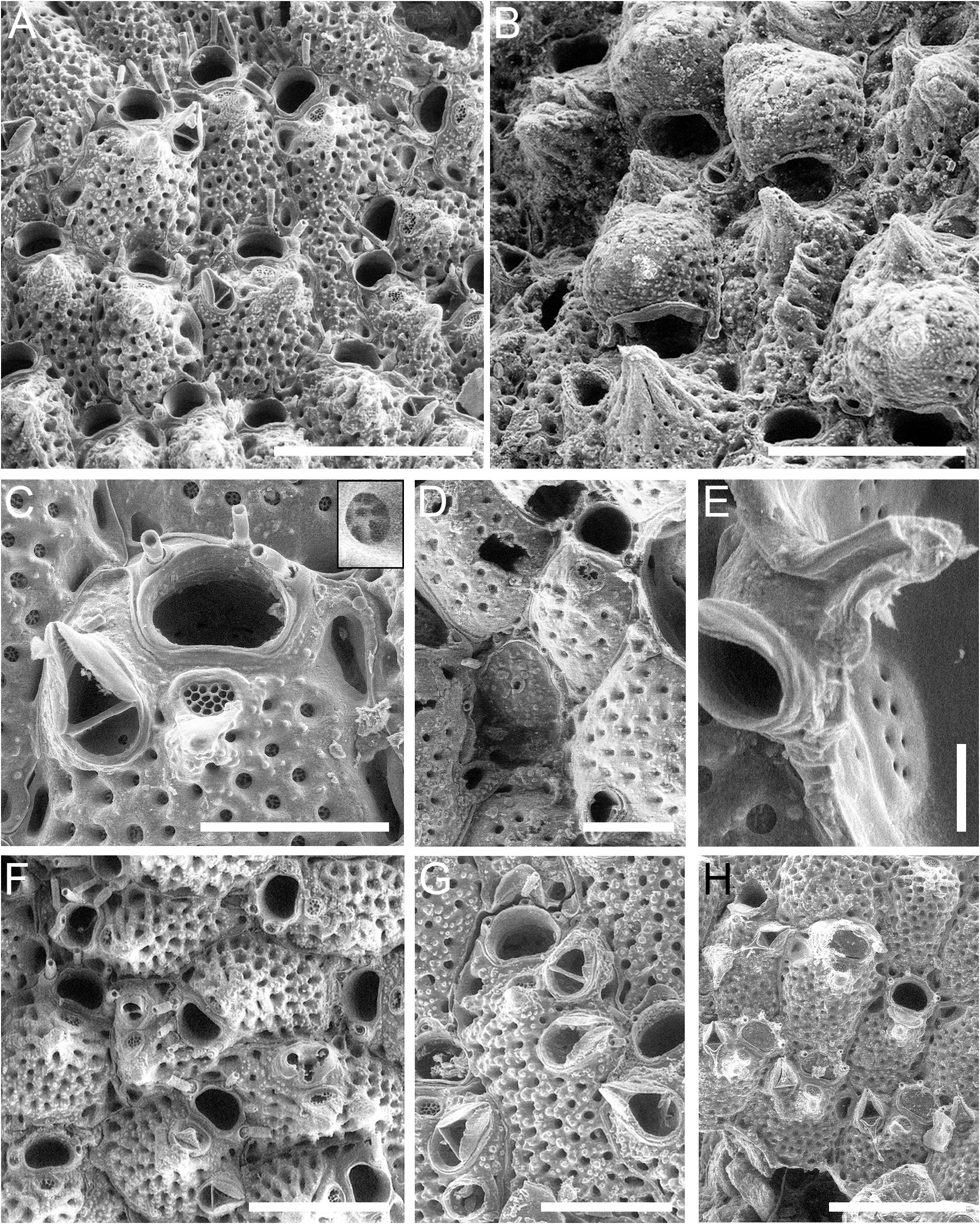
Microporella similis Chowdhury & Di Martino sp. nov. is similar to both M. cribrosa ( Fig. 5 View Fig ) and M. rota Chowdhury & Di Martino sp. nov. ( Fig. 4 View Fig ). Like M. cribrosa , it has robust spines ranging from five to seven and typically has paired avicularia; like M. rota , it has reticulate pseudopores. Nonetheless, there are notable distinguishing traits. Despite having a cribrate ascopore as the two aforementioned species, the one in M. similis differs from that of M. rota in having a distal projection expanding mid-proximally and a different shape of the cribrate plate pores, wich are mostly eight-shaped as well as trifoliate in M. similis but simply rounded in M. rota . In comparison to M. cribrosa , M. similis has a more delicate distal projection. Additionally, the frontal shield and ovicell surface of M. similis are tubercular and smooth, while in the other two species they are tubercular but also finely to coarsely granular. Ovicells of M. similis are larger, displaying a higher number of better defined ribs, while a lower number of pores are present in the grooves between ribs. In ovicellate zooids, the proximalmost pair of spines may either be retained, as observed in M. cribrosa , or obliterated, as seen in M. rota . The presence of a distinct, quadrangular umbo is another characteristic that sets this species apart.
Soule et al. (1995) reassigned to M. cribrosa specimens previously identified as M. californica by Robertson (1908). However, upon reviewing the description and illustration provided by Robertson (1908: 281, pl. 18 figs 32–34), particularly noting the depiction of the cribrate ascopore with delicate distal projection, it is likely that those specimens actually belong to M. similis Chowdhury & Di Martino sp. nov.
Following the SEM examination of the holotype of Microporella cribrosa , it becomes evident that it represents a complex of species. This evidence prompts the need for a comprehensive re-evaluation of prior records attributed to M. cribrosa , which may lead to the identification of additional colonies belonging to both M. rota Chowdhury & Di Martino sp. nov. and M. similis Chowdhury & Di Martino sp. nov., as well as potentially revealing the existence of more previously unidentified species. This might imply a more restricted distribution for M. cribrosa , suggesting also that it may not be among the commonest species of Microporella in California as previously thought, a notion supported by our sampling where we did not encounter any specimens of this particular species.
Distribution and ecology
To date, Microporella similis Chowdhury & Di Martino sp. nov. was recorded at three Pacific sites, two in California ( USA) waters (i.e., Dillon Beach and San Pedro Bay) down to 126 m depth ( Soule et al. 1995), and one in Mexican waters (i.e., Bahia Todos Santos) at 25–30 m depth. Studied specimens are usually detached from the substrate except in one instance in which the colony was encrusting an echinoid spine.
No known copyright restrictions apply. See Agosti, D., Egloff, W., 2009. Taxonomic information exchange and copyright: the Plazi approach. BMC Research Notes 2009, 2:53 for further explanation.
|
Kingdom |
|
|
Phylum |
|
|
Class |
|
|
Order |
|
|
SuperFamily |
Schizoporelloidea |
|
Family |
|
|
Genus |
Microporella similis Chowdhury & Di Martino
| Chowdhury, Ismael A., Martino, Emanuela Di, Lee, Hannah, Windecker, Claire C. & Craig, Sean 2024 |
Microporella cribrosa
| Soule D. F. & Soule J. D. & Chaney H. W. 1995: 144 |