
Microporella dentata Chowdhury & Di Martino, 2024
|
publication ID |
https://doi.org/ 10.5852/ejt.2024.932.2509 |
|
publication LSID |
lsid:zoobank.org:pub:231BF669-4E64-4EAD-8305-4AEA0481D807 |
|
DOI |
https://doi.org/10.5281/zenodo.11030372 |
|
persistent identifier |
https://treatment.plazi.org/id/8561E974-BC3F-FFB3-788F-CFEAFB38E347 |
|
treatment provided by |
Plazi |
|
scientific name |
Microporella dentata Chowdhury & Di Martino |
| status |
sp. nov. |
Microporella dentata Chowdhury & Di Martino sp. nov.
urn:lsid:zoobank.org:act:B70AFEDB-8620-4902-B464-17A652381A30
Fig. 2 View Fig , Table 2 View Table 2
Diagnosis
Encrusting Microporella ; zooids with single or paired avicularia with channelled rostrum; frontal shield tubercular, pseudoporous with indistinct marginal areolae and well developed central umbo; orifice with two prominent condyles and serrated proximal margin; four oral spines; ascopore cribrate, placed in a triangular depression, close to orifice; ovicells umbonate, with conspicuous radiating ridges, imperforate except for marginal pores.
Etymology
Latin ‘ dentatus ’, toothed, alluding to the denticles and prominent condyles present on the proximal margin of the orifice of this species.
Type material
Holotype USA • 1 colony of 50 zooids, several ovicellate, on inner surface of red abalone shell; California, MacKerricher State Park, Fort Bragg ; 39°29′25.764″ N, 123°48′8.748″ W; 2 Feb. 2020; I.A. Chowdhury and H. Lee leg.; SBMNH 704789 About SBMNH . GoogleMaps
Paratypes USA • 2 colonies of 40 zooids each, none ovicellate, on underside of small boulder; California, Greenwood; 39°7′45.0582″ N, 123°43′9.192″ W; 2 Feb. 2020; I.A. Chowdhury and H. Lee leg.; SBMNH 704790a–704790b GoogleMaps .
Description
Colony encrusting, multiserial, unilaminar, forming circular patches, found encrusting red abalone shells ( Haliotis rufescens Swainson, 1822 ) or small boulders.
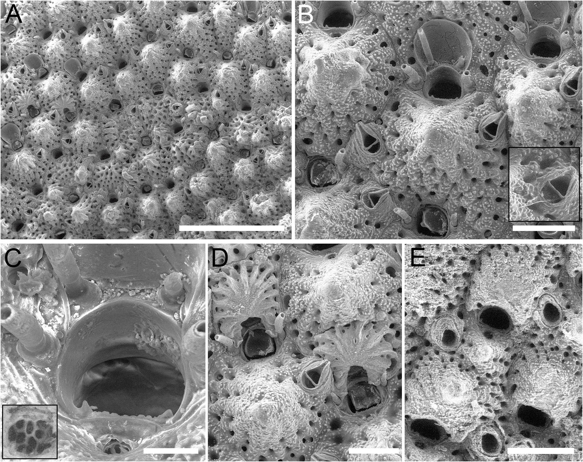
Autozooids rounded hexagonal to rectangular, ZL = 397–607 µm (494±54 µm, N = 20), ZW = 316– 522 µm (380±57 µm, N = 20), mean ZL/ZW = 1.30 ( Fig. 2A View Fig ), boundaries marked by grooves between slightly raised vertical walls. Frontal shield convex centrally, tubercular, coarsely granular, marked by radial ridges with circular pseudopores in grooves between ridges, 23–40 circular pseudopores, D = 7–20 µm; up to 10 circular to elliptical, marginal areolae per side, indistinguishable from pseudopores due to similar size.
Primary orifice transversely D-shaped, OL = 68–102 µm (86±8 µm, N = 20), OW = 84–116 µm (102±8 µm, N = 20), mean OL/OW = 0.84, mean ZL/OL = 5.74; hinge-line straight, serrated, with pair of sharply triangular condyles at corners ( Fig. 2C View Fig ). Four oral spines; proximalmost pair retained in ovicellate autozooids ( Fig. 2B, D View Fig ).
Ascopore in depressed triangular area outlined by distalmost ridges of frontal shield converging towards the centre ( Fig. 2C View Fig ) and forming the imperforate umbo proximal to ascopore ( Fig. 2B View Fig ); close to orifice, no more than 1–1.5 ascopore widths from proximal margin; ascopore opening 20–31 µm in diameter, cribrate, often obscured by proximal umbo ( Fig. 2C, E View Fig ).
Avicularium usually single, occasionally paired or absent, AvL = 58–112 µm (91±17 µm, N = 20), AvW = 35–55 µm (46±6 µm, N = 20), mean AvL/AvW = 1.98; located distolaterally on either side with rostrum tip at same level as ascopore or more proximally, proximolaterally to frontal umbo; crossbar complete; rostrum sharply triangular, narrowly channelled distally, with open, raised tip, directed distolaterally ( Fig. 2B, D View Fig ). Mandible triangular, slightly longer than rostrum (in Fig. 2B View Fig mandible length 90 µm, rostrum length 60 µm).
Ovicell prominent ( Fig. 2D–E View Fig ), rounded-quadrate, OvL = 172–201 µm (186±21 µm, N = 2), OvW = 200– 222 µm (211±15 µm, N = 2), mean OvL/OvW = 0.88, overlying frontal shield of next distal zooid, obscuring distal margin of orifice of maternal zooid; imperforate centrally, with a row of pseudopores all around the periphery inside the margin and areolae placed most-laterally; with radial ridges soon after formation but eventually covered by granulated frontal calcification from surrounding zooids and sometimes central, rounded, smooth umbo similar to that on the frontal shield.
Ancestrula not observed.
Remarks
The main character distinguishing this new species from the other species of Microporella is the serrated proximal margin of the orifice, which also bears two prominent, triangular condyles. The combination of a serrated orifice with a cribrate ascopore has been observed in Microporella elegans Suwa & Mawatari, 1998 from Japan. That species, however, differs in having reticulate pseudopores and avicularia directed laterally ( Suwa & Mawatari 1998). Microporella germana Dick & Ross, 1988 from Alaska is similar in the shape and position of the avicularium, in having a crenulated proximal oral margin, and in the number of spines; it differs in having an ascopore with lunate opening and smaller condyles ( Dick & Ross 1988). Additional similar species with a cribrate ascopore include: Microporella neocribroides Dick & Ross, 1988 from Alaska, which differs in having an orifice with smooth proximal margin and less prominent condyles ( Dick & Ross 1988); M. sanmiguelensis ( Soule, Chaney & Morris, 2004) which differs in having all zooids with paired avicularia ( Soule et al. 2004); M. santabarbarensis ( Soule, Chaney & Morris, 2004) , in which the ascopore is placed at the same level as the adjacent frontal shield rather than in a triangular depression ( Soule et al. 2004); and M. serrata Mawatari & Suwa, 1998 from Japan, in which the ascopore opening is reniform, rather than circular or elliptical ( Mawatari & Suwa 1998).
Distribution and ecology
Microporella dentata Chowdhur y & Di Martino sp. nov. is known from MacKerricher State Park beach to Pillar Point in California, USA. The colony in Fig. 2 View Fig encrusted a red abalone shell ( H. rufescens ), while the remaining colonies encrusted rocks, all in an exposed intertidal boulder field.
No known copyright restrictions apply. See Agosti, D., Egloff, W., 2009. Taxonomic information exchange and copyright: the Plazi approach. BMC Research Notes 2009, 2:53 for further explanation.
|
Kingdom |
|
|
Phylum |
|
|
Class |
|
|
Order |
|
|
SuperFamily |
Schizoporelloidea |
|
Family |
|
|
Genus |