
Heth, Cobb, 1898
|
publication ID |
https://doi.org/ 10.11646/zootaxa.4861.4.2 |
|
publication LSID |
lsid:zoobank.org:pub:8B330C85-5B40-48EF-8C17-48332637C1C9 |
|
DOI |
https://doi.org/10.5281/zenodo.4426644 |
|
persistent identifier |
https://treatment.plazi.org/id/8569BD6C-FF87-1274-31A9-1F37FA5FFEA5 |
|
treatment provided by |
Plazi |
|
scientific name |
Heth |
| status |
|
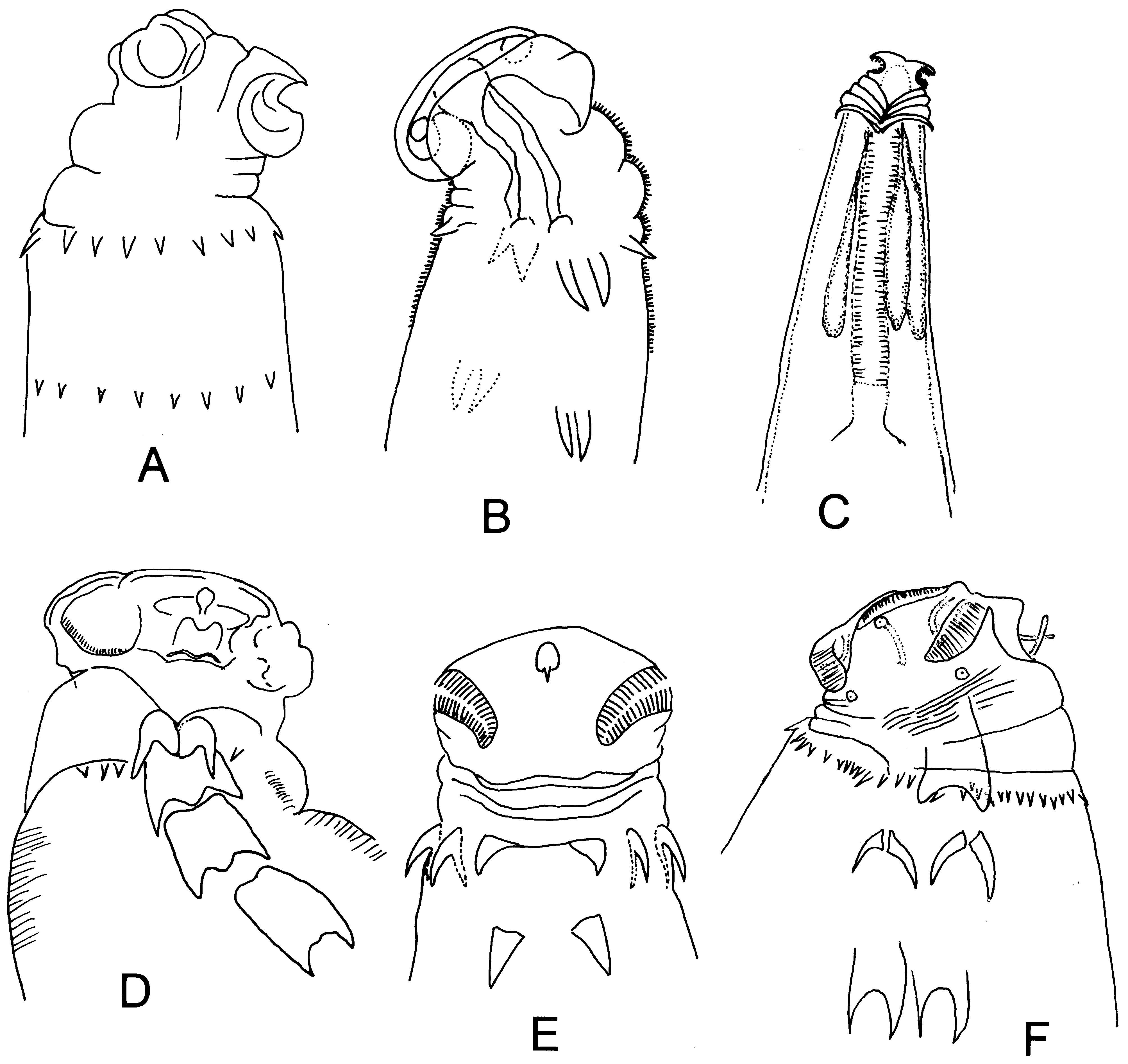
Key to group 3 female Heth spp.
1. Cervical collar with discontinuous spines; lateral spines present................................................ 2
- Cervical collar spines absent; base of most posterior fold in neck with small, single posteriorly curved spine ( Fig. 14C View FIGURE 14 ); females 1,140 –1,189 µm long......................................................................... H. spoliatus
2. Multiple somatic papillae present in anterior region; three pairs of lateral spines (cervical, anterior, posterior)............ 3
- Somatic papillae usually absent, rarely one or two in anterior region............................................ 5
3. Head about same width as anterior portion of body; each side of body with approximately 12 multi-cusped papillae flanking the cervical and lateral spines; cervical spines joined, with long, smooth sublateral extensions; lateral alae present ( Fig. 13E View FIGURE 13 ); females 1,800 –2,500 µm long..................................................................... H. imias
- Head narrower than anterior portion of body............................................................... 4
4. Sublateral cervical spine extensions short, smooth; anterior and posterior lateral spines fused at their bases; ( Fig 13C View FIGURE 13 ); females 1,640 –2,720 µm long......................................................................... H. baracoa
- Sublateral cervical spine extensions long, serrated; anterior and posterior spines slender, not fused at their bases ( Fig. 13D View FIGURE 13 ).......................................................................................... H. duvidosum
5. Head wide, about 1.5× width of anterior portion of body; cervical collar with about 12 small, well-separated spines of various sizes; one pair of lateral spines ( Fig. 13F View FIGURE 13 ); females 1,312 –1,600 µm long........................... H. macrocephala
- Head not enlarged, about same width as anterior portion of body............................................... 6
6. With cervical collar.................................................................................... 7
- With cervical collar of separate spines and a similar ring of spines posterior to collar ( Fig. 14A View FIGURE 14 ); females 2,142 –3,004 µm long..................................................................................... H. sinediscus
7. Four pairs of fused lateral spines on each side of body, increasing in length from anterior to posterior, approximately 10, 16, 23 and 32 µm long, respectively ( Fig. 14D View FIGURE 14 ); females 1,685 –1,890 µm .................................... H. travassosi
- Fewer than four pairs of lateral spines on each side.......................................................... 8
8. Cervical collar with more than 40 non-contiguous spines of similar length around circumference of neck; posterior to cervical collar ( Fig. 14F View FIGURE 14 ); females 2,400 –3,090 µm long..................................................... H. tuzetae
- Cervical collar with fewer than 40 discontinuous spines....................................................... 9
9. Transverse rows of minute cuticular spines encompassing anterior esophageal region; two pairs of lateral spines, their bases separate, 11-12 µm long ( Fig. 14B View FIGURE 14 ); females 1,831 –2,126 µm long................................... H. spinalatum
- Esophageal region minute cuticular spines................................................................ 10
10. Lateral spines separate; cervical collar with 8 spines around circumference of neck, interrupted by two lateral spines in the same plane as cervical spines; ( Fig. 14E View FIGURE 14 ); females 1,550 –1,830 µm long................................... H. travofilhoi
- Lateral spine bases touching or clearly fused............................................................... 11
11. Cervical collar formed by 10 large spines around circumference of neck; one pair of lateral spines ( Fig. 13G View FIGURE 13 ); female length 2,062 –2,186 µm ............................................................................ H. multiplus
- Lateral collar region plate-like with strong spine at each end.................................................. 12
12. One pair of lateral spines posterior to collar, broadly fused at base ( Figs. 13A, B View FIGURE 13 ); females 1,550 –1,830 µm long. H. artigasi
- Posterior lateral spines not broadly fused, bases meeting at a point............................................. 13
13. Vulva with overlapping anterior flap; area rugosa absent....................................... H. magnavulvaris
- Vulva without an anterior vulval flap; area rugosa present anterior to vulva ( Fig. 13H View FIGURE 13 )................... H. parartigasi

No known copyright restrictions apply. See Agosti, D., Egloff, W., 2009. Taxonomic information exchange and copyright: the Plazi approach. BMC Research Notes 2009, 2:53 for further explanation.
|
Kingdom |
|
|
Phylum |
|
|
Class |
|
|
Order |
|
|
InfraOrder |
Rhigonematomorpha |
|
SuperFamily |
Ransomnematoidea |
|
Family |