
Meterythrops, Smith, 1879
|
publication ID |
https://doi.org/ 10.1080/00222930600956858 |
|
persistent identifier |
https://treatment.plazi.org/id/860DB34F-2715-D520-CDC1-1CBDFBBEFD7A |
|
treatment provided by |
Carolina |
|
scientific name |
Meterythrops |
| status |
|
Meterythrops View in CoL sp. Murano, 1977
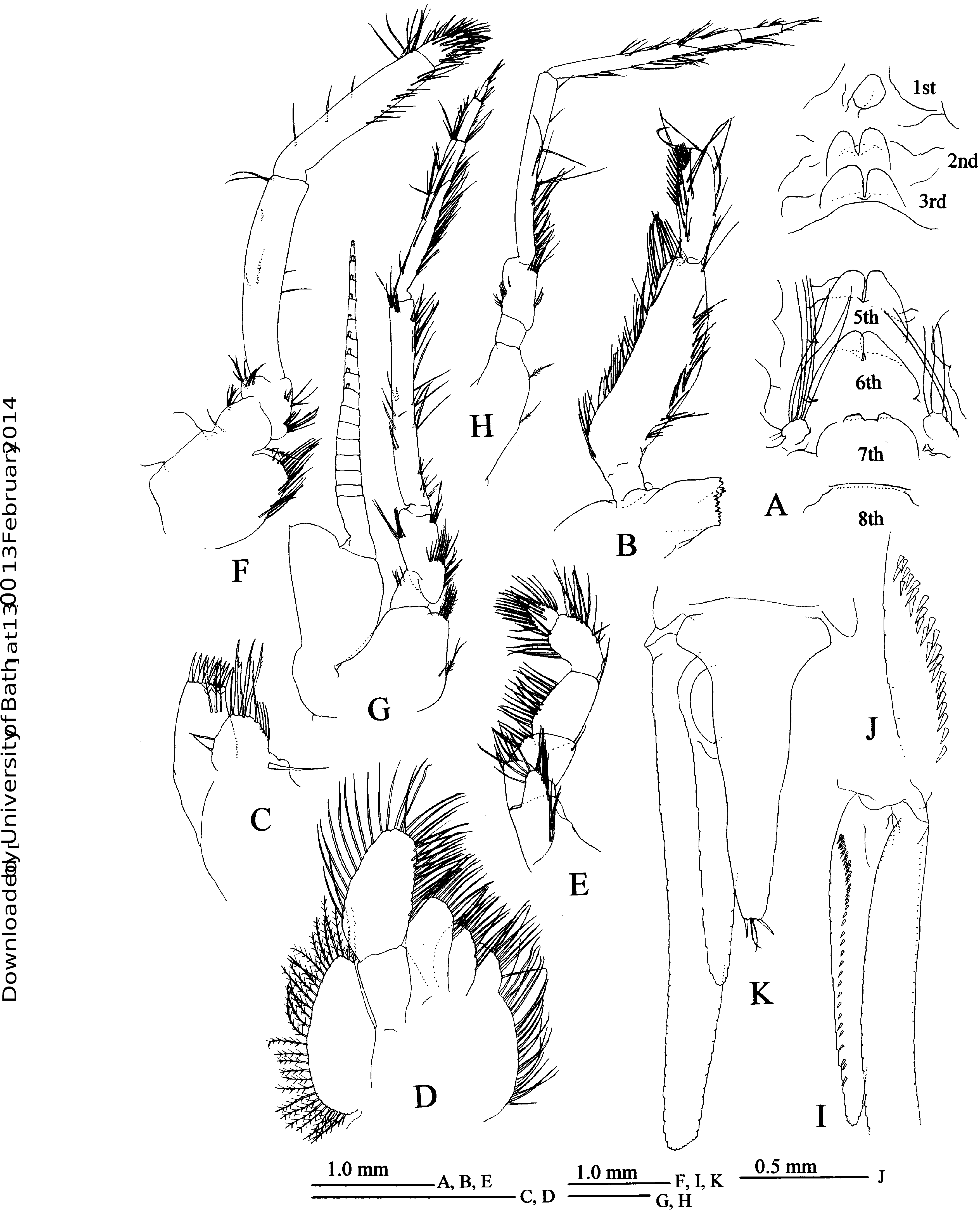
( Figure 7 View Figure 7 )
Meterythrops sp. Murano, 1977, p 177, Figure 24.
Material examined
Material reported by Murano (1977). NSMT-Cr 16813, two females (damaged, ca 25 mm), Stn TR 8, the Japan Sea (39 ° 29.39N, 134 ° 44.69E), from stomach of a tadpole sculpin, Malacocottus gibber , collected by a trawl from the sea floor at a depth of 1035 m, 2 June 1970, coll. M. Okiyama. GoogleMaps
Description of female
First to seventh thoracic somites with sternal process; process on first somite knob-shaped, those of second to seventh somites bilobed and flattened, those of fifth to seventh gradually becoming smaller towards posterior somite ( Figure 7A View Figure 7 ). Abdominal somites smooth; first somite broken, second to fifth somites subequal in length, sixth somite 1.4 times longer than fifth.
Carapace produced anteriorly into low triangular rostral plate with obtuse apex extending to base of eyes; anterolateral corner acute; posterior margin emarginate.
Eyes globular, as long as broad; cornea occupying half of eye in dorsal view.
Antennular peduncle robust, first segment slightly longer than broad, third segment 1.2 times as long as first.
Antennal scale extending beyond apex of antennular peduncle by two-fifths of its length, four times as long as broad; lateral margin smooth, terminating in spiniform process; apical lobe occupying three-eighths of scale length, three times as long as terminal spine of lateral margin, with indistinct subapical suture.
Labrum without frontal acute process.
Mandibular palp: second segment three times as long as broad; third segment more than half of second segment in length ( Figure 7B View Figure 7 ).
Maxillule: lateral lobe armed with 13 robust spines on distal margin and with three setae on ventral surface ( Figure 7C View Figure 7 ).
Maxilla: second segment of endopod 1.8 times as long as broad, armed with numerous long setae on margin; exopod extending to distal margin of proximal segment of endopod ( Figure 7D View Figure 7 ).
First thoracopodal endopod short and robust ( Figure 7E View Figure 7 ). Second thoracopodal endopod long, carpopropodus slightly shorter than merus, dactylus armed densely with setae and with claw on distal end ( Figure 7F View Figure 7 ). Third to eighth thoracopodal endopods long, slender; carpopropodus divided into three-subsegments, proximal subsegment occupying three-fifths of carpopropodus in length and articulated obliquely from middle subsegment, distal subsegment slightly shorter than middle and articulated transversely from middle one ( Figure 7G, H View Figure 7 ). Exopod of thoracopod with flagellum 14-segmented in first pair, 16- segmented in second to seventh pairs, and 15-segmented in eighth pair. Sixth thoracopod with reduced oostegite; seventh and eighth thoracopods with developed oostegite.
All pleopods reduced to unsegmented single lobe.
Uropodal endopod extending beyond apex of telson by one-fifth of its length, armed with 42 spines on mesial ventral margin from statocyst region to distal one-seventh; spines in statocyst region lined in double rows ( Figure 7 View Figure 7 I–K). Uropodal exopod 1.5 times as long as endopod ( Figure 7K View Figure 7 ).
Telson 1.3 times as long as last abdominal somite, twice as long as maximum width, abruptly narrowing near base, and then becoming gradually narrower toward apex; lateral margin naked throughout but finely serrated in distal half; apex narrowly truncated, armed with two pairs of spines and median pair of plumose setae ( Figure 7K View Figure 7 ).
Remarks
Murano (1977) differentiated Meterythrops sp. from M. robustus based on morphological differences in the antennal scale, uropodal endopod, and telson. It was ascertained through the present study that two characters previously reported by Murano (1977) for Meterythrops sp. correspond with M. robustus : the lateral margin of the telson is finely serrated in the distal half, and the uropodal endopod has two rows of spines in the statocyst region. However, the specimens differ from M. robustus by the large body size, globular eyes, length–width ratio of the antennal scale, and shape of the telson.
It is important to acknowledge that no male specimens have been collected to date, and thus this classification should be verified by future studies on male characters.
No known copyright restrictions apply. See Agosti, D., Egloff, W., 2009. Taxonomic information exchange and copyright: the Plazi approach. BMC Research Notes 2009, 2:53 for further explanation.