
Trichotrigona Camargo & Moure, 1983
|
publication ID |
https://doi.org/ 10.11646/zootaxa.3956.3.4 |
|
publication LSID |
lsid:zoobank.org:pub:6A8CA309-D8F4-44B6-BE13-7C53A23E62FB |
|
DOI |
https://doi.org/10.5281/zenodo.6118159 |
|
persistent identifier |
https://treatment.plazi.org/id/874DD440-2D2F-D97E-9189-AFAB7F90DFF5 |
|
treatment provided by |
Plazi |
|
scientific name |
Trichotrigona Camargo & Moure, 1983 |
| status |
|
Trichotrigona Camargo & Moure, 1983 View in CoL
Trichotrigona Camargo & Moure 1983: 421 View in CoL ; Michener 1990: 128; 1997: 59; 2000: 799; 2007: 823; Camargo & Pedro 2007a: 73; 2007b: 524
Type species: Trichotrigona extranea Camargo & Moure, 1983: 424 (by original designation).
Diagnosis. A diagnosis of the genus Trichotrigona is presented following Camargo & Moure, 1983 and Camargo & Pedro, 2007a, including modifications, in order to accommodate the new species, Trichotrigona camargoiana sp. nov. The main characters are highlighted in boldface lettering. The system of Moure (1961) was slightly modified in order to include new diagnostic characters unknown prior to the discovery of Trichotrigona , but the sequence of the structures was maintained as far as possible. The characters mentioned here, which are invariable among the species, are not repeated in the descriptions.
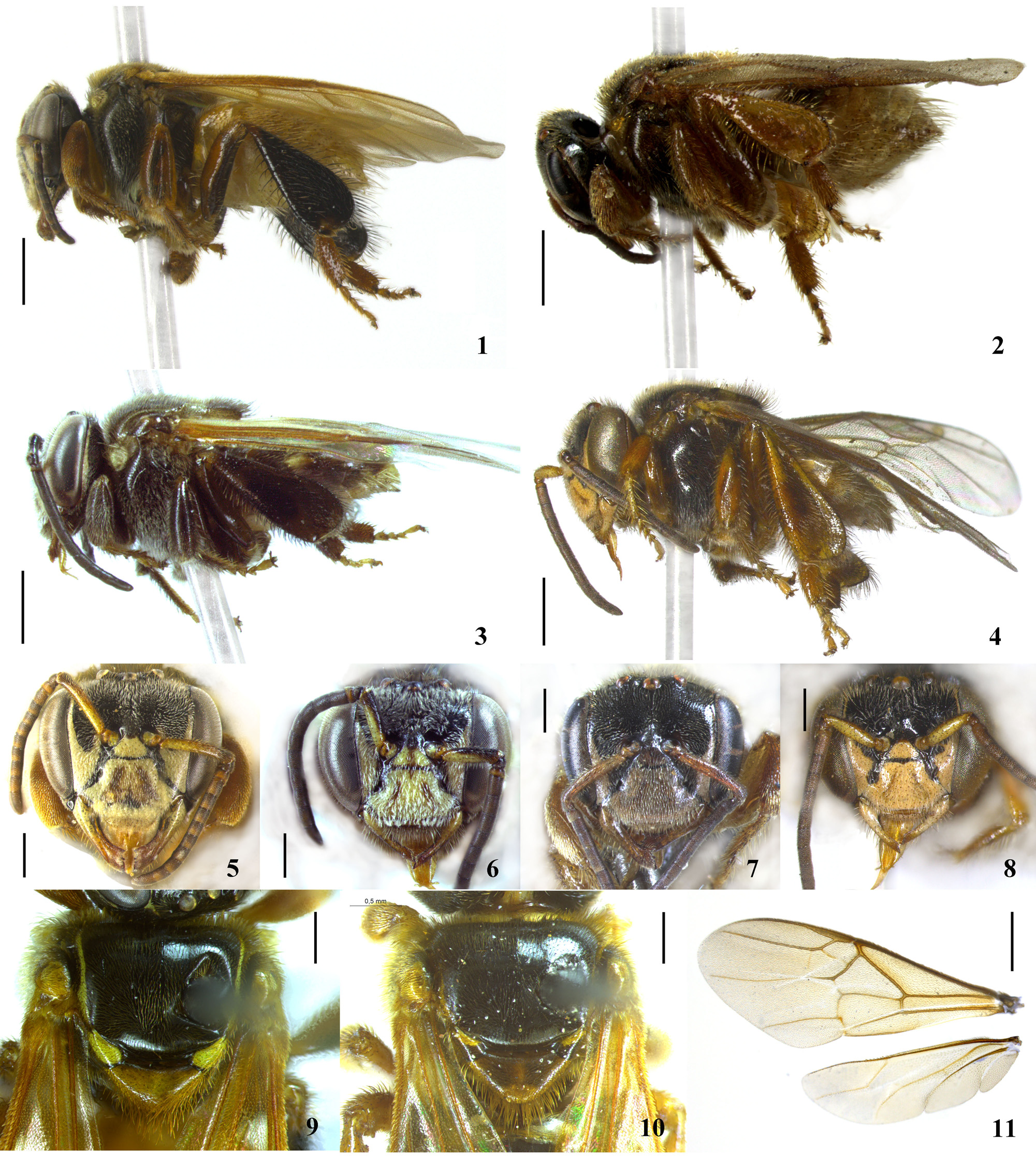
a) Integument smooth and shiny; piligerous punctures small and sparse, the space between punctures 3 to 4 x their diameter ( Figs. 9, 10 View FIGURES 1 – 11 ); yellow maculation pronounced on head ( Figs. 5–8 View FIGURES 1 – 11 ), standing out on supraclypeal area, clypeus and lower paraocular areas extending as a fragmentary stripe rounding the ocular orbits; on thorax, well defined on pronotum, pronotal lobes and metepisternum, variable (pronounced or vestigial, depending on the species) on sides of mesoscutum, axillae and posterior edge of scutellum ( Figs. 9, 10 View FIGURES 1 – 11 ).
b) Compound eyes with erect setae well evident (0.04–0.05 mm length, Fig. 18 View FIGURES 12 – 21 ), almost as long as those of the paraocular areas.
c) Head slightly narrower than thorax; maximum interorbital distance longer than length of compound eye; inner orbits slightly sinuous and convergent below; superior alveolar tangent clearly above the middle of face ( Figs. 5, 7 View FIGURES 1 – 11 ); interalveolar distance ca. 2/3 of alveolorbital; frons slightly depressed medially and above, elevated between the antennal alveoli; frontal carina absent.
d) Clypeus slightly arched, twice as wide as long; covered only with short, semi-erect pilosity; epistomal sulcus slightly curved on sides.
e) Mandibles with two small acute denticles in upper third of apical margin, the space between them semicircular.
f) Vertex convex, little elevated above the superior orbital tangent, depressed medially and with two arches, one behind each lateral ocellus; preoccipital carina absent; ocelloccipital distance, 1.5x diameter of median ocellus; orbitoccipital 1.3x ocelloccipital; interocellar distance a little more than 2x ocellar diameter; ocellorbital 2/3 of interocellar ( Figs. 12, 13 View FIGURES 12 – 21 ).
g) Antennal scape 1.5x longer than alveolocellar distance ( Figs. 12, 13 View FIGURES 12 – 21 ); flagellomeres slightly longer than their diameters; fl.2 longer than fl.1 and approximately as long as fl.3.
h) Prescutal sutures and median sulcus weakly impressed; scutellum clearly projected beyond the metanotum, approximately ogive shaped ( Figs. 9, 10 View FIGURES 1 – 11 ).
i) Forewings longer than body ( Figs. 1, 2 View FIGURES 1 – 11 ), remarkably hirsute ( Fig. 11 View FIGURES 1 – 11 ); pterostigma small, parastigma shorter than width of pterostigma; marginal cell 4.5x longer than wide, narrowed at base and almost closed at apex; bifurcation between M and Cu anterior to cu-anal vein; submarginal angle between Rs and Rs+M obtuse; M strong, angled on 1st m-cu almost reaching distal edge of wing; submarginal cells absent; Cubital vein evident; hamuli 6; jugal lobe half length of anal; M and cu-anal veins absent in hind wings ( Fig. 11 View FIGURES 1 – 11 ).
j) Tibia III approximately club shaped, enlarged; posterior border slightly concave on basal third, the anterior weakly convex; postero-distal corner entirely rounded ( Figs. 19, 20 View FIGURES 12 – 21 ); setae on posterior edge simple, long and short intercalated; some plumose setae on distal border; corbicular area large, hairy, approximately 3/5 tibia length; elevated keirotrichiate area narrower than the anterior lowered zone and almost as wide as the posterior; both anterior and posterior lowered zones with longer and sparser pilosity ( Fig. 20 View FIGURES 12 – 21 ); penicillum reduced; rastellum absent, instead, slender tapering soft setae ( Fig. 20 View FIGURES 12 – 21 ).
k) Basitarsus II long and thin, about 5x as long as wide; basitarsus III flat, 1/3 wide as long with parallel sides; distal edge obtuse angled with rounded vertex ( Fig. 19 View FIGURES 12 – 21 ); inner surface uniformly setose ( Fig. 20 View FIGURES 12 – 21 ).
l) Tibia I of worker swollen, external surface covered with long, stiff, spatulate setae ( Figs. 12, 16, 17 View FIGURES 12 – 21 )
m) Metapostnotum well developed, convex; basal area glabrous, smooth, shiny, with some small setae on sides; abdomen robust, as wide as the thorax, approximately ogived ( Figs. 9, 10 View FIGURES 1 – 11 ); last tergum (TVII) acuminate at apex.

No known copyright restrictions apply. See Agosti, D., Egloff, W., 2009. Taxonomic information exchange and copyright: the Plazi approach. BMC Research Notes 2009, 2:53 for further explanation.
|
Kingdom |
|
|
Phylum |
|
|
Class |
|
|
Order |
|
|
Family |
Trichotrigona Camargo & Moure, 1983
| Pedro, Silvia R. M. & Cordeiro, Guaraci Duran 2015 |
Trichotrigona
| Camargo 2007: 73 |
| Michener 1990: 128 |
| Camargo 1983: 421 |