
Crenubiotus crenulatus (Richters, 1904) Lisi & Londoño & Quiroga, 2020
|
publication ID |
https://doi.org/ 10.11646/zootaxa.4822.4.4 |
|
publication LSID |
lsid:zoobank.org:pub:E19991A3-2DFA-4127-8A5E-DCA6687533A0 |
|
DOI |
https://doi.org/10.5281/zenodo.4450334 |
|
persistent identifier |
https://treatment.plazi.org/id/876987F7-8A64-FFBD-FF05-779AFAFC42CE |
|
treatment provided by |
Plazi |
|
scientific name |
Crenubiotus crenulatus |
| status |
comb. nov. |
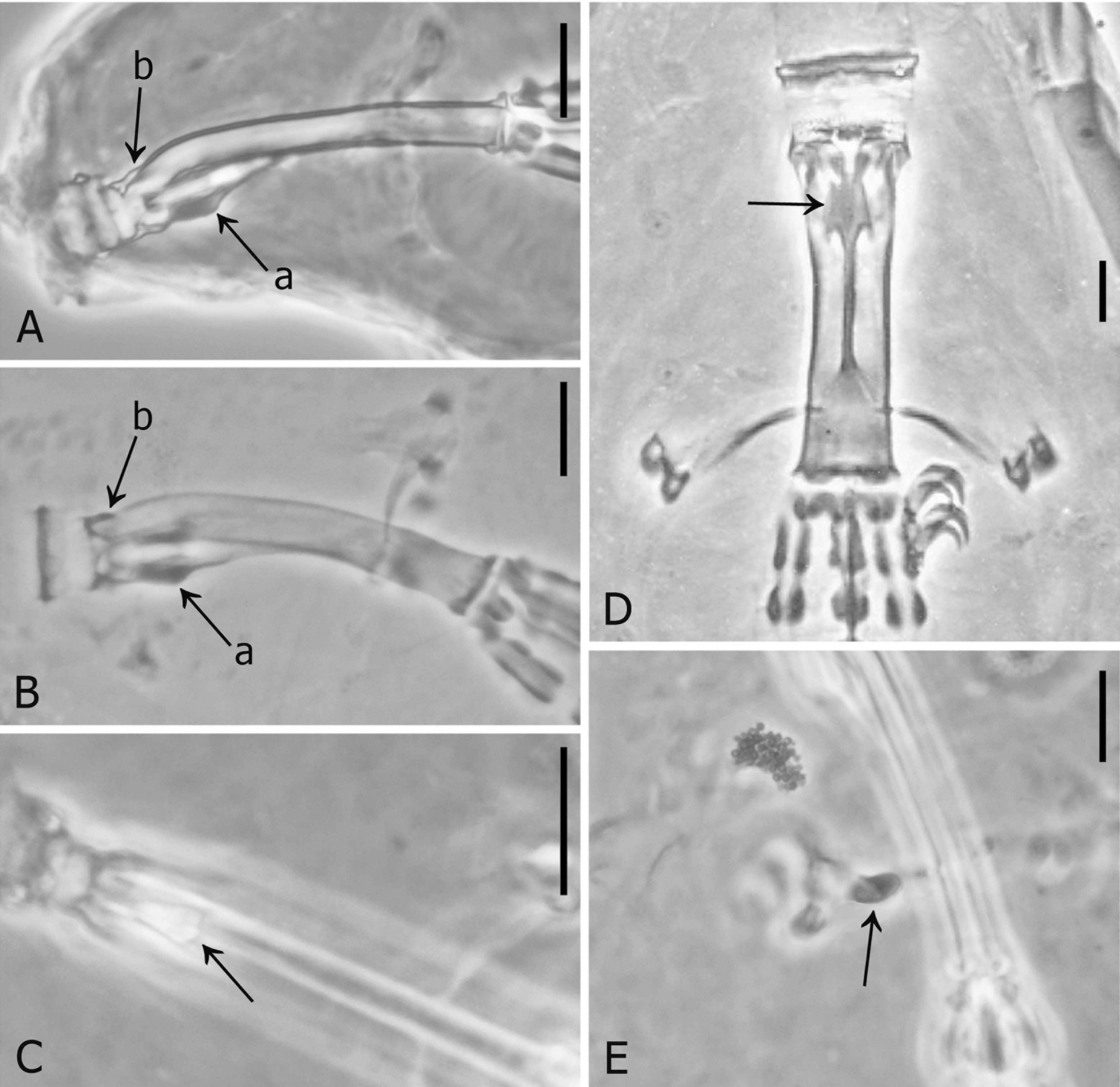
Redescription of Crenubiotus crenulatus comb. nov. ( Richters, 1904a)
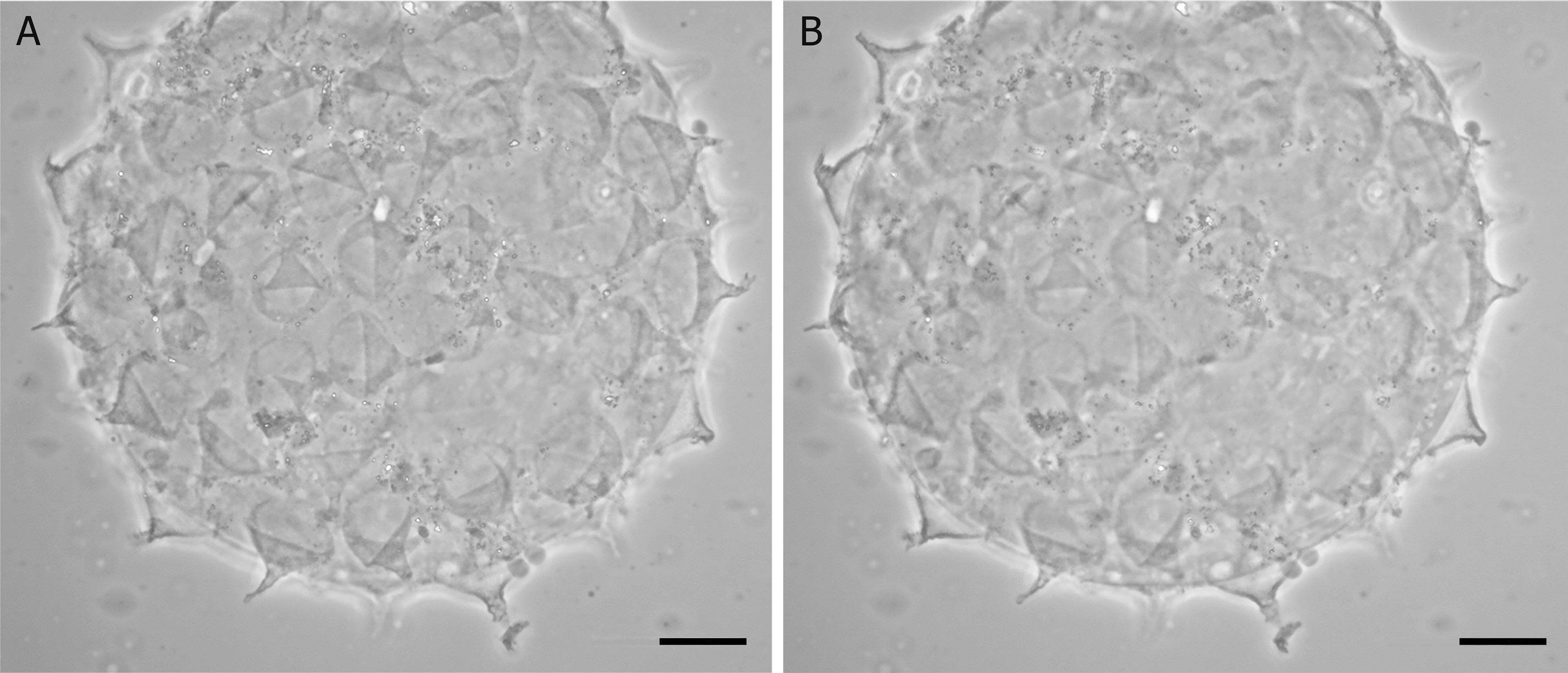
( Figs 6–7 View FIGURE 6 View FIGURE 7 , Table 2)
Material examined: neotype, 4 neoparatypes and 3 eggs, deposited in the Pilato and Binda collection (slide Nos. 2284 and 2285) at the University of Catania. Type locality: Valtellina (Northern Italy), Palabione, 1700 m asl (these are the only data available from Binda 1974) .
Description: Body length up to 320 µm, colourless after mounting. Eye-spots present before mounting. Cuticle with circular and elliptical pores probably present in all life stages, global diameter pt range about 0.7–9.2; the largest pores are a very cephalic pair, constantly present in all specimens lateral to the mouth, which are elliptical (minor × major diameter of these pores 2.5 µm × 2.9 µm; pt = 7.4 × 8.5, measurable only in one specimen 254 µm long); apart from that pair of cephalic pores, the largest, elliptical, are present in the caudal portion of the body (major diameter range 2.9–3.1 µm, pt = 9.1–9.2); the smallest pores, instead, sparsely distributed on the cuticle, are rounded and have diameter starting from more than about 0.7 µm (it was difficult to measure precisely such small structures; pt more than about 2.1). Examining the available material, it seems the pores are present in all life stages and do not show allometric growth. Very small cuticular tubercles present in a dorso-lateral caudal band just anterior to the hind legs ( Fig. 6E View FIGURE 6 in the evidenced polygon), as well as on the dorsal cuticle of legs IV ( Fig. 6E View FIGURE 6 arrow); legs very probably also provided with smaller tubercles/granules (Pilato, pers. comm.) but no longer visible due to the micropreparation age.
Bucco-pharyngeal apparatus ( Fig. 6A View FIGURE 6 ) of modified ‘ Macrobiotus type’, i.e. with ten peribuccal lamellae, rigid buccal tube with ventral lamina which is provided with an additional ventral thickening on its anterior portion ( Fig. 6A View FIGURE 6 arrow, 6D arrow); dorsal apophysis absent or very reduced into a small thickening ( Fig. 1B View FIGURE 1 arrow b, but this detail should be ascertained and also depends on interpretation).
Oral cavity armature ( Fig. 6B, C View FIGURE 6 ) seemingly without anterior and posterior bands of teeth (not visible, at least in light microscopy), but provided of the crest system, which is represented only by the lateral crests (no medial crests or teeth present); each latero-dorsal crest forms at its medial extremity a rounded thickening appearing like a tooth ( Fig. 6B View FIGURE 6 arrow); the ventral crests ( Fig. 6C View FIGURE 6 , arrow indicating one crest), instead, simply continue medially in the ventral lamina.
Big stylet furcae with the thickened, swollen apices of the branches very developed and elongated laterally as described for the genus.
Two rod-shaped macroplacoids plus a microplacoid in the pharynx; the first macroplacoid is longer and shows a medial incision, the second, shorter, with subterminal incision ( Fig. 6A View FIGURE 6 ).
Symmetric, slightly robust doubled claw Y-shaped ( Fig. 6F, G View FIGURE 6 ), with a long stalk system, proximally forming a laminar peduncle while distally it has a round section, separated by a septum from a subsequent intermediate tract; from this, a distal incomplete septum, closing only the main branch base, leads to a following common tract of the two branches where their suture is visible; claw branches diverging quite high in the whole claw height and forming a nearly acute angle. Large lunules on all legs, indented in a regular pattern by long, narrow teeth equal to one another ( Fig. 6 View FIGURE 6 F–G).
Egg ( Fig. 7 View FIGURE 7 ), non-areolated, with conical processes (about 15–18 in the optical section, 46–47 in the hemisphere). The processes are usually short and wide cones which form abruptly a long, narrower terminal portion quite variable in shape (from relatively thick to narrow) and irregular either: only in some cases it is a simple slender cone, while in the others it is variously branched and/or show an uneven surface which gives an irregular shape, though remaining relatively transparent.
We noticed also the following details under the microscope, but they are barely visible due to the slide age and cannot be shown clearly on photos: only in few processes, there can be observed a single internal “bubble” at the base of this distal portion (with similar appearance to Fig. 8G View FIGURE 8 arrow, in Crenubiotus revelator sp. nov.). The process tips usually end with a very delicate filament variously frayed. Very fine, delicate reticulation on the main body of the processes (distal portion excluded), difficult to see (drawn by Binda 1974); the process base forms short, narrow projections on the egg shell, drawn as dots by Binda (1974), which are not long enough to continue on the egg shell between the processes and connect to one another ( Binda 1988 described the egg shell as “perfectly smooth”).


No known copyright restrictions apply. See Agosti, D., Egloff, W., 2009. Taxonomic information exchange and copyright: the Plazi approach. BMC Research Notes 2009, 2:53 for further explanation.