
Parasmittina Osburn, 1952
|
publication ID |
https://doi.org/ 10.1080/00222930701391773 |
|
persistent identifier |
https://treatment.plazi.org/id/877A7251-CC74-DE09-FE62-24BAD44E1FDB |
|
treatment provided by |
Felipe |
|
scientific name |
Parasmittina Osburn, 1952 |
| status |
|
Genus Parasmittina Osburn, 1952 View in CoL
Parasmittina avicularissima ( Gontar, 1982) View in CoL
( Figure 23 View Figure 23 ) Parasmittina jeffreisii avicularissima Gontar 1982, p 548 View in CoL , Figures 1 View Figure 1 , 5a, b View Figure 5 .
Material examined
ANC, colony on rock (NHM 2006.2.27.72), colony on rock (NHM 2006.2.27.73), extensive colony on rock (NHM 2006.2.27.74). Additional material: 23 specimens.
Description
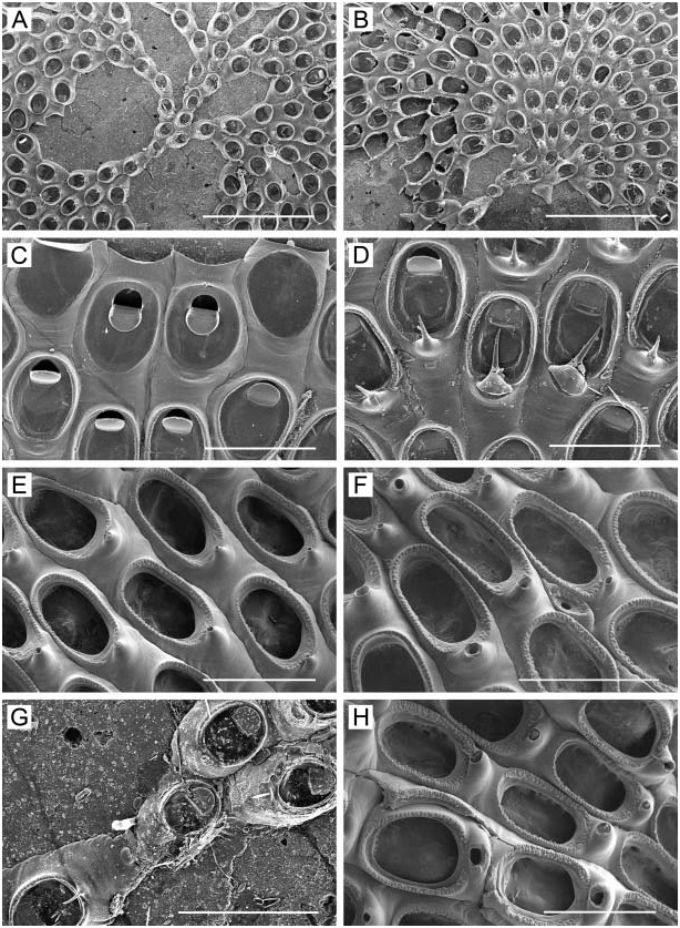
Colony encrusting, coherent, unilaminar, but sporadically building up frontally budded layer of irregularly orientated zooids; irregularly circular, largest observed 3.8 cm in maximum dimension; bright yellow when alive, with lemon-yellow membranous growing edge one or two zooids deep. Zooids ( Figure 23B, C, E View Figure 23 ) oval to irregularly hexagonal, rounded distally, 0.40–0.63 mm long (0.49¡ 0.06 mm), 0.27–0.43 mm wide (0.34¡ 0.04 mm), separated by shallow groove, with appressed adjacent vertical walls forming a thick line of calcification flanked by rows of areolar pores. Frontal wall convex, vitreous; smooth, or rugose with coarse granulation; imperforate centrally, with 7–10 conspicuous areolar pores along each lateral margin, separated by short buttresses; with large conical or nodular suboral umbo variable in size, occasionally with one to three additional protuberances scattered elsewhere on frontal surface. Primary orifice ( Figure 23B View Figure 23 ) subcircular, typically slightly longer than broad, 0.10–0.14 mm long (0.12¡ 0.01 mm), 0.10–0.13 mm wide (0.11¡ 0.01 mm), with a low, narrow, truncate lyrula and long, pointed condyles directed proximomedially. Young zooids ( Figure 23A View Figure 23 ) have two short, ephemeral distal spines. Peristome formed by narrow, raised lip proximally and laterally in young zooids, or rarely by two lateral lappets separated by sinus; primary orifice becomes sunken with increased secondary calcification. Zooids with or without a large avicularium ( Figure 23A–C View Figure 23 ), about 0.12–0.15 mm long, lateral to orifice, abutting peristome, the rostrum raised distally, with an acute, slightly long-triangular mandible directed medially, approximately in line with proximal margin of orifice; cross-bar thin, complete; sometimes the lateral-oral avicularium is paired. The lateral-oral avicularium may be replaced by a larger avicularium ( Figure 23D, E View Figure 23 ) of similar shape occupying central or proximal area of frontal wall and pointing distally, distolaterally, or sometimes proximally. Small avicularia ( Figure 23D, F View Figure 23 ), about 0.07–0.10 mm long, pyriform or oval in shape, with semicircular mandible directed laterally or proximally, can also occur anywhere along the proximal or lateral margins proximal to the orifice, with or without the larger, acute types; sometimes an oval avicularium overlaps the margin of an ovicell. Ovicell ( Figure 23F, G View Figure 23 ) spherical, broader than long, 0.20–0.28 mm long (0.24¡ 0.02 mm), 0.23– 0.33 mm wide (0.28¡ 0.02 mm), overhanging the orifice; smooth, flattened on top and bearing a single large, circular or transversely elliptical pore; recumbent and sunken in frontal wall of distal zooid, with contributions of ectocystal calcification from that zooid and laterally flanking zooids delineated by raised suture lines; ovicell lacking ornamentation, or with one to three conical, tuberculate processes, one per sector of secondary calcification. Interzooidal communication via uniporous septula. Ancestrula not observed; ancestrular region we observed ( Figure 23H View Figure 23 ) appears entirely covered by a frontally budded layer of irregularly orientated zooids.
Remarks
The stable character that distinguishes this species from any other Parasmittina reported from the northwestern Pacific, including P. jeffreysii ( Norman, 1903) , P. trispinosa ( Johnston, 1838) , and P. macroavicularia ( Androsova, 1958) , is the presence of only a single, large pore in the ovicell.
Gontar (1982) described and illustrated two types of avicularia in her original description, but only briefly mentioned their arrangement, which we found to be quite variable. Most zooids located marginally or peripherally lack avicularia or have only the lateral-oral avicularium with a triangular mandible pointed medially. In contrast, zooids situated near the colony centre tend to have several different combinations of avicularia, such as: (1) a triangular lateral-oral avicularium on both sides of orifice, mandibles pointing medially; (2) an oval avicularium on each side of the orifice; (3) both triangular and oval avicularia lateral to orifice; (4) a single triangular avicularium located proximally on the frontal wall, with the mandible directed distally, laterally, or proximally; (5) a single oval avicularium lateral to orifice; (6) one or two oval avicularia in the proximal half of the frontal wall; or (7) one oval avicularium lateral to the orifice and another more proximally.
Distribution
Parasmittina avicularissima was originally described from Crabovaya Bay, Shikotan Island, southern Kuril Islands. Akkeshi Bay is the second and southernmost known locality.

No known copyright restrictions apply. See Agosti, D., Egloff, W., 2009. Taxonomic information exchange and copyright: the Plazi approach. BMC Research Notes 2009, 2:53 for further explanation.
|
Kingdom |
|
|
Phylum |
|
|
Class |
|
|
Order |
|
|
Family |
Parasmittina Osburn, 1952
| Grischenko, Andrei V., Dick, Matthew H. & Mawatari, Shunsuke F. 2007 |
Parasmittina jeffreisii avicularissima
| Gontar 1982: 548 |