
Pseudomma maasakii, Meland, Kenneth & Brattegard, Torleiv, 2007
|
publication ID |
https://doi.org/10.5281/zenodo.179324 |
|
DOI |
https://doi.org/10.5281/zenodo.5667043 |
|
persistent identifier |
https://treatment.plazi.org/id/882387EC-C73B-0452-C8AB-66E00C6DFEA8 |
|
treatment provided by |
Plazi |
|
scientific name |
Pseudomma maasakii |
| status |
sp. nov. |
Pseudomma maasakii n. sp.
Pseudomma View in CoL sp. Murano and Mauchline 1999: 279 –280.
Pseudomma View in CoL sp. M&M Meland 2004:3; Meland and Willassen 2004: 18 View Cited Treatment S rRNA ( AY624301 View Materials ), COI mtDNA ( AY624281 View Materials ).
Material examined
Type material. Holotype (adult male, 16 mm), IMNH-2183, stn BIOICE 2860. Allotype (adult female, 19 mm), ZMBM-68270, stn BIOICE 3167. Paratypes, Stn BIOICE 2856, 2 females ( 18 mm), IMNH-2184. Stn BIOICE 2859, 1 adult male ( 15 mm), 1 immature female ( 16 mm), IMNH-2185. Stn BIOICE 2860, 1 adult female (thorax), 1 juvenile ( 8 mm) IMNH-2186. Stn BIOICE 2864, 1 male ( 14 mm), IMNH-2187. Stn BIO- ICE 3162, adult female ( 19 mm), IMNH-2188. Stn BIOICE 3164, adult female ( 18 mm), 2 juveniles, IMNH- 2189. Stn BIOICE 3167, 1 adult female ( 19 mm), IMNH-2190.
Description
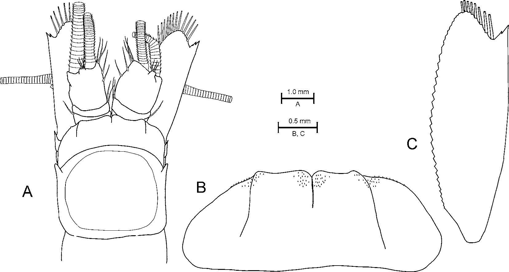
Carapace ( Fig. 3 View FIGURE 3 A) with anterior margin evenly rounded and anteriorly produced lateral corners.
Ocular plate ( Fig. 3 View FIGURE 3 A, B) extending to mid-portion of first segment of antennular peduncle; plate deeply cleft, medial section slightly produced in males, anterior portion of dorsal surface finely serrated with minute spinules; antero-lateral margins armed with eight minute setae.
Antennal scale ( Fig. 3 View FIGURE 3 C) three times longer than broad; distal terminal denticle on outer margin consisting of two to three spines; apex extending beyond terminal denticle, without suture, outer margin of apex armed with five setae.
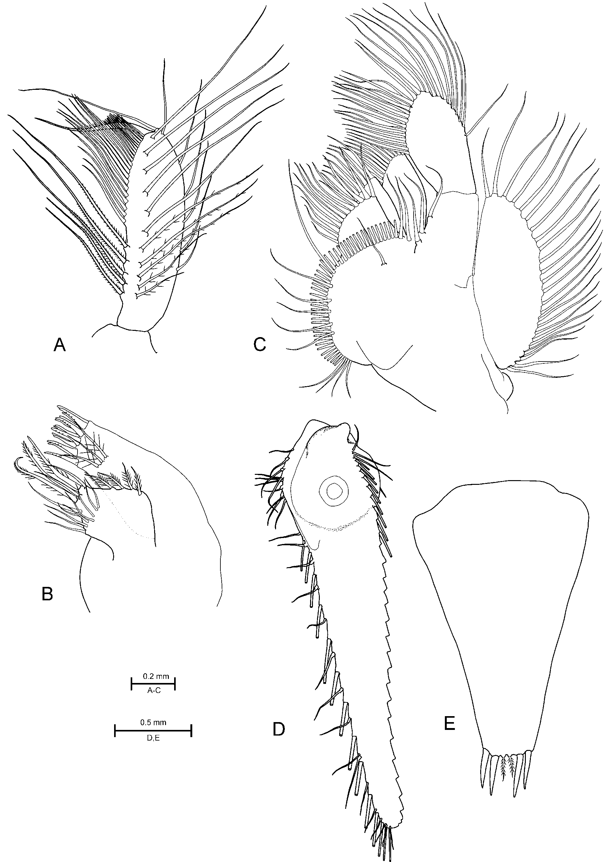
Left mandible setal row consisting of three hirsute spines and right mandible setal row consisting of eight entire spines. Distal segment of mandible palp ( Fig. 4 View FIGURE 4 A) with five enlarged proximal ventral setae, medial margin with row of eight to 11 setae, dorsal margin with five to six setae.
Maxillule ( Fig. 4 View FIGURE 4 B), apex of coxal lobe armed with three strong setae bordered by three smaller setae placed distal-posterior, distal-ventral, and distal-anterior; anterior lateral margin armed with four setae in medial region; ventral surface and posterior lateral margin supporting eight to ten setae; ventral surface of maxillule basis supporting three setae, posterior lateral margin armed with robust setae, apex supporting 14 strong cuspidate setae.
Maxilla ( Fig. 4 View FIGURE 4 C) with three setae on proximal inner margin of endopod; exopod supporting 24-28 lateral setae; coxa with one or two setae on dorsal surface, coxal surface without spines or denticles, lateral margin of coxa armed with two rows of setae, dorsal row consisting of one large and four or five smaller setae.
First and second thoracic appendages formed as maxillipeds; first maxilliped with large nail bearing five denticles, dactylus fringed with four to five large setulate setae on each lateral margin; second maxilliped with small nail, dactylus fringed with nine to ten large setulate setae on each lateral margin. Third to eighth thoracic appendages take on the form of long and slender pereopods. Female marsupium consists of three pairs of oostegites arising from the sixth to eighth pereopods, increasing in size posteriorly. Male genital organ extending beyond seventh pereopod, bearing two apical setae.
Sixth abdominal somite two times longer than fifth. Pleopods of male biramous; first pleopod with unsegmented endopod and 12–segmented exopod; second to fourth pleopods with 10-segmented endo- and exopods; fifth pleopod with 9-segmented endo- and exopods; distal setae on third and fourth pleopods not modified. Female pleopods uniramous, taking on the form of unsegmented plates and set with apical and ventral surface setae.
Uropod endopod ( Fig. 4 View FIGURE 4 D) with one ventrally placed strong spinose seta on inner margin near statocyst, outer margin of endopod armed with 13 large setae, and 16 small setae.
Telson ( Fig. 4 View FIGURE 4 E), lateral margins entire, apex armed with two pairs of spinose setae; apex with one pair of median plumose setae.
Etymology
The species was first recognized by Murano and Mauchline (1999) and is named in honour of Dr. Maasaki Murano for his huge contribution to mysid taxonomy.
Distribution
Three specimens of Pseudomma maasakii have earlier been recorded from the Rockall Trough as stomach content in the deep-water grenadier species Nematonurus armatus (Hector, 1875) and Coryphaenoides guentheri (Vailant, 1888) ( Murano and Mauchline 1999) . The present BIOICE material extends its species range into 2300 m depths of the Iceland Basin.
Remarks
Due to the shared telson characters with two pairs of apex setose setae and the absence of lateral spinose setae, P. maasakii resembles P. m a t s u i Murano, 1966. However, it is easily distinguishable from all Pseudomma species based on the antennal terminal denticle consisting of two to three spines ( Fig. 3 View FIGURE 3 C), and also the anterior lateral margin of the maxillule coxal lobe that is armed with four setae in the medial region ( Fig. 4 View FIGURE 4 B).
No known copyright restrictions apply. See Agosti, D., Egloff, W., 2009. Taxonomic information exchange and copyright: the Plazi approach. BMC Research Notes 2009, 2:53 for further explanation.
|
Kingdom |
|
|
Phylum |
|
|
Class |
|
|
Order |
|
|
Family |
|
|
Genus |
Pseudomma maasakii
| Meland, Kenneth & Brattegard, Torleiv 2007 |
Pseudomma
| Murano 1999: 279 |