
Amblyops spinifera Nouvel and Lagardère, 1976
|
publication ID |
https://doi.org/ 10.5281/zenodo.179324 |
|
DOI |
https://doi.org/10.5281/zenodo.5667041 |
|
persistent identifier |
https://treatment.plazi.org/id/882387EC-C73D-045F-C8AB-61380EC1FCE0 |
|
treatment provided by |
Plazi |
|
scientific name |
Amblyops spinifera Nouvel and Lagardère, 1976 |
| status |
|
Amblyops spinifera Nouvel and Lagardère, 1976 View in CoL
Amblyops spinifera Nouvel and Lagardère 1976: 1275 View in CoL –1281.
Material examined
Type material. Holotype (adult female, 13.0 mm), MNHN-My43, stn Gch 70 ( Nouvel and Lagardère, 1976).
Additional material. Stn BIOICE 2844, 1 immature male (12 mm), 2 immature males (9 mm), 2 adult females (13 mm), 1 adult female (12 mm), 1 adult female (11 mm), 2 immature females (8 mm), IMNH- 2181. Stn BIOICE 3181, 1 immature males (10 mm), 2 immature females (10 mm, damaged), juvenile (8 mm), IMNH-2182.
Description
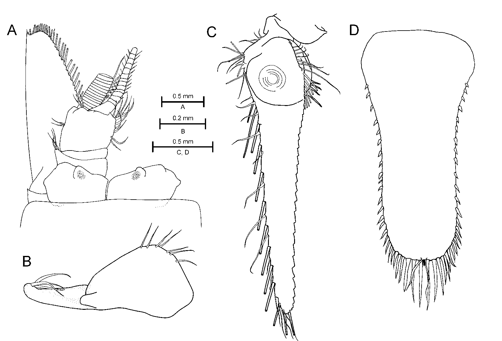
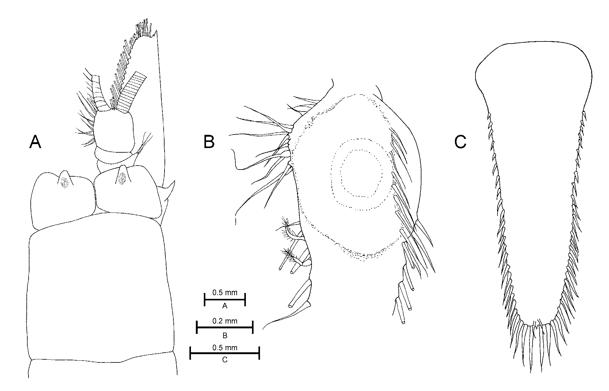
Carapace ( Fig. 2 View FIGURE 2 A) with anterior margin evenly rounded, without rostrum, lateral margin straight, posterior dorsal margin exposing last two thoracic somites.
Ocular plates ( Fig. 2 View FIGURE 2 A) separated, each plate with a anteriorly produced papilla on medial anterior margins, plate is densely set with minute spinules on anterior and anterolateral margins.
Antennal scale ( Fig. 2 View FIGURE 2 A) three times longer than broad, distal terminal denticle on outer margin consisting of two spines, apex small, not extending beyond terminal denticle, small suture present.
Left mandible setal row consisting of six hirsute spines and right mandible setal row consisting of eight entire spines. Distal segment of mandible palp with one enlarged proximal ventral seta, medial margin with row of ten setae, dorsal margin with seven setae.
Maxillule, apex of coxal lobe armed with three strong setae bordered by three smaller setae placed distalposterior, distal-ventral, and distal-anterior; anteriolateral margin armed with two setae in medial region; ventral surface and posteriolateral margin supporting five setae; ventral surface of maxillule basis supporting three setae, posteriolateral with small setae, apex supporting 14 cuspidate setae.
Maxilla with three setae on proximal inner margin of endopod; exopod supporting 28 lateral setae; coxa with five setae on dorsal surface, coxal surface covered with minute spines and denticles, lateral margin of coxa armed with two rows of setae, dorsal row consisting of one large and five smaller setae, setae in ventral row entire.
First and second thoracic appendages formed as maxillipeds; first maxilliped with large nail, dactylus fringed with four large setulate setae on each lateral margin; second maxilliped with long nail (0.5 of dactylus length) armed with five denticles, dactylus fringed with three to four large setulate setae on each lateral margin. Third to eighth thoracic appendages take on the form of long and slender pereopods. Female marsupium consists of three pairs of oostegites arising from the sixth to eighth pereopods, increasing in size posteriorly. Male genital organ short, barely extending beyond eighth pereopod, bearing two apical setae.
Sixth abdominal somite 1.5 times longer than fifth. Male pleopods biramous (following characters are in an immature male); first pleopod with unsegmented endopod and 13-segmented exopod; second to fourth pleopods with 12-segmented endo- and 13-segmented exopods; fifth pleopod with 11-segmented endo- and 12- segmented exopod. Female pleopods uniramous, taking on the form of unsegmented plates and set with apical and ventral surface setae.
Uropod exopod length 1.4 times endopod length, endopod with two or three ventrally placed strong spinose seta on inner margin near statocyst ( Fig. 2 View FIGURE 2 B), outer margin of endopod armed with 13 large setae, and 15 small setae.
Telson ( Fig. 2 View FIGURE 2 C) linguiform, distal ¾ of lateral margins armed with 23–26 spinose setae on each side, apex armed with three pairs of spinose setae and one medial spine, pair of median plumose setae slightly displaced dorsally on the apex.
Remarks
The resemblance of Amblyops spinifera Nouvel and Lagardère, 1976 to A. kempi ( Holt and Tattersall, 1905) is so close that identifying the Iceland Basin specimens proved very difficult. Nonetheless, based on what seems to be a single medial spine on the apex of the telson, opposed to two in A. kempi , and the more medially placed papilla on the ocular plates, we are quite sure that we are dealing with A. spinifera . We have also noted that A. spinifera has a small, but distinct oval structure situated within the ocular papilla ( Fig. 2 View FIGURE 2 A), also seen in A. trisetosa ( Fig. 1 View FIGURE 1 A), and described by Tattersall (1923) as “a central mass of nerve cells” for A. tattersalli Zimmer, 1914 . Possibly overlooked in past descriptions, we suspect this structure to be present in several others, if not all Amblyops species.
Although we do not doubt their taxonomic status, in a phylogenetic context the similiar resemblance in morphology among A. spinifera , A. kempi , A. durbani , A. trisetosa , and A. longisquamosus indicate a close relationship. It is interesting to note a pattern of small morphological variation among these deep-sea species, comparable to that found in the genus Pseudomma (Meland 2004) .
No known copyright restrictions apply. See Agosti, D., Egloff, W., 2009. Taxonomic information exchange and copyright: the Plazi approach. BMC Research Notes 2009, 2:53 for further explanation.
|
Kingdom |
|
|
Phylum |
|
|
Class |
|
|
Order |
|
|
Family |
|
|
Genus |
Amblyops spinifera Nouvel and Lagardère, 1976
| Meland, Kenneth & Brattegard, Torleiv 2007 |
Amblyops spinifera Nouvel and Lagardère 1976 : 1275
| Nouvel 1976: 1275 |