
Anastrepha bivittata (Macquart)
|
publication ID |
https://doi.org/10.11646/zootaxa.3911.3.7 |
|
publication LSID |
lsid:zoobank.org:pub:75CC87E7-799B-4EC4-BEB1-30F22912F721 |
|
DOI |
https://doi.org/10.5281/zenodo.6104290 |
|
persistent identifier |
https://treatment.plazi.org/id/884F8781-7E4C-FFF8-FF19-E46FD7CAA65F |
|
treatment provided by |
Plazi |
|
scientific name |
Anastrepha bivittata (Macquart) |
| status |
|
Anastrepha bivittata (Macquart) View in CoL
Figs. 1–20
Urophora bivittata Macquart 1843: 379 View in CoL , Tab. 30, Fig. 3 View FIGURES 2 – 9 .
Acrotoxa bivittata: Loew 1873: 231 (classification).
Anastrepha bivittata: Bezzi 1909: 284 View in CoL (list), 286 (in key); Hendel 1914: 15 (in key, list); Greene 1934: 134 (in key), 157; Lima 1934: 499; Stone 1942: 22 (unrecognized species); Foote 1967: 8 (catalog); Steyskal 1977: 33 (list of species not in key); Norrbom et al. 1999a: 77 (catalog, type data); Norrbom et al. 1999b: 322, 335 (classification).
Anastrepha fumipennis Lima 1934: 499 View in CoL . Stone 1942: 18 (in key), 90 (revision); Foote 1967: 11 (catalog); Steyskal 1977: 25 (in key); Norrbom et al. 1999a: 79 (catalog); Zucchi 2000: 22 (in key). New synonymy.
Diagnosis. This species can be recognized from all other species of Anastrepha by the following combination of characters: wing pattern mostly dark brown; cells br and bm entirely infuscated; aculeus tip nonserrate, slightly tapered to blunt apex.
Description. [Information in brackets from lectotype] Mostly orange. Setae dark brown to black. Body length 8.25–11.75 [9.9] mm.
Head. Yellow except ocellar tubercle brown. Facial carina, in profile, straight on dorsal 3/4, lower part protruding. 4–6 [4] frontal setae. 2 orbital setae. Ocellar seta weak, as long as ocellar tubercle. Antenna not extended to ventral facial margin. Palpus in lateral view dorsally curved, evenly setulose.
Thorax. Mostly dark orange; with following areas white to pale yellow: postpronotal lobe and lateral margin of scutum bordering it, not extending onto notopleuron; medial scutal vitta, slender anteriorly, extended to level of postpronotal lobe, posteriorly slightly broadened, extended lateral to acrostichal seta (similar in shape to A. atrigona, Norrbom 1991 , fig. 3A); sublateral scutal vitta from transverse suture to posterior margin, including base of intra-alar seta; scutellum; dorsal margins of anepisternum and katepisternum; katepimeron; and most of anatergite and katatergite [pleuron of lectotype mostly covered by mold, markings not visible]. Scutum without brown markings. Subscutellum and mediotergite dark orange, mediotergite narrowly dark brown laterally. Mesonotum 2.65–3.90 [3.17] mm long. Postpronotal lobe, notopleuron, scutum and scutellum entirely microtrichose [scutum largely covered by debris in lectotype, but mostly if not entirely microtrichose]. Scutal setulae brown except on and bordering medial vitta. Chaetotaxy typical for genus [in lectotype both postpronotal, right presutural supra-alar and posterior notopleural, both postsutural supra-alar, except base of left one, left acrostichal, dorsocentral, and intra-alar, both postalar, except base of left one, absent, but sockets large]. Katepisternal seta ca. half as long and much weaker than anepisternal seta, yellowish.
Legs. Entirely yellow to orange. Fore femur with posterodorsal and ventral rows of well developed setae.
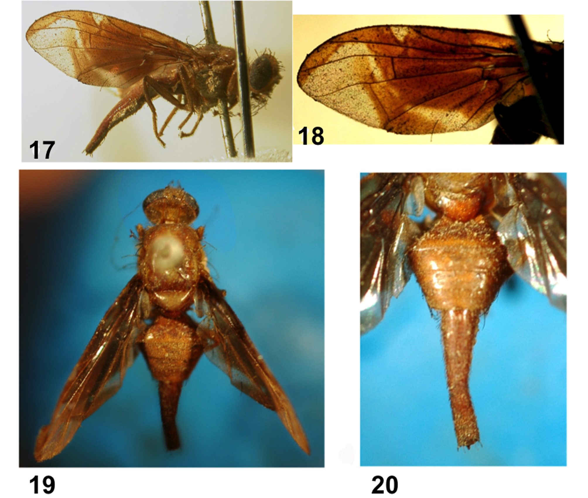
Wing ( Figs. 1. a–h, 17, 18 View FIGURES 17 – 20 ). Length 7.10–9.47 [7.28] mm, width 2.72–3.97 [2.93] mm, ratio 2.38–2.61 [2.48]. Apex of vein R1 at 0.56–0.60 [0.60] wing length, proximal to level of anterior end of crossvein r-m. Cell c 0.98–1.20 [1.13] times as long as pterostigma; pterostigma 2.67–3.75 [3.13] times as long as wide. Vein R2+3 nearly straight. Crossvein r-m at 0.74–0.78 [0.77] distance from bm-cu to dm-cu on vein M. Vein M very strongly curved apically; cell r4+5 0.64–0.75 [0.67] times as wide at apex as at level of dm-cu. Cell bcu with distal lobe long, length of bcu 1.52–1.75 [1.52] times as long as anterior margin, lobe 0.68–0.80 times as long as vein A1+Cu2. Pattern mostly dark brown. C-band often with elongate paler area posteriorly in cell c. S-band distal to r-m and anterior part of proximal arm of V-band (from anterior half or more of dm-cu) orange with narrow brown margins. C- and S- bands completely fused basally, cells br, bm and bcu entirely infuscated although bm and bcu usually paler medially. Base of S-band without posterior extensions in middle of cell cu1 or cell a1. Hyaline mark distal to apex of vein R1 variable, often triangular, small and not reaching vein R2+3 [e.g., in lectotype], or triangular with vertex rounded and reaching vein R 4+5, or interrupted at R 2+3 ( Figs. 1, 18 View FIGURES 17 – 20 ). S-band with distal section of medium width, at apex of vein R2+3 0.51–0.69 [0.56] times width of cell r2+3; not extended to apex of vein M. Vband with proximal arm connected to (usually) or separated from S-band anteriorly, but fused to varying extent to Sband in cell dm; extended basally along posterior wing margin almost to vein A1+Cu2 but not connected to base of S-band along wing margin. Distal arm absent or reduced to small spot anterior to vein M ( Lima 1934, Est. LXV, fig. 10; Fig. 1h). Wing entirely microtrichose except posterobasal half of cell bc, sometimes basal half of cell bm [entirely microtrichose in lectotype], and most of alula [bare on anterior 2/ 3 in lectotype]; all of cells br, dm, bcu, cu1, and a1+cu2 microtrichose. Area surrounding apex of lobe of cell bcu with microtrichia similar in density to area anterodistal to it along vein Cu1.
Abdomen ( Figs. 2 View FIGURES 2 – 9 , 19, 20 View FIGURES 17 – 20 ). Entirely orange, without brown markings.
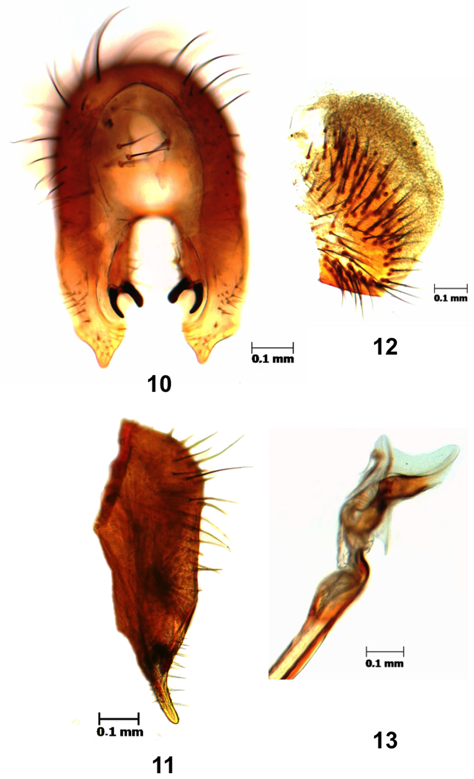
Male terminalia. Epandrium ( Fig. 10 View FIGURES 10 – 13 ) with posterodorsal margin with moderate medial V-shaped indentation. Lateral surstylus medium ( Fig. 11 View FIGURES 10 – 13 ), extended beyond prensisetae by approximately 1.5 times length of prensiseta; in lateral view elongate, triangular and strongly tapered apically; in posterior view, apex acute, medial margin concave, lateral margin concave. Proctiger ( Fig. 12 View FIGURES 10 – 13 ) with ventral and lateral sclerotized areas connected. Phallus 5.22 mm long, 1.49 times as long as mesonotum; glans ( Fig. 13 View FIGURES 10 – 13 ) 0.61 mm long.
Female terminalia. Oviscape ( Figs. 2 View FIGURES 2 – 9 , 19, 20 View FIGURES 17 – 20 ) 3.10–4.20 [3.28] mm long, 1.03–1.17 [1.03] times as long as mesonotum, entirely orange; spiracle at basal 0.27–0.33 [0.33]. Eversible membrane ( Figs. 2, 3, 8 View FIGURES 2 – 9 ) with about 30 long, slender, hook-like dorsobasal denticles in triangular pattern. Aculeus ( Figs. 4, 6 View FIGURES 2 – 9 ) 3.13–4.26 [3.43] mm long, 0.96–1.07 [1.05] times oviscape length; base 0.21–0.28 [0.21] mm wide; shaft 0.095–0.14 [0.12] mm wide at midlength; tip ( Figs. 5, 7 View FIGURES 2 – 9 ) 0.22–0.30 [0.23] mm long, 0.07 [0.07] times aculeus length, 0.09–0.12 [0.11] mm wide, 2.09–2.73 [2.09] times as long as wide; in ventral view gradually tapered (angle changes very slightly near midlength), nonserrate, extreme apex relatively blunt; 0.06 mm wide in lateral view, 0.55 times ventral width. Spermathecae elongate ovoid (similar to Norrbom 1991, Fig. F), normally sclerotized. Egg white, slightly curved, 1.71 mm long in straight line, posterior third slender ( Fig. 9 View FIGURES 2 – 9 ).
Hosts. The only reported host plant is Geissospermum laeve (Vell.) Miers (Apocynaceae) (Uramoto et al. 2008).
Distribution. Brazil (Espírito Santo, Rio de Janeiro).
Type data. Urophora bivittata was described from an unstated number of female specimens from “Patria inconnue [country unknown]; je la dois á l’obligeance de M. de la Fresnaye [I owe it to the kindness of Monsieur de la Fresnaye].” Bezzi (1909) suggested that it may have originated in Brazil and may be deposited in the Museum National d’Histoire Naturelle, Paris (as “? Museo Parigi”, which Greene (1934) mistakenly interpreted as the “Museum at Para, Brazil ”), however, no specimens were located there during visits by Norrbom or other tephritid specialists. Although the types of many species described from Macquart’s personal collection, which is deposited in the Museum d'Histoire Naturelle, Lille, have apparently been lost, a single female in relatively good condition is present ( Norrbom e t a l. 1999a) in a box labeled “B Tephritidae ” and “Dipteres, C. Macquart, B", in the third column beneath a blue bordered label with “ U. bivittata ” in Macquart’s writing ( Figs.15–17 View FIGURES 14 – 16 View FIGURES 17 – 20 ). This female ( Figs. 18–21 View FIGURES 17 – 20 View FIGURES 21 – 24 ), here designated as lectotype to fix and stabilize the concept of this name, was examined briefly by Norrbom in 1990 and more extensively in 2006. It was mounted on a slender pin, which was deteriorating, and therefore was double-mounted on a block of styrofoam..
Anastrepha fumipennis was described from two female syntypes in the Fundação Instituto Oswaldo Cruz, from Brazil: Rio de Janeiro: Manguinhos, collected 24 Apr 1917 and 7 Apr 1930; one from vial 475, with wing slide 1059, and the other from vial 837, with wing slide 1710 and the abdomen on slide 1887. The FIOC collection was examined by Zucchi in 1978, and only t h e syntype in vial 837 and the abdomen slide 1887 were found.
Other specimens examined. BRAZIL: Espírito Santo: Linhares, Mata Atlântica (Reserva Natural da Vale), McPhail trap, 9.IV.2002, D. S. Martins 1♀ ( ESALQ). Idem, 11.III.2003, D. S. Martins, 2♀ ( ESALQ). Idem, 18.III.2003, D. S. Martins, 1♀ ( ESALQ). Idem, 22.II.2005, D. S. Martins, 1♀ ( ESALQ). Idem, 25.IV.2006, D. S. Martins, 1♀ ( ESALQ). Idem, 12.II.2008, D. S. Martins, 1♀ ( ESALQ). Idem, 23.VIII.2010, D. S. Martins, 2♀ ( ESALQ). Idem, 13.VIII.2013, D. S. Martins, 1♀ ( ESALQ). Idem, 22.IV.2006, D. S. Martins, 1♂ ( ESALQ).
Comments. The description of Urophora bivittata is brief and not very detailed, but it is accompanied by a dorsal habitus illustration including the wing, and a lateral view of the head (Tab 30, Fig. 3 View FIGURES 2 – 9 ). Loew (1873) considered this species to probably belong in Acrotoxa Loew , now considered a synonym of Anastrepha , and it has been treated as an unrecognized species of the latter genus by subsequent specialists. Allowing for the usual inaccuracies in Macquart’s illustrations (e.g., in the wing venation), the habitus is consistent with the species that has been known as A. fumipennis Lima. The wing pattern in particular is similar, including the entirely infuscated basal cells and the absence of the distal arm of the V-band, and the long, tubular oviscape is consistent with Anastrepha species. Lima (1934), in placing the description of A. fumipennis immediately after A. bivittata , recognized this similarity, but considered them distinct based on oviscape length (shorter than the abdomen in A. bivittata , longer than abdomen in A. fumipennis ). Examination of the lectotype female of A. bivittata , however, showed there to be no significant differences, and we therefore consider A. fumipennis to be a junior synonym of A. bivittata . We have not reexamined the syntypes of A. fumipennis , but Lima’s description is sufficiently detailed to recognize their identity.


| FIOC |
Fundacao Instituto Oswaldo Cruz |
No known copyright restrictions apply. See Agosti, D., Egloff, W., 2009. Taxonomic information exchange and copyright: the Plazi approach. BMC Research Notes 2009, 2:53 for further explanation.
|
Kingdom |
|
|
Phylum |
|
|
Class |
|
|
Order |
|
|
Family |
|
|
Genus |
Anastrepha bivittata (Macquart)
| Uramoto, Keiko, Zucchi, Roberto A. & Norrbom, Allen L. 2015 |
Anastrepha fumipennis
| Zucchi 2000: 22 |
| Norrbom 1999: 79 |
| Steyskal 1977: 25 |
| Foote 1967: 11 |
| Stone 1942: 18 |
| Lima 1934: 499 |
Anastrepha bivittata :
| Norrbom 1999: 77 |
| Norrbom 1999: 322 |
| Steyskal 1977: 33 |
| Foote 1967: 8 |
| Stone 1942: 22 |
| Greene 1934: 134 |
| Lima 1934: 499 |
| Hendel 1914: 15 |
| Bezzi 1909: 284 |
Acrotoxa bivittata :
| Loew 1873: 231 |
Urophora bivittata
| Macquart 1843: 379 |