
Antodendrina ligula, Wisshak, 2017
|
publication ID |
https://doi.org/10.5852/ejt.2017.390 |
|
publication LSID |
lsid:zoobank.org:pub:4D1D1CA3-8345-4BA3-9C7C-5EBDD40752CE |
|
DOI |
https://doi.org/10.5281/zenodo.3853665 |
|
persistent identifier |
https://treatment.plazi.org/id/12CB3E8D-D5D7-4C07-B99D-C581C0BED15A |
|
taxon LSID |
lsid:zoobank.org:act:12CB3E8D-D5D7-4C07-B99D-C581C0BED15A |
|
treatment provided by |
Carolina |
|
scientific name |
Antodendrina ligula |
| status |
igen. et isp. nov. |
Antodendrina ligula igen. et isp. nov.
urn:lsid:zoobank.org:act:
Fig. 31 View Fig
Diagnosis
Initial depression tear-shaped or elongate, with thin connections leading to up to six radiating and distinctly widening lobes with rounded termination, from which thin, ramifying, tapering filaments may emerge.
Etymology
From the Latin ‘ligula’, spoon, referring to the shape of the lobes of this dendrinid.
Type material, locality and horizon
The holotype ( Fig. 31 View Fig A–C) and the paratype ( Fig. 31D View Fig ), together with numerous further specimens in all stages of ontogeny, are cast in epoxy resin from two fragments of Inoceramus sp. bivalve shells from the Upper Campanian ( grimmensis / granulosus Zone), sampled in the Saturn Quarry near Kronsmoor , Germany. The casts are deposited in the trace fossil collection of the Senckenberg Institute in Frankfurt, Germany (SMF XXX 873, including the holotype, and SMF XXX 874, including the paratype).
Description
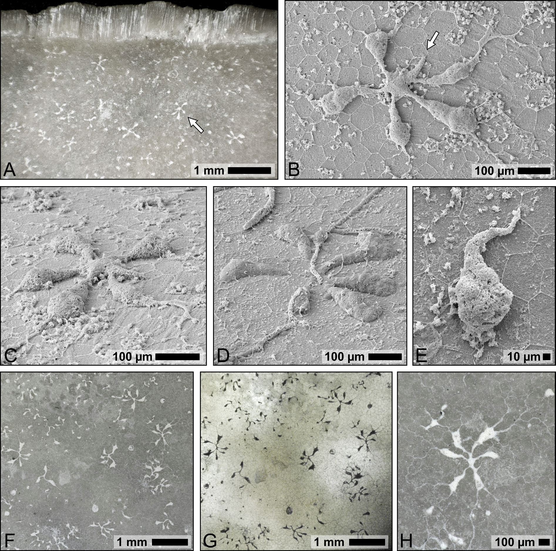
The characteristic blossom-shaped outline of this dendrinid trace is most conspicuous when filled with white chalk sediment, contrasting the darker colour of the inoceramid bivalve substrate ( Fig. 31A, F, H View Fig ), or the inverse pattern in transmission light micrographs of transparent substrates ( Fig. 31G View Fig ). These traces have so far been found, often clustered in high numbers ( Fig. 31A View Fig , F–G), exclusively in shells of inoceramid bivalves, preferably on their inner surface, where they show various degrees of xenoglyphic patterns caused by the polygonal crystallites of the prismatic shell microstructure (e.g., Fig. 31B, H View Fig ). From a small initial point of entry, the ontogeny of the trace starts out with a single tear-shaped depression or elongate cavity ( Fig. 31E View Fig ). From this cavity, several radiating and rather rapidly widening (laterally and vertically) lobes emerge, one after another, until a maximum of six lobes are formed. This process partly obscures the original shape of the initial cavity, so that the initial point of entry ( Fig. 31B View Fig ) is not always visible. The lobes are straight or bent and end in blunt, club-shaped terminations, from which ramifying thin galleries may emerge that spread and taper along the delineations of the calcite prisms ( Fig. 31B View Fig ). In some cases, the cavities are shallower and the thin connections of the lobes to the central depression may be almost or actually intermittent. This morphological expression is represented by the paratype ( Fig. 31D View Fig ). In any case, the deepest relief is reached near the terminations of the lobes. Surface texture is uniformly smooth to slightly bulged.
Concerning morphometry, a total of 36 individuals with at least three lobes were measured (including the types). The length of these intermediate or late ontogenetic stages varies from 289 to 675 µm (mean = 502 ± 95 µm) and the central cavity was measured to be 67 to 275 µm (mean = 162 ± 42 µm) in length. Note that for practical reasons the maximum diameter was measured as the distance between opposing terminations of the lobes, even though thin filaments with indistinct terminations may extend a couple of hundred microns further along the substrate surface, leading to an actual extent of the trace of up to around a millimetre. There is a maximum of 6 lobes (mean = 4 ± 1; n = 160), with a length of 83 to 308 µm (mean = 191 ± 49 µm; n = 160), a minimum width of 7 to 55 µm (mean = 21 ± 8 µm; n = 160) and a maximum width of 16 to 164 µm (mean = 73 ± 26 µm; n = 160).
No known copyright restrictions apply. See Agosti, D., Egloff, W., 2009. Taxonomic information exchange and copyright: the Plazi approach. BMC Research Notes 2009, 2:53 for further explanation.