
Lissosabinea christofferseni, Tavares & Mendonça & Colavite, 2022
|
publication ID |
https://doi.org/ 10.11646/zootaxa.5182.1.5 |
|
publication LSID |
lsid:zoobank.org:pub:0D0DD2A5-7CC7-44B8-89F8-35D3E5668B6F |
|
DOI |
https://doi.org/10.5281/zenodo.7065546 |
|
persistent identifier |
https://treatment.plazi.org/id/887A878C-6E29-FF86-50FA-FA62FC7A9A6D |
|
treatment provided by |
Plazi |
|
scientific name |
Lissosabinea christofferseni |
| status |
sp. nov. |
Lissosabinea christofferseni sp. nov.
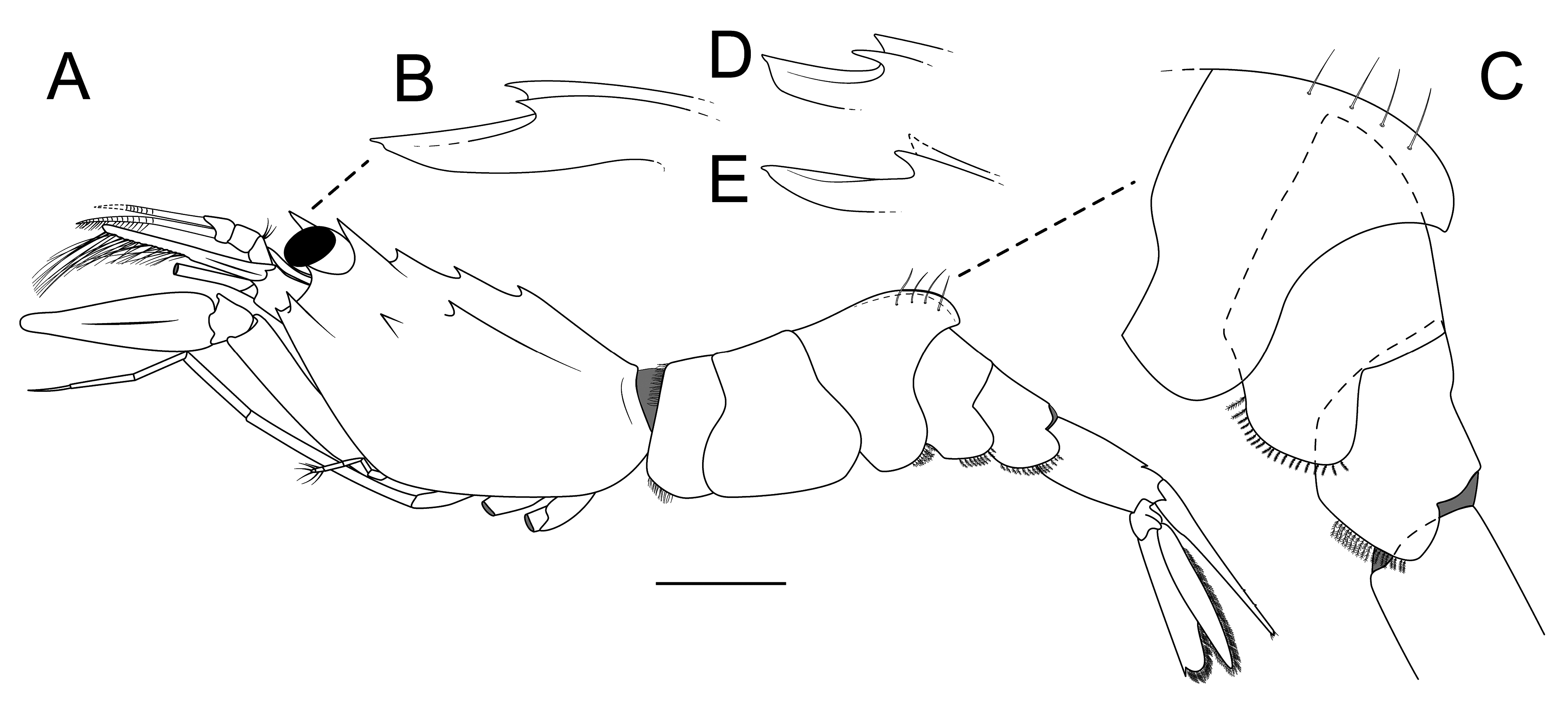
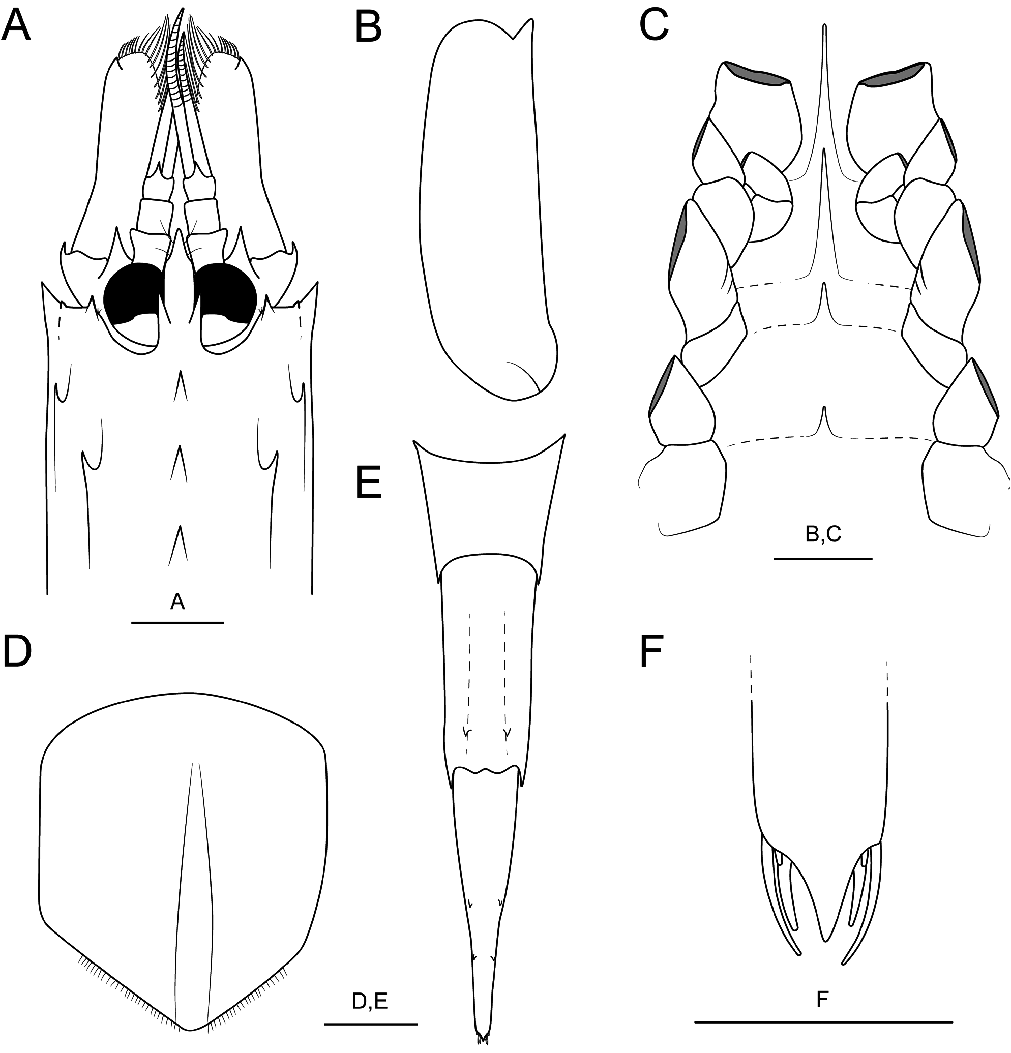
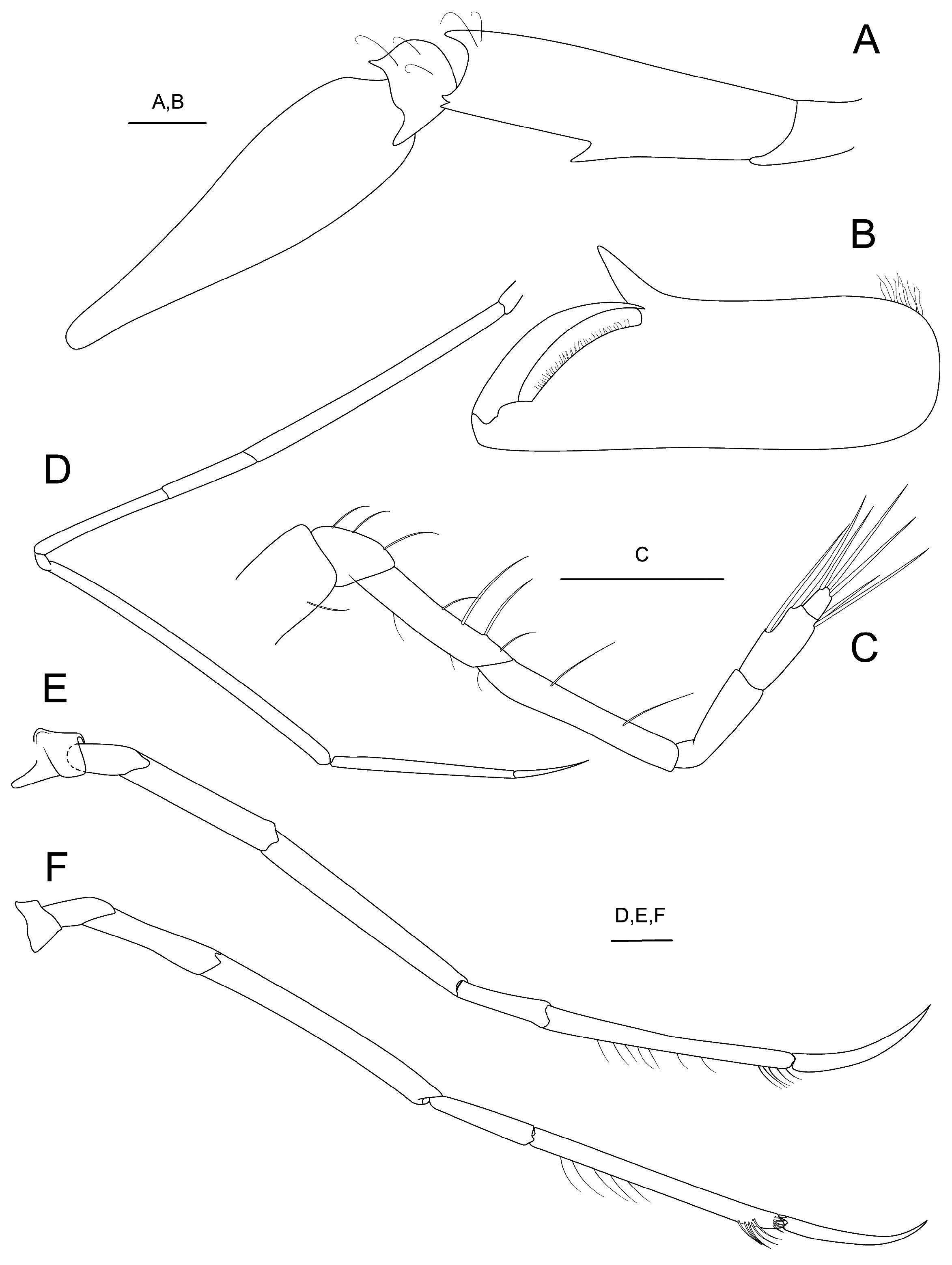
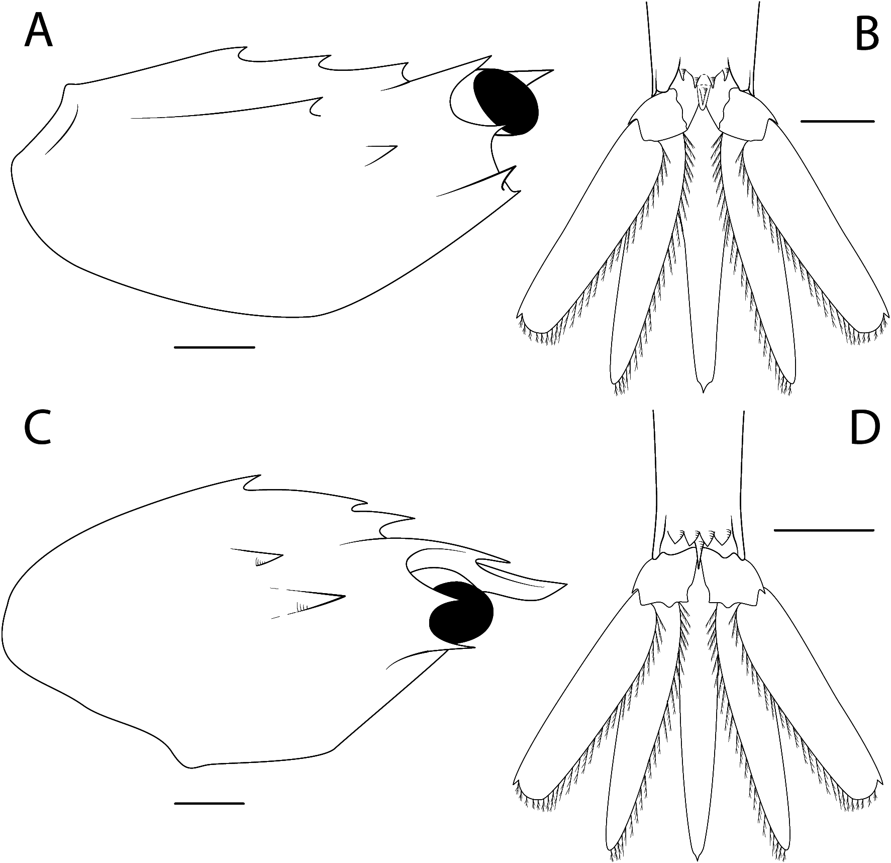
( Figures 1A–E View FIGURE 1 ; 2A–F View FIGURE 2 ; 3A–F View FIGURE 3 ; 4A, B View FIGURE 4 )
Lissosabinea cf. tridentata View in CoL — Christoffersen, 1988: 53, 54; Komai 2006: 36, 44; Spivak et al. 2019: 48 [non Lissosabinea tridentata ( Pequegnat, 1970) View in CoL ]
Lissosabinea tridentata View in CoL — Spivak 1997: 79, 80.
Type material. Holotype, female, cl 4.7 mm (MZUSP 22798), R/ V “Marion Dufresne”, TAAF MD 55/ Brésil Expedition , off Espírito Santo, Abrolhos Bank, stn 53 CB92 , 19°34’S, 38°55’W, M. Tavares coll., 29.v.1987, 340– 360 m, on fluid mud with foraminiferans and pteropods GoogleMaps . Paratypes: 1 male, cl 3.5 mm (MZUSP 22799) and 1 female, cl 4.2 mm (MZUSP 29971), same data as holotype GoogleMaps .
Type locality. Off the coast of Espírito Santo, Abrolhos Bank , 19°34’S, 38°55’W, 340–360m. GoogleMaps
Distribution. So far only known from Brazil: Abrolhos Bank (off the coast of Espírito Santo in fluid mud with foraminiferans and pteropods, and sand with foraminiferans) and Chuí (off the coast of Rio Grande do Sul, in sand calcareous rock and corals, and sand and gravel), between 166 and 360 m.
Etymology. The new species is named after our colleague, Martin Christoffersen (Universidade Federal da Paraíba), in recognition for his numerous contributions to the Brazilian zoology.
Description. Rostrum slightly upcurved distally, broad proximally, distal part laterally compressed, leaf-like, abruptly narrowing at about distal 0.25 in dorsal view; tip almost reaching distal margin of first article of antennular peduncle ( Figs. 1A, B View FIGURE 1 ; 2A View FIGURE 2 ); dorsal surface deeply concave with low, blunt median carina extending to level of base of first epigastric tooth, shallow sulcus on either side of median carina; rostral lateral margin forming strongly upturned lateral carina extending to about distal 0.50 of rostrum, ending in strong dorsolateral tooth ( Fig. 1B, D, E View FIGURE 1 ); rostral ventral face compressed, forming gently convex longitudinal keel ending abruptly near tip of rostrum ( Fig. 1B, D, E View FIGURE 1 ).
Carapace ( Figs. 1A View FIGURE 1 , 2A View FIGURE 2 , 4A View FIGURE 4 ) middorsal carina sharply delimited, extending to 0.80 of carapace length, armed with three strong nearly equidistant teeth; first epigastric tooth strongest, directed upwards, almost reaching base of rostrum; second epigastric and cardiac teeth nearly equal in size, directed forward.
Cornea of eye ( Fig. 2A View FIGURE 2 ) spherical, maximum diameter 0.21 of carapace length.
Antennal tooth small, almost reaching to midlength of cornea. Branchiostegal tooth straight, slightly directed upward, short, reaching slightly beyond midlength of antennal basicerite only ( Figs. 1A View FIGURE 1 , 2A View FIGURE 2 ). Pterygostomial angle armed with acute tooth; hepatic and epibranchial teeth of about same size, carinae low, epibranchial carina longest ( Figs. 1A View FIGURE 1 , 4A View FIGURE 4 ).
Antennular peduncle ( Fig. 2A View FIGURE 2 ) reaching 0.50 of antennal scale; stylocerite spiniform, laterally compressed in distal half, reaching nearly distal margin of first peduncular segment; lateral flagellum of 8–13 articles in females; mesial flagellum of 12–15 articles in females.
Antennal scale ( Fig. 2A, B View FIGURE 2 ) about 0.57 of carapace length, about 3.30 times as long as wide, lateral margin slightly concave, distal blade rounded; basicerite with small ventrolateral tooth; carpocerite reaching about midlength of antennal scale.
Fourth thoracic sternite with long median spur; IV-VIII sternites with acute median spine each, spines decreasing progressively in size posteriorly ( Fig. 2C View FIGURE 2 ).
Mouthparts not dissected.
Third maxilliped slightly overreaching distal end of antennal scale; ultimate segment longer than penultimate segment; antepenultimate segment moderately lateraly compressed.
First pereopod ( Fig. 3A, B View FIGURE 3 ) with palm about 3 times as long as wide; cutting edge of palm strongly oblique; pollex relatively large, triangular, slightly recurved; carpus with one moderately large spine on lateral margin; merus with strong dorsodistal spine not reaching distal margin of anteriorly extended carpus, distolateral margin without tooth; ventral lamina terminating in acute tooth.
Second pereopod ( Fig. 3C View FIGURE 3 ) falling far short of midlength of merus of first pereopod; dactylus about 0.36 length of propodus; propodus not widened distally; carpus as long as dactylus and propodus together; merus almost twice as long as carpus; ischium distinctly shorter tha merus.
Third pereopod ( Fig. 3D View FIGURE 3 ) very slender; ischium about 0.63 length of merus.
Fourth pereopod ( Fig. 3E View FIGURE 3 ) moderately slender, overreaching antennal scale by length of dactylus and about 0.40 of propodus; dactylus compressed dorsoventrally, about 0.58 as long as propodus; propodus with distal tuft of setae; carpus 0.37 as long as propodus; merus nearly 8.0 times as long as wide, unarmed on dorsodistal margin; ischium about 0.66 times as long as merus.
Fifth pereopod ( Fig. 3F View FIGURE 3 ) similar to fourth, overreaching antennal scale by slightly more than length of dactylus; ischium 0.40 times as long as merus.
Pleonal somites 1–5 pleura all rounded marginally. Second pleonal somite smooth, dorsally ( Fig. 1A View FIGURE 1 ). Third somite somewhat gibbous with distinct middorsal carina stretching from back to front almost 4/5 over entire length of somite, strongly convex in lateral view, posterodorsal margin of somite strongly produced posteriorly ( Figs. 1A, C View FIGURE 1 ; 2D View FIGURE 2 ). Fourth and fifth somites also smooth, non-carinate dorsally. Sixth somite about 1.3 times as long as high; dorsal surface with shallow sulcus along midline flanked by two submedian carinae, each ending in acute spine at some distance from posterior margin ( Figs. 1A, C View FIGURE 1 ; 2E View FIGURE 2 ),ventral surface with transverse row of four acute spines adjacent to posterior margin succeeded medially by distinct preanal spine ( Fig. 4B View FIGURE 4 ).
Telson tapering to small posteromedial spine, with 2 pairs of dorsolateral movable spines; 3 pairs of posterolateral blunt spines: lateralmost longest, second very short, mesialmost about 4/5 the length of the lateralmost spine; terminal process blunt ( Fig. 2E, F View FIGURE 2 ).
Remarks. Lissosabinea christofferseni sp. nov. immediately differs from the other described species of Lissosabinea by having two longitudinal, dorso-submedian carinae on the sixth pleonal somite, each terminating in a small acute submarginal spine ( Fig. 2E View FIGURE 2 ), whereas in all its congeners the sixth pleonal somite is devoid of such carinae and spines ( Komai 2006; Taylor & Collins 2009).
Lissosabinea tridentata , L. armata and L. christofferseni sp. nov. are the only species in the genus with three teeth on dorsal midline of the carapace and in that they stand apart from L. arthuri , L. beresfordi , L. ecarina , L. indica , and L. lynseyae , whose carapace bears two teeth on dorsal midline (cf. Komai 2006; Taylor & Collins, 2009), and from L. unispinosa , which is unique in having only one tooth on the dorsal midline of the carapace ( Komai 2006).
Lissosabinea christofferseni sp. nov. can be separated from its northwestern Atlantic counterpart L. tridentata by having the (characters for L. tridentata within brackets): (1) distal part of the rostrum leaf-like, abruptly narrowing at about distal 0.25 in dorsal view ( Fig. 2A View FIGURE 2 ) (vs distal part of rostrum spiniform, tapering progressively distally in dorsal view) (see Komai 2006: fig. 6B); (2) constriction of rostrum subdistally, well beyond the pair of rostral lateral spines in dorsal view ( Fig. 2A View FIGURE 2 ) (vs rostral constriction starting at the level of the base of the pair of rostral lateral spines in dorsal view (see Komai 2006: fig. 6B); (3) pair of rostral lateral spines relatively shorter and broader in L. christofferseni sp. nov. than in L. tridentata ( Fig. 2A View FIGURE 2 vs Komai 2006: fig. 6B); (4) rostrum’s ventral keel not reaching to rostral tip ( Fig. 1B, D, E View FIGURE 1 ) (vs ventral keel gently joining the rostral tip ( Fig. 4C View FIGURE 4 ) (see also Komai 2006: fig. 6A); (5) carapace cardiac, second and first epigastric teeth nearly equidistant in lateral view ( Fig. 4A View FIGURE 4 ) (vs first and second epigastric teeth distinctly closer to each other than to the cardiac tooth ( Fig. 4C View FIGURE 4 ) (see also paratype of L. tridentata in Komai 2006: 5 B); (6) carapace epibranchial carina well defined and epibranchial and hepatic spines well separated from each other ( Figs. 2A View FIGURE 2 , 4A View FIGURE 4 ) (vs epibranchial carina poorly defined and epibranchial and hepatic spines much closer to each other) ( Fig. 4C View FIGURE 4 ); (7) third maxilliped almost reaching to distal end of antennal scale (vs third maxilliped overreaching antennal scale by half length of ultimate segment); (8) telson with 2 pairs of dorsolateral spines; 3 pairs of posterolateral blunt spines: lateralmost longest, second very short, mesialmost about 4/5 the length of the lateralmost spine; terminal process blunt ( Fig. 2E, F View FIGURE 2 ) (vs 2 pairs of dorsolateral spines; 1 pair of posterolateral minute spines, 3 long simple setae; terminal process acutely pointed (see Komai 2006: fig. 5E, F).
Lissosabinea armata differs from L. christofferseni sp. nov. by having the shape of the rostrum much more similar to that of L. tridentata (see Komai 2006: fig. 8A); in the antennal tooth elongate, slightly overreaching the anterior margin of eye and in the first epigastric tooth also elongate, overhanging the base of the rostrum (vs antennal and first epigastric teeth much shorter in L. christofferseni sp. nov.). Lissosabinea armata yet differs in possessing much longer branchiostegal spines, the median carina on the third pleonal somite extremely high, the distal margin of the antennal blade obliquely truncate and the sixth pleonal somite provided with numerous minute dorsoproximal spines ( Komai 2006: fig. 8E) (vs distal margin of blade broadly rounded and sixth pleonal somite devoid of dorsoproximal spines in L. christofferseni sp. nov.) ( Figs. 1A, C View FIGURE 1 ; 2E View FIGURE 2 ; 4A View FIGURE 4 ). Lissosabinea armata also differs in the arrangement of the posterolateral spines in the telson, which are as follows: lateralmost spine short, blunt; second spine slender; mesialmost spine stout, longest; terminal process rounded (see Komai 2006: fig. 8E, F) (see above for L. christofferseni sp. nov.) ( Fig. 2E, F View FIGURE 2 ).
Although the two specimens from Chuí assigned to Lissosabinea cf. tridentata by Christoffersen (1988) were not located for study, they are referred herein to Lissosabinea christofferseni sp. nov. on account of the sixth pleonal somite being armed with one pair of submedian dorsal carinae and submarginal spines, a character unique to the present new species ( Fig. 2E View FIGURE 2 ).
Lissosabinea christofferseni sp. nov. has two arthrobranchs at the base of the third maxilliped, one large and a much smaller one. Such arrangement of the arthrobranchs was also found for L. indica by Kim & Natsukari (2000) and is reported herein for L. tridentata (see below). Christoffersen (1988) reported only one arthrobranch in the third maxilliped in the specimens from Chuí (as far it was possible to ascertain in the exuviae and the damaged specimen available to him) and may well have overlooked the smaller arthrobranch.
| V |
Royal British Columbia Museum - Herbarium |
No known copyright restrictions apply. See Agosti, D., Egloff, W., 2009. Taxonomic information exchange and copyright: the Plazi approach. BMC Research Notes 2009, 2:53 for further explanation.
|
Kingdom |
|
|
Phylum |
|
|
Class |
|
|
Order |
|
|
InfraOrder |
Caridea |
|
Family |
|
|
Genus |
Lissosabinea christofferseni
| Tavares, Marcos, Mendonça, Joel Braga De & Colavite, Jéssica 2022 |
Lissosabinea tridentata
| Spivak, E. D. 1997: 79 |
Lissosabinea cf. tridentata
| Spivak, E. D. & Farias, N. E. & Ocampo, E. H. & Lovrich, G. & Luppi, T. 2019: 48 |
| Komai, T. 2006: 36 |
| Christoffersen, M. L. 1988: 53 |