
Urocythereis ilariae, Aiello & Barra & Parisi, 2016
|
publication ID |
https://doi.org/ 10.5852/ejt.2016.193 |
|
DOI |
https://doi.org/10.5281/zenodo.3852156 |
|
persistent identifier |
https://treatment.plazi.org/id/887FB10D-607B-B455-213A-FD0DFC4C5EB1 |
|
treatment provided by |
Valdenar |
|
scientific name |
Urocythereis ilariae |
| status |
sp. nov. |
Urocythereis ilariae View in CoL sp. nov.
urn:lsid:zoobank.org:act:607A810F-773C-4F8F-91E7-D2EB5223CB7D
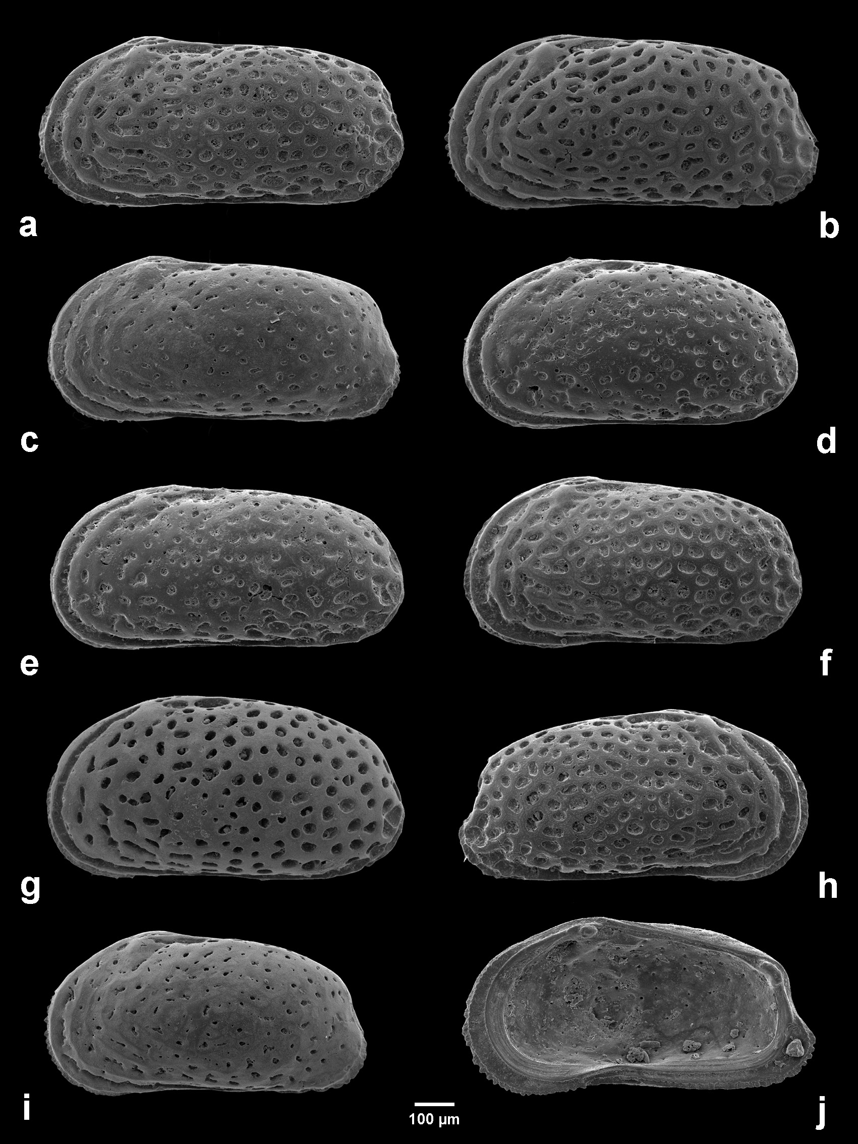
Figs 2B View Fig ; 3 View Fig G–H; 4G–H; 5G–H; 6G–H; 14; 18A–J; 19A–C, E–J
Urocythereis favosa (Roemer) View in CoL n. ssp. Bassiouni, 1965: pl. 40, figs 8–9.
Urocythereis favosa (Roemer) View in CoL subsp. – Wouters 1973: 385, pl. 2, fig. 7.
Urocythereis View in CoL sp. – Bonaduce, Ciampo & Masoli 1976: 46, pl. 22, fig. 9.
? Urocythereis View in CoL sp. – Athersuch 1977: pl. 17, fig. 2.
Urocythereis View in CoL aff. U. favosa (Roemer) – Arbulla, Pugliese & Russo 2001: fig. 3t.
? Urocythereis View in CoL sp.1 – Barra 1997: 82, pl. 4, fig. 6.
Urocythereis View in CoL sp.1 – Aiello et al. 2006: tables 3, 10. — Aiello, Barra & Parisi 2013: fig. 1d.
Diagnosis
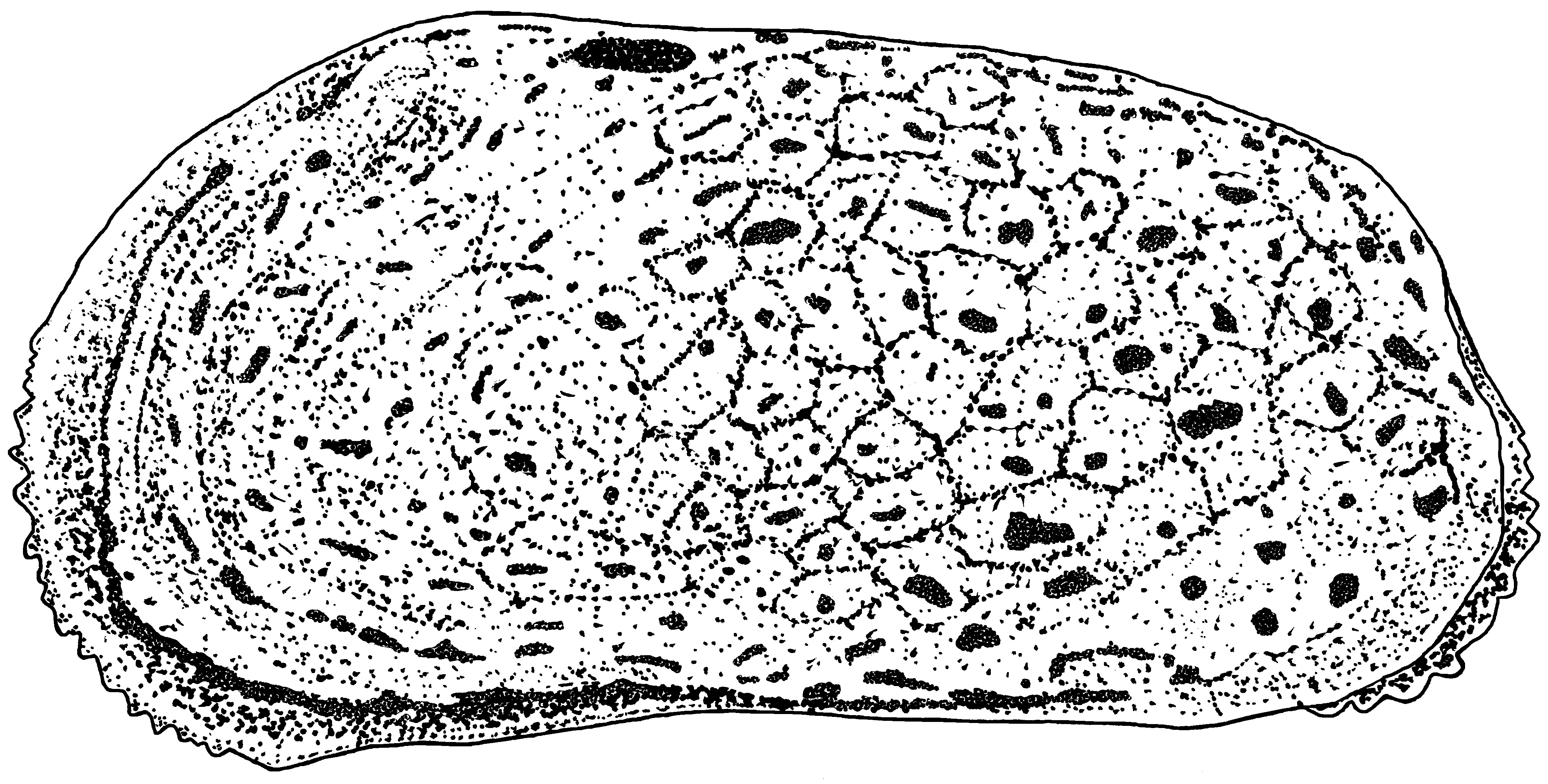
A large reticulate species of Urocythereis , subrectangular in lateral view, inflated-ovate in dorsal view. Reticulum with large polygonal-rounded, frequently coalescing, large fossae separated by broad muri. In the anteroventral area the muri form distinct riblets running parallel to the margin.
Etymology
In honour of our friend and collegue Ilaria Mazzini, in recognition of her important contribution to ostracodology.
Type material (4 carapaces, 43 valves: 29 adults and 14 juveniles)
Holotype
IONIAN SEA: ABMC 2014 /03
Paratypes
IONIAN SEA: ABMC2 014/026–036, ABMC 2014/038, ABMC 2014/042, ABMC 2014/044, ABMC 2014/046–049, ABMC 2014/063–064, ABMC 2014/069, ABMC 2014/072–073, ABMC 2014/080, ABMC 2014/097–103, ABMC 2014/120–135.
Stratum typicum
Recent.
Locus typicus
La Strea Bay (Porto Cesareo Lagoon), Southern Italy, Ionian Sea, sampling station E4, 17°54'25" N, 40°15'59" E, depth 1.5 m bsl.
Description
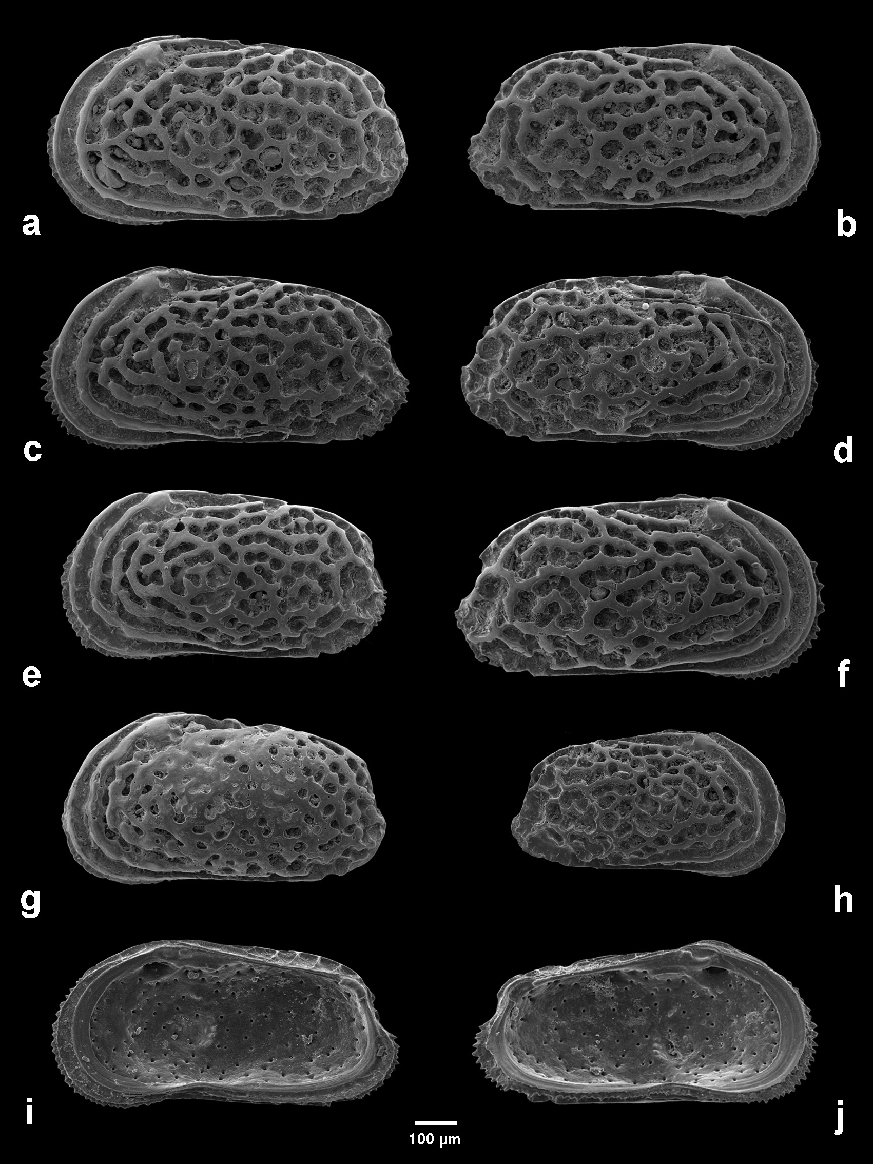
Measurements (holotype): LV: L = 0.85 mm, H = 0.44 mm ( Fig. 18A View Fig ).
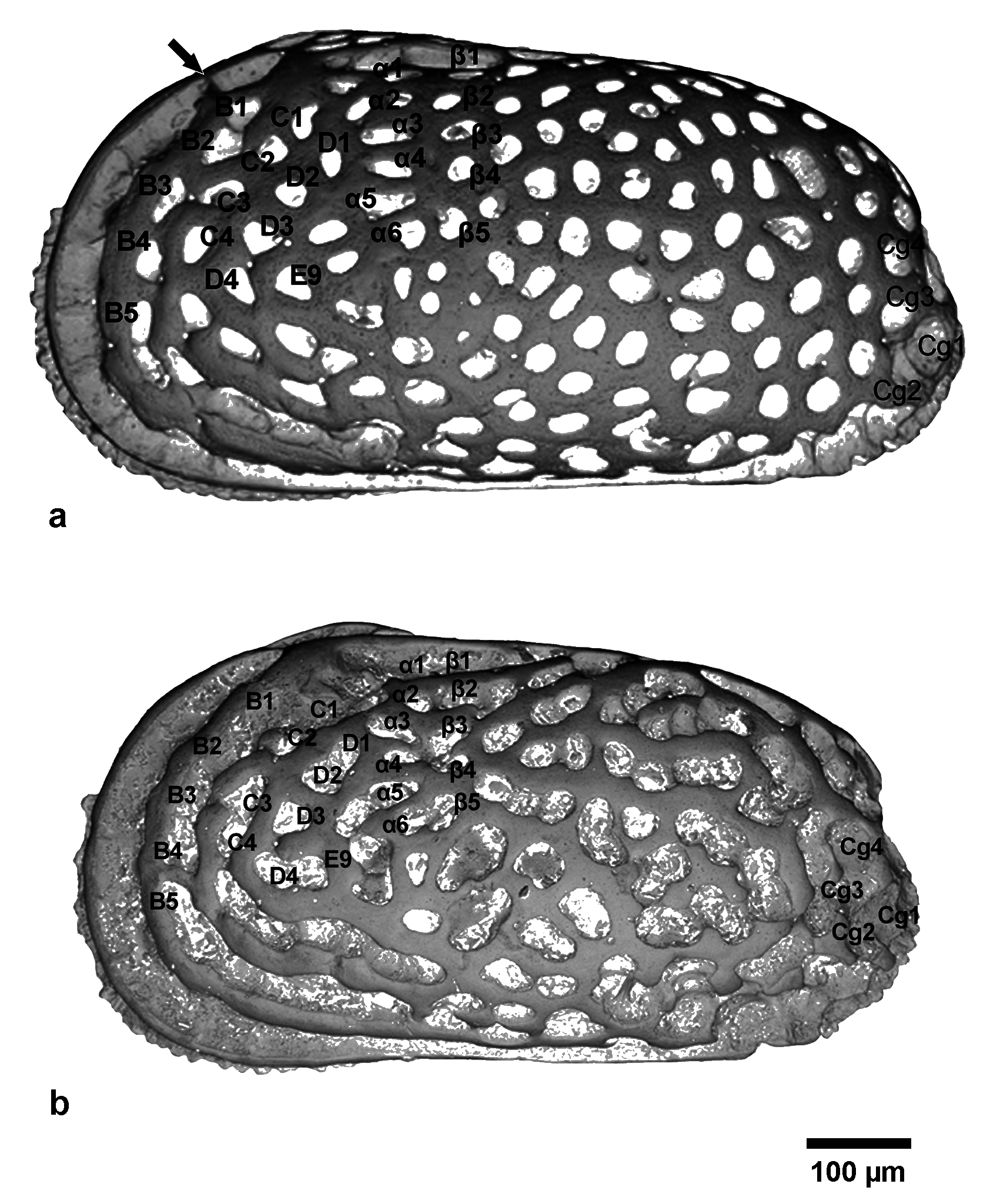
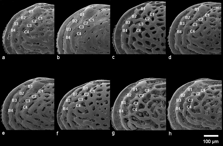
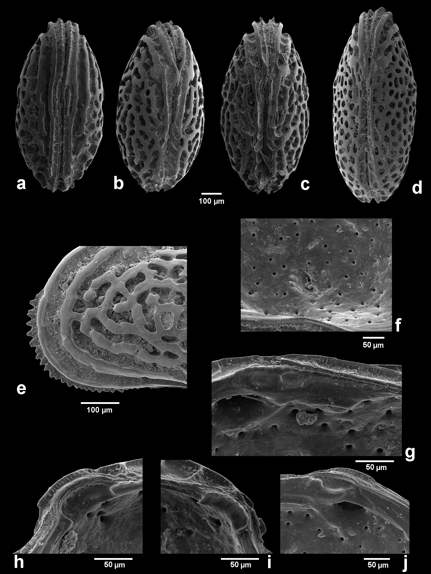
Large (L = 0.85–0.90 mm) species of Urocythereis , characterized by large fossae and strongly developed muri, subrectangular in lateral view, inflated-ovate in dorsal view. Valves strongly calcified and thick. Dorsal margin gently, unevenly convex, ventral margin weakly sinuous; anterior end broadly rounded, denticulate in the lower part; upper part of the posterior margin concave, lower part of the posterior margin convex, variably denticulate, forming short blunt caudal process located below mid-height. Maximum height at anterior cardinal angle, greatest length below mid-height. Surface of valves coarsely reticulate. Fossae, showing subrounded or irregular shape, coalesce, especially in marginal areas, forming both multiple anastomized elongated fossae and deep sulci parallel to margin. The corresponding muri tend to form a system of concentric riblets. Marginal rim starts from anterior part of dorsal margin, behind eye tubercle ( Fig. 19C View Fig ), and ends in posteroventral angle. Second riblet, constantly well developed, runs parallel to margin of valve except posterior end. This ocular riblet is connected with eye tubercle and rises above dorsal margin. Marginal rim and second riblet not connected. Third riblet, irregularly developed, delimits anteriorly the reticulum stricto sensu from subocular area to posterior part of ventral area and is connected with second riblet anteriorly, at mid height, through single radial murus; in the ventral area second and third riblets converge and, in lateral view, they seem apparently to be connected, but ventral view ( Fig. 19A View Fig ) shows they remain separate. The fossae between second and third riblet mainly anastomized. Fourth riblet fully part of reticulum, and shows a rather regular parallel trend only in anterocentral area. Surface of central area irregularly reticulate with subrounded/polygonal fossae with a low degree of anastomosis. Conversely, fossae located in proximity of caudal process coalesce following a longitudinal trend. Rare specimens show celation, never fully developed. Muri smooth, not papillate ( Fig. 19E View Fig ).
Hinge holamphidont (sensu Scott 1961): in left valve posterior hinge socket elongate and curved; anterior element formed by ovate-rounded (or elongate) tooth and elongate socket; median bar smooth; its posterior thickening forms, in some cases, barely defined toothlet; right valve hinge complementary, with faintly crenulate teeth ( Figs 18 View Fig I–J; 19G–J).
Inner lamella, marginal pore canals and muscle scar pattern ( Fig. 19F View Fig ) characteristic of genus (details in Athersuch 1977).
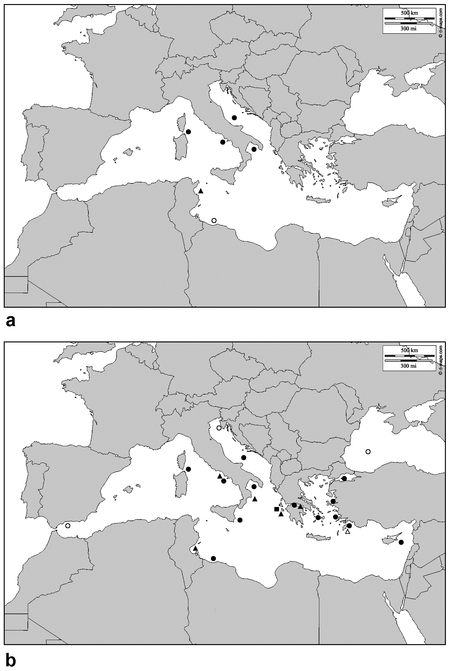
Distribution
The species occurs in the Recent of the Mediterranean: Gulf of Naples ( Bassiouni 1965), Sardinia ( Arbulla et al. 2001), South Adriatic Sea ( Bonaduce et al. 1976) and possibly Libya (see section Remarks); it has previously been recorded in fossil associations from the Tyrrhenian (upper Pleistocene) of Tunisia only ( Wouters 1973). Distribution data are summarized in Fig. 14 View Fig .
Remarks
U. ilariae sp. nov. has previously been assigned to U. favosa ( Bassiouni 1965; Wouters 1973), type species of the genus Urocythereis (neotype figured by Athersuch 1977). The reticulation of U. favosa differs from that of U. ilariae sp. nov. in the different style of fossal anastomosis. This is mostly evident, for example, in the anterodorsal zone, where the continuous depressed area formed by the fossal pattern C1-C2/B1-B4 is present in U. ilariae sp. nov. and absent in the Pliocene species.
The shell characters of U. exedata , described by Uliczny (1969) as a subspecies of U. favosa (SEM micrographs in Mostafawi & Matzke-Karasz 2006), show a close resemblance to those of U. ilariae sp. nov., especially in the structure of the ocular riblet, homologous to Bradleya ’s “ocular ridge” ( Benson 1972). The Pliocene species probably represents an ancestor of the living form. The two species differ in some reticulum features. In the anteroventral area of U. ilariae sp. nov. the third and the fourth riblets are connected ventrally and anteriorly; consequently they delimit the merged C fossae, forming an anteroventral furrow enclosed by muri. Conversely, in U. exedata the anteroventral area is characterized by a segment of the third riblet encircled by an elongated ring made up of B and C anastomized fossae, anteriorly and ventrally connected. In the anterodorsal area of U. ilariae sp. nov., the third concentric riblet is more or less developed in different specimens ( Figs 2B View Fig ; 18D, F View Fig ), while in U. exedata in the anterodorsal area the fossae of the B group coalesce with C and D fossae, the muri follow a radial trend and consequently the third concentric riblet is virtually absent.
The assignment of the North-African form, figured by Athersuch (1977) as Urocythereis sp. and by Barra (1997) as Urocythereis sp. 1, to U. ilariae sp. nov. needs further investigation. At the current state of knowledge we are inclined to interpret the morphological differences between the central Mediterranean species and the Lybian deme as the beginning of an allopatric speciation.
Urocythereis margaritifera (G.W. Müller, 1894) Figs 2A View Fig ; 3 View Fig A–F; 4A–F; 5A–F; 6A–F; 16A–K; 17A–J; 19D
Cythere oblonga Brady, 1866: 353 View in CoL , pl. 59, figs 5a–d (non C. oblonga M’Coy, 1844 View in CoL ).
Cythereis margaritifera G.W. Müller, 1894: 368 View in CoL , pl. 32, figs 26, 29, 32, 35–37.
Cythereis (Auris) distinguenda Neviani, 1928: 105 (synonymy only) (non p. 105 description and pl. 2, figs 91–93).
Urocythereis margaritifera alba Uliczny, 1969: 65 View in CoL , pl. 15, fig. 9.
Urocythereis View in CoL sp. Athersuch, 1977: pl. 17, fig. 5.
Urocythereis View in CoL sp. 2 Barra, 1997: 82-83, pl. 4, fig.8.
Urocythereis View in CoL sp. 3 Barra, 1997: 83, pl. 4, fig. 11.
Hemicythere (Urocythereis) margaritifera View in CoL – Ruggieri 1953: 94, pl. 6, fig. 1.
Urocythereis britannica Athersuch View in CoL – Kubanc 1995: 32–33, pl. 8, figs 4a–b.
Urocythereis crenulosa (Terquem) – Mostafawi & Matzke-Karasz 2006: pl. 6, fig. 9 (non pl. 8, fig. 1; non Cythere crenulosa Terquem, 1878 View in CoL ).
Urocythereis distinguenda View in CoL – Athersuch 1977: 257, 259, pl. 7, figs 1–6; pl. 8, figs 1–6; pl. 9, figs 1–5; pl. 12, figs 5–6; figs 3c–d. — Athersuch 1979: fig. 2.19. — Aiello et al. 2006: tabs. 3, 7, 10. — Aiello, Barra & Parisi 2013: fig. 1b.
Urocythereis favosa (Roemer) View in CoL – Barbeito-Gonzalez 1971: 279, pl. 13, figs 1b, 3b, 4b, 6b, pl. 46, figs 24- 27 (non pl. 13, figs 2b, 5b, pl. 46, figs 28–29). — Doruk 1974: pl. 38, fig. 3, pl. 40, figs 1–3 (non pl. 34, figs 1-2, pl. 38, figs 1-2). — Puri 1974: pl. 13, fig. 3. — Tunoglu 1999: pl. 7, fig. 1.
Urocythereis View in CoL aff. U. favosa View in CoL – Bonaduce, Ciampo & Masoli 1976: 45, pl. 22, fig. 8 (sic fig. 7).
? Urocythereis favosa View in CoL – Triantaphyllou, Tsourou, Koukousioura & Dermitzakis 2005: pl. 3, fig 11.
Urocythereis margaritifera View in CoL – Athersuch 1977: 260, 262, pl. 12, figs 1–4; pl. 13, figs 1–6; pl. 14, figs 1–5; figs 3e–f. — Tsapralis 1981: 100, pl. 1, fig. 1. — Lachenal 1989: 175–176, pl. 3, fig. 14. — Kubanç 1995: 31–32, pl. 8, figs 3a–c. — Aiello et al. 2006: tabs. 3, 5. — Perçin-Paçal & Balkis 2012: pl. 2, fig. 3. — Aiello, Barra & Parisi 2013: fig. 1a.
? Urocythereis margaritifera View in CoL – Aranki 1987: 72, pl. 19, figs 5–7. — Stancheva 1989: pl. 2, fig. 9. — Şafak, Avşar & Meriç 1999: pl. 3, fig. 12.
Urocythereis View in CoL cf. U. margaritifera View in CoL – Arbulla, Pugliese & Russo 2001: fig. 3s.
Urocythereis View in CoL ? margaritifera – Aiello, Barra & Parisi 2013: fig. 1c.
Urocythereis margaritifera alba View in CoL – Breman 1976: 63-64, pl. 9, fig. 124. — Aiello, Barra, De Pippo & Donadio 2012: pl. 2, fig. 8.
? Urocythereis margaritifera alba View in CoL – Uffenorde 1972: 79, pl. 8, fig. 9.
Urocythereis margaritifera margaritifera View in CoL – Uliczny 1969: 65, pl. 15, fig. 8.
? Urocythereis margaritifera margaritifera View in CoL – Sissingh 1972: 128, pl. 10, fig. 8.
Urocythereis seminulum (Seguenza) View in CoL – Şafak, Avşar & Meriç 1999: pl. 3, fig. 11.
Urocythereis View in CoL sp. – Mostafawi, 1994: 107, pl. 7, fig. 6.
Distribution
The species is widely distributed in the infralittoral waters of the Eastern Mediterranean ( Brady 1866; Barbeito-Gonzalez 1971; Doruk 1974; Athersuch 1977, 1979; Kubanç 1995; Tunoglu 1999; Perçin- Paçal & Balkis 2012), the Tyrrhenian Sea (G.W. Müller 1894; Puri 1974) and the southern Mediterranean ( Athersuch 1977; Lachenal 1989; Barra 1997). Recordings from the Black Sea are uncertain: the specimen figured by Stancheva (1989) is a young instar, and Schornikov (1969) reported Müller’s original drawings. The species is present in the southern part of the Adriatic Sea; the findings in the central and northern Adriatic are doubtful ( Uffenorde 1972; Bonaduce et al. 1976; Breman 1976).
Fossil specimens have been reported from the Upper Pleistocene–Holocene of the Gulf of Gabès ( Lachenal 1989), the Pleistocene of Southern Italy ( Ruggieri 1953; Aiello et al. 2012), Zakynthos ( Tsapralis 1981), the Northern Peloponnesus ( Mostafawi 1994) and, possibly, Rhodes ( Sissingh 1972) and from the Pliocene of Cephalonia ( Uliczny 1969). Distribution data are summarized in Fig. 14 View Fig .
The presence of the species in Miocene sediments ( Şafak et al. 1999) has to be confirmed by further studies.
Remarks
The analysis of the shell features of the Urocythereis population in the La Strea Bay and comparisons with the literature have convinced us that U. margaritifera and U. distinguenda (= U. oblonga ) are two morphotypes of the same species. In particular, we consider the latter “species” as the celated variation of the former. Celation is not expressed homogeneously on the valves in all the specimens; consequently, also “transitional” shells show different morphs.
The original illustration by Müller (1894: pl. 32, fig. 26) shows anteroventral fossae horizontally merged; the lectotype reported by Athersuch (1977: pl. 13, fig. 2) and the Libyan specimen figured by Barra (1997, as U. sp. 2) shows the same feature. Presently, we do not regard this character as diagnostic, due to the observed variability.
Athersuch (1977) figured some appendages, including the right copulatory appendage, of U. distinguenda and the left copulatory appendage of U. margaritifera . Regarding the discrimination of the two species, the author stated that in U. margaritifera the ductus ejaculatorius is short and is contained within the area of the appendage, whereas in U. distinguenda the duct is much longer and passes beyond the ventral margin. Examination of Athersuch’s illustrations (figs 4.d; 5.i) and a comparison with Müller’s drawing of the male copulatory appendage of Cythereis margaritifera (pl. 32, fig. 32) show that only very subtle differences are present and they represent, in our opinion, intraspecific variations.
The maximum mesh size is reached in the subspecies Urocythereis margaritifera alba Uliczny, 1969 . This form, in which celation is not developed, does not occur in the La Strea Bay. In some specimens the SEM micrographs revealed a feeble trace of the muri underlying the secondary calcification, as shown in Fig. 15 View Fig . The comparison of this hidden reticulation with the specimens figured by Uliczny (1969), Breman (1976) and Aiello et al. (2012) suggests that U. m. alba is a non-celate morphotype of U. margaritifera . The North Adriatic form figured by Uffenorde (1972) (very similar to the Pliocene specimen figured by Şafak et al. 1999 as U. margaritifera ) shows some tiny differences in the reticulation pattern and the assignment to U. margaritifera is queried.
The Libyan form figured by Athersuch (1977: pl. 17, fig. 5) as U. sp. and by Barra (1997) as U. sp. 3 fits the U. margaritifera morph c ( Fig. 17E View Fig ) well.
The left valve, figured by Aranki (1987) from the western Mediterranean shallow waters, shows some minor differences in the reniform outline and in some details of the reticulum. The relationships between Mediterranean and Atlantic forms (i.e., between U. margaritifera and U. britannica , frequently reported as U. oblonga ) need further investigations.
Ruggieri (1953) hypothesized that U. favosa and U. margaritifera might be conspecific, the latter species representing a subspecies of the former. In spite of the similarity of the two forms, some features of the reticulum seem to allow a separation of the two species. In U. favosa (neotype figured in Athersuch 1977) the fossae B3 and B4 are merged, as well as C3 and C4; in U. margaritifera they are distinct; in U. favosa the fossae D2-D1 and α4 coalesce while in U. margaritifera they are distinct. In the caudal group the fossa Cg 4 in U. margaritifera is separate, whereas in U. favosa the arrangement of the fossae is similar to that in U. ilariae sp. nov. In the La Strea Bay, and possibly in the Recent of the Mediterranean, U. favosa s.s. is not recorded and we prefer to retain them as separate species.
Some Urocythereis spp. from the Pliocene of Rhodes have been described by Terquem (1878) and figured by Mostafawi (1989). They are distinct from U. margaritifera in some characters of the reticulum. By contrast, the specimen from Cephalonia figured by Mostafawi & Matzke-Karasz (2006) as U. crenulosa fits well with some specimens of U. margaritifera from Porto Cesareo.
The Atlantic forms reported as U. britannica and (erroneously) as U. oblonga (e.g., Guillaume et al. 1985; Ruiz et al. 2006) show a high variability and a complex affinity with U. margaritifera and U. favosa and they have not been considered in the present study.
No known copyright restrictions apply. See Agosti, D., Egloff, W., 2009. Taxonomic information exchange and copyright: the Plazi approach. BMC Research Notes 2009, 2:53 for further explanation.
|
Kingdom |
|
|
Phylum |
|
|
Class |
|
|
Order |
|
|
Family |
|
|
Genus |
Urocythereis ilariae
| Aiello, Giuseppe, Barra, Diana & Parisi, Roberta 2016 |
Urocythereis
| Barra D. 1997: 82 |
Urocythereis
| Barra D. 1997: 82 |
Urocythereis
| Barra D. 1997: 83 |
Urocythereis britannica
| Kubanc C. 1995: 32 |
Urocythereis
| Mostafawi N. 1994: 107 |
Urocythereis margaritifera
| Aranki J. F. 1987: 72 |
| Şafak, Avşar & Meriç 1999 : pl. 3 |
Urocythereis
| Bonaduce G. & Ciampo G. & Masoli M. 1976: 46 |
Urocythereis
| Bonaduce G. & Ciampo G. & Masoli M. 1976: 45 |
Urocythereis margaritifera alba
| Breman E. 1976: 63 |
| Aiello, Barra, De Pippo & Donadio 2012 : pl. 2, fig. 8. |
Urocythereis favosa (Roemer)
| Wouters K. 1973: 385 |
Urocythereis margaritifera alba
| Uffenorde H. 1972: 79 |
Urocythereis margaritifera margaritifera
| Sissingh W. 1972: 128 |
Urocythereis margaritifera alba
| Uliczny F. 1969: 65 |
Urocythereis margaritifera margaritifera
| Uliczny F. 1969: 65 |
Hemicythere (Urocythereis) margaritifera
| Ruggieri G. 1953: 94 |
Cythereis (Auris) distinguenda
| Neviani A. 1928: 105 |
Cythereis margaritifera G.W. Müller, 1894: 368
| Muller G. W. 1894: 368 |
Cythere oblonga
| Brady G. S. 1866: 353 |
Urocythereis favosa (Roemer)
| Bassiouni, 1965 : pl. 40, figs 8–9 |
Urocythereis
| Athersuch 1977 : pl. 17 |
Urocythereis
| Arbulla, Pugliese & Russo 2001 |
Urocythereis
| Aiello et al. 2006 : tables 3, 10 |
| Aiello, Barra & Parisi 2013 : fig. 1d. |
Urocythereis
| Athersuch, 1977 : pl. 17, fig. 5 |
Urocythereis crenulosa (Terquem)
| Mostafawi & Matzke-Karasz 2006 : pl. 6 |
Urocythereis distinguenda
| Athersuch 1977: 257 |
| Athersuch 1979 : fig. 2.19 |
| Aiello et al. 2006 : tabs. 3, 7, 10 |
| Aiello, Barra & Parisi 2013 : fig. 1b |
Urocythereis favosa (Roemer)
| Barbeito-Gonzalez 1971: 279 |
| Doruk 1974 : pl. 38, |
| Puri 1974 : pl. 13, fig. 3 |
| Tunoglu 1999 : pl. 7, fig. 1 |
Urocythereis favosa
| Triantaphyllou, Tsourou, Koukousioura & Dermitzakis 2005 : pl. 3, fig 11 |
Urocythereis margaritifera
| Athersuch 1977: 260 , 262 |
| Tsapralis 1981: 100 |
| Lachenal 1989: 175–176 |
| Kubanç 1995: 31–32 |
| Aiello et al. 2006 : tabs. 3, 5 |
| Perçin-Paçal & Balkis 2012 : pl. 2, |
| Aiello, Barra & Parisi 2013 : fig. 1a. |
Urocythereis
| Arbulla, Pugliese & Russo 2001 : fig. 3s |
Urocythereis
| Aiello, Barra & Parisi 2013 : fig. 1c. |
Urocythereis seminulum (Seguenza)
| Şafak, Avşar & Meriç 1999 : pl. 3 |