
Bonnetina tenuiverpis, Ortiz & Francke, 2014
|
publication ID |
https://doi.org/ 10.1080/00222933.2014.924770 |
|
DOI |
https://doi.org/10.5281/zenodo.4327867 |
|
persistent identifier |
https://treatment.plazi.org/id/891E87F1-FF9A-FF9C-FE44-817FFCCDFAF8 |
|
treatment provided by |
Carolina |
|
scientific name |
Bonnetina tenuiverpis |
| status |
sp. nov. |
Bonnetina tenuiverpis View in CoL sp. nov.
urn:lsid:zoobank.org:act:B1F7E8B8-F542-430B-BEE3-9F1349419DCA
( Figures 1–8 View Figure 1 View Figure 2 View Figure 3 View Figure 4 View Figure 5 View Figure 6 View Figure 7 View Figure 8 ; Tables 1, 2)
Holotype. ♂ ( CNAN-T0755 ). MEXICO: Mexico state: Otzoloapan municipality: 3 km NNE of Zuluapan town: 19.1627°, −100.3085°: 1250 m asl. 7 August 2012. David Ortiz, Carlos Santibáñez, Diego Barrales & Rodrigo Monjaraz, cols. Under a stone. Matured in captivity 11 October 2012.
Allotype. ♀ ( CNAN-T0756 ): same collecting data as holotype. Other paratypes. ♀ ( AMNH): same collecting data as holotype. ♀ ( CNAN-T0757 ): same locality as holotype. 16 December 2001. Oscar F. Francke and Edmundo González, cols.
Etymology
The specific name is composed by the Latin adjective tenuis (slender) and plural noun verpis (penes). It makes reference to the attenuate condition of the male pedipalpal bulbs of this species.
Diagnosis
Males differ from those of all known species of the genus by the shape of its palpal bulbs, that are also remarkably slender compared to those of the other species. It additionally has much better developed tibiae I accessory apophyses than B. alagoni and B. aviae . It is a distinctly smaller species than B. cyaneifemur , B. rudloffi , B. papalutlensis and B. tanzeri . Females differ from those of other species by having subdigitiform spermatheca instead of domiform (as in B. alagoni and B. aviae ), flattened ( B. tanzeri ), subtriangular ( B. papalutlensis ) or digitiform ( B. cyaneifemur ). Females of B. rudloffi have not been described.
Description
Male holotype
Morphology
Some quantitative characters are given in Table 1
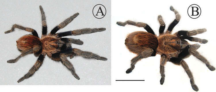
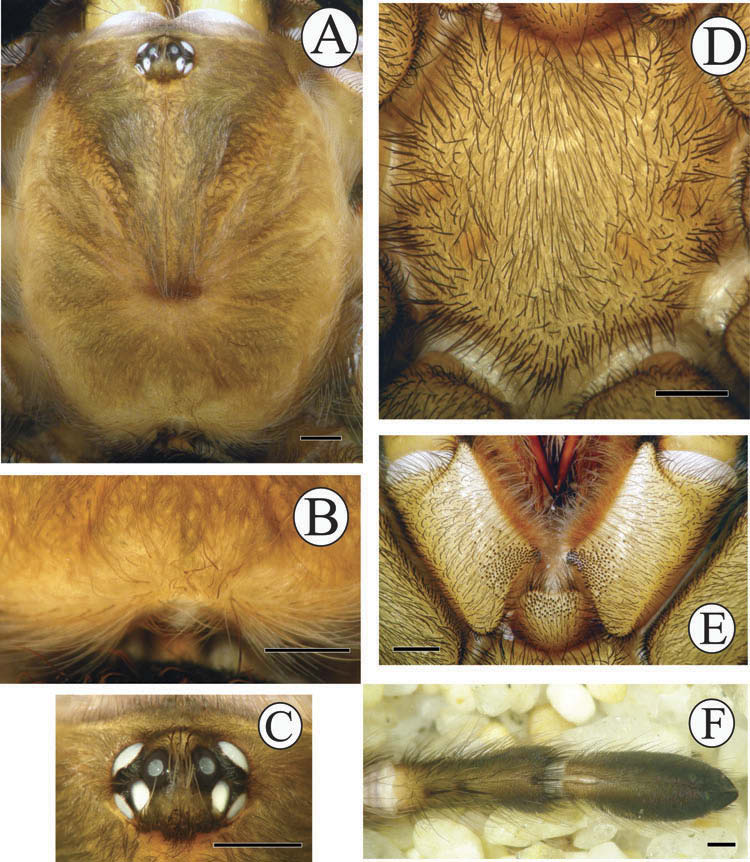
Coloration and pilosity. Carapace covered by dense shiny coppery pubescence, that masks partially the dark brown colour of the integument ( Figures 1A View Figure 1 , 2A View Figure 2 ). Posterior area of carapace bears distinctly thick erect setae ( Figure 2B View Figure 2 ). Leg and pedipalpal segments (except femora) with intermixed copper and light brown setae. Femora black, with iridescent blue tones. Abdomen dark brown, with long and thick red setae and dense short dark brown setae.
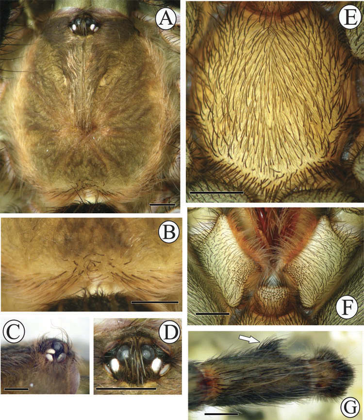
Carapace with caput nearly flat and fovea broad and procurved. Ocular area: eight eyes disposed in two rows on markedly elevated tubercle (PLE almost perpendicular to carapace) ( Figure 2C View Figure 2 ); anterior eye row procurved; posterior row, recurved ( Figure 2D View Figure 2 ). Ocular quadrangle width, 1.14; length, 0.86. Clypeus, 0.14 wide. AME circular, diameter, 0.28; ALE ovoid, greater diameter, 0.36; PME ovoid, greater diameter, 0.26; PLE ovoid, greater diameter, 0.26. Row of erect hairs present between fovea and ocular area.
Chelicerae with seven teeth (left appendage), and seven teeth plus one tiny basalmost tooth (right), close and parallel to the promargin on ventral side.
Sternum ( Figure 2E View Figure 2 ) slightly convex to its centre, covered uniformly by erect thick hairs and other hairs much smaller; with three pairs of sigillae, placed opposite to coxae I, II and III. Labium subtrapezoidal ( Figure 2F View Figure 2 ); middle length, 0.90; anterior width, 0.80; posterior width, 1.25.
Appendage segment lengths (left limbs). Palp: femur, 3.9; patella, 2.5; tibia, 3.0; total, 9.4. Leg I: femur, 5.8; patella, 3.5; tibia, 4.1; metatarsus, 3.6; tarsus, 2.6; total, 19.6. Leg II: femur, 5.1; patella, 2.9; tibia, 3.6; metatarsus, 3.3; tarsus, 2.6; total, 17.5. Leg III: femur, 4.1; patella, 2.5; tibia, 3.0; metatarsus, 4.0; tarsus, 2.4; total, 16.0. Leg IV: femur, 5.5; patella, 3.0; tibia, 4.8; metatarsus, 6.1; tarsus, 2.9; total, 22.3 (Leg formula: leg IV> I> II> III).
Retrolateral face of palpal tibiae with prominent, apically inclined conicshaped nodule near apex, densely covered by long and thick setae ( Figure 2G View Figure 2 ).
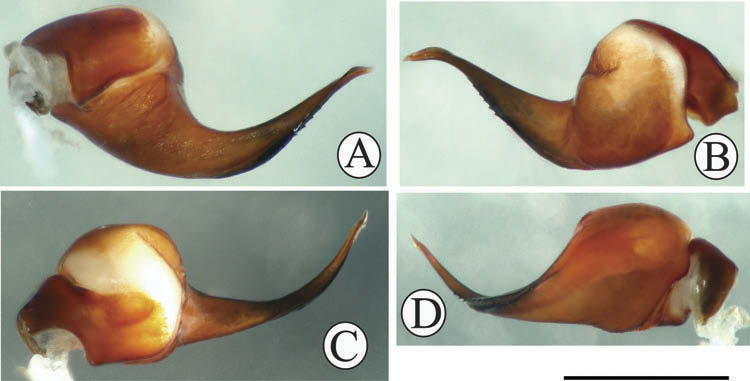
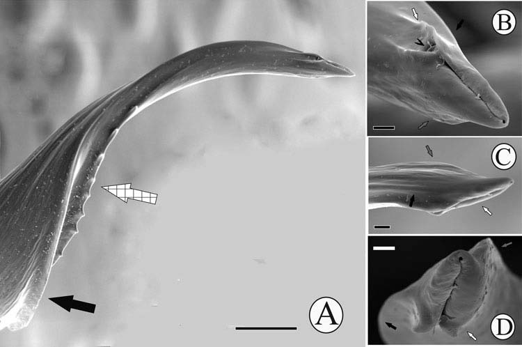
Palpal bulbs ( Figures 3 View Figure 3 , 4 View Figure 4 ) with embolus very slender, with its apical half markedly curved and twisted dorsally and retrolaterally (from base to apex) to the point that in the apex, the ventral structures of the bulb become prolateral. Prolateral inferior, prolateral superior, sperm pore and dorsal keels present. PI keel is mostly serrated and extends from the base of the embolus to half of the space between its most apical denticle and the sperm pore. PS keel extends from close to the base of the embolus to sperm pore level. SP keels extend from the bulb apex to the sperm pore and are folded onto each other, except in the basalmost region, forming the sperm pore; they are not completely fused at the apex, allowing a small pore at embolus tip. D keel, about as long as SP keels, is subapical to the embolus.
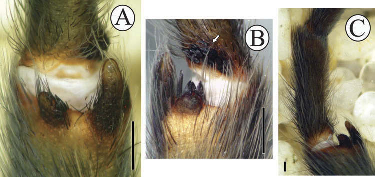
Legs I holding organ. Three apophyses near the apex of tibiae I, with separated bases ( Figure 5A, B View Figure 5 ). Prolateral apophysis conical and bent prolaterally, bearing a megaspine on its internal border; length, 0.34/0.36 (left/right). Retrolateral apophysis subdigitiform, parallel to tibial axis; length, 0.80/0.90. Accessory apophysis semicircular and well developed, bearing three subtriangular megaspines at its apex and a spine on the internal border; length, 0.24/0.20. When flexed, the moderately curved metatarsus I ( Figure 5C View Figure 5 ) folds between prolateral and retrolateral apophyses.
Nodule of 17 granules on basal ventro-retrolateral region of both metatarsi I ( Figure 5B View Figure 5 ).
Femora of palps and legs I and II prolaterally and femora of legs IV retrolaterally covered by pad of simple and ciliated hairs.
Palpal coxae and trochanters with non-plumose setae pro- and retrolaterally.
Scopulae Metatarsi. On legs I entire segment except apex; on legs II apical 5/6; on legs III distal half of segment and on legs IV apical 1/5. Tarsi. On legs I and II entire with few dispersed type B hairs; on legs III divided completely by a band of 1–3 fine hairs; on tarsi IV divided full length by band of 3–4 very thick hairs.
Very dense claw tufts on every leg.
Abdominal urticating hairs. Type III, in dorsal oval patch, located mostly in posterior half of abdomen, covering 0.42 of its length.
Spination pattern (left limbs). Palp: femur p0-0-1. Leg I: femur p0-0-1; tibia v2-2-0 p0-1-1; metatarsus v0-0-1. Leg II: femur p0-0-1; tibia v2-2-3 p0-1-1; metatarsus v2-0- 1. Leg III: femur p0-1-1 r0-0-1; tibia v1-1-3 p1-0-1 r0-0-1; metatarsus v1-3-4 p1-1-1 r0-1-1. Leg IV: femur r0-0-1; tibia v2-2-3 r1-0-1; metatarsus v1-2-4 p0-0-2 r0-0-1.
Molecular characterization
Mitochondrial CO1 partial sequence (GenBank accession: KC807369 View Materials ):
g ttg cct cct tcg ttg ttt tta tta gta tta tct tct ttg act gat gta ggg gtt ggg gct ggg tga act att tat ccg cct tta tct tct ttt atg ggt cat tct ggt gga ggg atg gat ttt gct att ttt tct ttg cat ttg gct ggg gct tcg tct att atg ggg tct gta aat ttt att act act gtt ata aat atg cgt gca tct gga ata tcg atg gaa cgt att cct ttg ttt gtt tga tct gta gtg att aca act gta tta ttg tta tta tcg tta ccg gtg ttg gct ggg gct att act ata tta ttg tcg gat cgg aat ttt aat act tct ttt ttt gac ccg gct ggt ggt ggg gat cct att ttg ttt cag cat tta ttt tga ttt ttt gga cat ccg gag gta tat att ttg att tta cca gga ttt gga atg gta tct cat att att agt tct tct gtt ggt aaa cgt gaa cct ttt gga tct tta gga ata att tat gct atg gtt aga att ggt gga ata gga ttt gtt gtg tga gca cat cat atg ttt tct gtt ggt ata gat gtt gat act cgt gca tat ttt act gcg gca act ata gtg att gct gta cct acg gga att aag gtt ttt aga tga ata gca acg ttg tat gga tca tat ttt aag ttg gat gtg tct ttg atg tgg tgt att gga ttt gta ttt tta ttt act atg ggt gga ttg act ggg gta gta tta gct aat tct tct ttg gat att gtg ttg cat gat acg tat tat gtt gtt gct cat ttt cat tat gtg ttg aga atg gga gct gta ttt gct att atg ggg ggt ttg gct tat tga ttt cct tta ttt ttt gga act ata ata aat ccn nnn tta atg aag ttg cag ttt gtt ata atg ttt gtt ggt gta aat tta act
Preservation state
The specimen is in optimal conditions, stored in a flask of 80% ethanol. Right leg III preserved in 96% ethanol at –20°C for molecular studies. Left pedipalpal bulb is housed apart, coated with gold.
Female allotype
Morphology Some quantitative characters are given in Table 1
Coloration and pilosity. As for holotype, but the carapace pubescence is less dense and the femora lack blue tones ( Figures 1B View Figure 1 , 6A View Figure 6 ). Posterior area of carapace bears distinct setae, considerably thinner than those on the holotype ( Figure 6B View Figure 6 ).
Carapace with caput elevated. Fovea procurved. Anterior eye row procurved; posterior row recurved ( Figure 6C View Figure 6 ). Ocular quadrangle width, 1.56; length, 0.92. Clypeus, 0.22 wide. AME circular, diameter, 0.40; ALE ovoid, greater diameter, 0.52; PME ovoid, greater diameter, 0.42; PLE ovoid, greater diameter, 0.40. Row of erect hairs present between fovea and ocular area.
Chelicerae with nine teeth plus one tiny basalmost tooth (left appendage) and eight teeth plus one tiny basalmost tooth (right), close and parallel to the promargin on ventral side.
Sternum ( Figure 6D View Figure 6 ) slightly convex, covered uniformly by thick erect hairs and other hairs much smaller; with three pairs of sigillae, placed opposite to coxae I, II and III. Labium subtrapezoidal; middle length, 1.25; anterior width, 1.15; posterior width, 2.05 ( Figure 6E View Figure 6 ).
Appendage segment lengths (left limbs). Palp: femur, 5.7; patella, 3.8; tibia, 3.6; tarsus, 3.8; total, 16.9. Leg I: femur, 7.5; patella, 5.0; tibia, 3.9; metatarsus, 4.2; tarsus, 2.9; total, 23.5. Leg II: femur, 6.0; patella, 4.0; tibia, 3.7; metatarsus, 4.3; tarsus, 3.1; total, 21.1. Leg III: femur, 5.7; patella, 4.2; tibia, 3.8; metatarsus, 5.3; tarsus, 3.1; total, 22.1. Leg IV: femur, 8.0; patella, 4.8; tibia, 5.9; metatarsus, 7.4; tarsus, 4.0; total, 30.1 (Leg formula: leg IV> I> III> II).
Scopulae. Metatarsi. On legs I entire; on legs II entire except base; on legs III apical half prolaterally and apical third retrolaterally ( Figure 6F View Figure 6 ); on legs IV apical fourth. Tarsal scopulae and femoral pads as on holotype.
Coxae and trochanters of palps and legs I and II as in holotype, covered by nonplumose hairs .
Abdominal urticating hairs. Type III, in oval dorsal patch (with rear part distinctly narrower), located mostly in posterior half of abdomen, covering 0.43 of its length.
Spination pattern (left limbs). Palp: femur, p0-0-1 r0-0-1; tibia, v0-1-4. p0-1-0 r0-1-0. Leg I: femur, p0-0-1; tibia, v0-1-0 p0-2-0; metatarsus, v1-1-1. Leg II: femur, p0-0-1; tibia, v0-1-1 p0-1-1; metatarsus, v2-0-2. Leg III: femur, p0-0-1 r0-0-1; tibia, v3-2-2 p2- 2-1 r1-0-1; metatarsus, v1-1-3 p2-2-2 r0-1-1. Leg IV: femur, r0-0-1; tibia, v2-2-3 r0-1- 1; metatarsus, v8 p1-1-3 r1-0-1.
Only one spermatheca subdigitiform ( Figure 7A, B View Figure 7 ), symmetrical (in dorsal and ventral views) with well-defined base, neck and fundus; length, 1.02; base width, 0.72. A wide poorly sclerotized atrium is at its base.
Molecular characterization
Mitochondrial CO1 partial sequence (GenBank accession: KC807370 View Materials ):
ttg ttg ccn cct tcg ttg ttt tta tta nta tta tct tct ttn act gat gta ggg gtt ggg gct ggg tga act att tat ccg cct tta tct tct ttt atg ggt cat tct ggt gga ggg atg gat ttt gct att ttt tct ttg cat ttg gct ggg gct tcg tct att atg ggg tct gta aat ttt att act act gtt ata aat atg cgt gca tct gga ata tcg atg gaa cgt att cct ttg ttt gtt tga tct gta gtg att aca act gta tta ttg tta tta tcg tta ccg gtg ttg gct ggg gct att act ata tta ttg tcg gat cgg aat ttt aat act tct ttt ttt gac ccg gct ggt ggt ggg gat cct att ttg ttt cag cat tta ttt tga ttt ttt gga cat ccg gag gta tat att ttg att tta cca gga ttt gga atg gta tct cat att att agt tct tct gtt ggt aaa cgt gaa cct ttt gga tct tta gga ata att tat gct atg gtt aga att ggt gga ata gga ttt gtt gtg tga gca cat cat atg ttt tct gtt ggt ata gat gtt gat act cgt gca tat ttt act gcg gca act ata gtg att gct gta cct acg gga att aag gtt ttt aga tga ata gca acg ttg tat gga tca tat ttt aag ttg gat gtg tct ttg atg tgg tgt att gga ttt gta ttt tta ttt act atg ggt gga ttg act ggg gta gta tta gct aat tct tct ttg gat att gtg ttg cat gat acg tat tat gtt gtt gct cat ttt cat tat gtg ttg aga atg gga gct gta ttt gct att atg ggg ggt ttg gct tat tga ttt cct tta ttt ttt gga act ata ata aat ccn nnn tta atg aag ttn cng ttt gtt ata atn ttt gtt ggn gta aat ata act
Preservation state
The specimen is in optimal conditions, stored in a flask with 80% ethanol. Right leg III preserved in 96% ethanol at –20°C for molecular studies. Genital area is housed in a plastic vial inside the flask.
Variation
The variation of some quantitative characters in the three females examined is summarized in Tables 1 and 2. None of the females has thick erect setae on the posterior margin of carapace, although setation in this area is distinct from the rest of carapace.
All have a row of erect hairs that extends between ocular area and fovea.
The shape of the urticating hairs patch varies, although it is oval for all specimens.
Coxae and trochanters lack plumose setae and hairs in all specimens.
The pubescence of the carapace is considerably denser on the male than on the three females. The coloration is very similar in the two females that were examined alive.
Spermathecae of the three specimens share the same structural pattern ( Figure 7 View Figure 7 ), although that of AMNH paratype is wider. The AMNH paratype also has tarsi on all the legs divided, probably due to its smaller size ( Pérez-Miles 1994).
The holotype and allotype differ only by one base pair of the CO1 sequenced region (0.1% uncorrected pairwise sequence divergence).
Distribution
Bonnetina tenuiverpis is known only from the vicinity of Zuluapan town, situated in the State of Mexico and geographically at 1250 m asl in the Trans-Mexican Volcanic Belt (Eje Neovolcánico), which extends from Pacific to Atlantic coast of Central– Southern Mexico.
Natural history
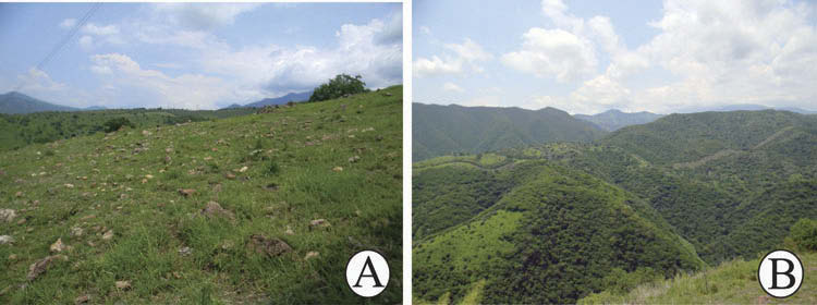
The specimens looked faded when collected in August 2012, and all moulted in captivity in September and October. The only male matured on 11 October, suggesting that the breeding season might include fall. The only known population lives in an absolutely open, very rocky grassland ( Figure 8 View Figure 8 ) that is frequently used by cattle. Dominant vegetation of the area is tropical deciduous forest. The specimens were found mainly on the edges of middle sized rocks (20–150 cm diameter), in shallow depressions bordered by silk, presumably dug by them. Two specimens of the tarantula Brachypelma cf. albiceps, three of another Bonnetina species and few of Diplocentrus scorpions were also found syntopically. These three species could be competing for resources or even preying on B. tenuiverpis .

| AMNH |
American Museum of Natural History |
No known copyright restrictions apply. See Agosti, D., Egloff, W., 2009. Taxonomic information exchange and copyright: the Plazi approach. BMC Research Notes 2009, 2:53 for further explanation.
|
Kingdom |
|
|
Phylum |
|
|
Class |
|
|
Order |
|
|
Family |
|
|
SubFamily |
Theraphosinae |
|
Genus |