
Chaetonotus (Chaetonotus) invitatus, 2019
|
publication ID |
https://doi.org/ 10.5852/ejt.2019.511 |
|
publication LSID |
lsid:zoobank.org:pub:8FDAD45D-1B7D-446F-8B34-026EDF192210 |
|
DOI |
https://doi.org/10.5281/zenodo.5620059 |
|
persistent identifier |
https://treatment.plazi.org/id/5350662C-4565-4EB6-96B0-4742ED038286 |
|
taxon LSID |
lsid:zoobank.org:act:5350662C-4565-4EB6-96B0-4742ED038286 |
|
treatment provided by |
Plazi |
|
scientific name |
Chaetonotus (Chaetonotus) invitatus |
| status |
sp. nov. |
Chaetonotus (Chaetonotus) invitatus View in CoL sp. nov.
urn:lsid:zoobank.org:act:5350662C-4565-4EB6-96B0-4742ED038286
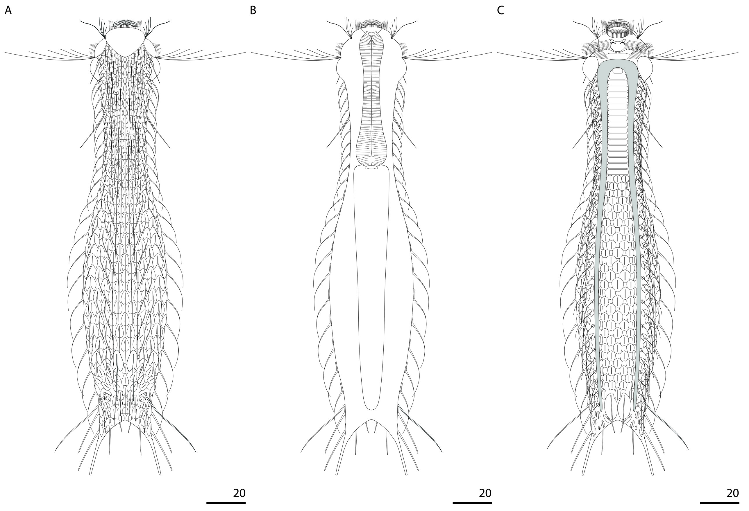
Figs 2–11 View Fig View Fig View Fig View Fig View Fig View Fig View Fig View Fig View Fig View Fig , A 1–A View Fig 2 View Fig ; Tables 2, A 1–A 2
Diagnosis
Slender, bottle-shaped, body measuring from 174.6 to 194.7 μm in length. Head five-lobed, cephalion, epipleurae and hypopleurae clearly demarcated in head outline. Hypostomium kidney-shaped with horn-like protuberances anterolaterally and pair of reinforcements near anterior edge. Two additional plates beyond posterior hypostomium edge. Ocellar granules absent. In mouth ring, two weak cuticular teeth arising from anterior pharynx region. All scales one-lobed, with shallow to deep posterior notches, keeled and spined. Scales distributed in 23–25 single longitudinal rows (5–7D +4DL+6L+4LV+ 4V) with 25–27 scales in central row. Scales strongly differing morphologically in body areas. Scales located close to one another and with overlapping edges on furcal base and furcal appendages. Spines basally bent and thick, subsequently strongly and gradually tapering towards hair-like end, ventral spines hairlike along entire length. Lateral spines more strongly curved than dorsal and dorsolateral ones. Length of spines gradually increases from dorsal, dorsolateral, lateral and ventrolateral surfaces towards ventral surface and from head towards widest trunk region. Dorsally on posterior trunk region, two or three scales with longer and thicker spines. Dorsally and dorsolaterally on posterior trunk region, on furcal base and furcal appendages, scales with shorter spines, with rudimentary spines or without any spine. Dorsolaterally on furcal appendages, one pair of scales with long, rigid and spike-like spines reaching to furca inner indentation. Furcal appendages with two pairs of scales with long, thick parafurcal spines tapering to their ends. Ventral interciliary field covered with semirectangular plates on pharyngeal region and one-lobed, keeled scales on intestine region. Four pairs of ventral interciliary field terminal scales. Pharynx narrow, with anterior and posterior dilatations. Intestine straight without anterior section differing in form and morphology.
Etymology
From the Latin ʻ invitatus ʼ = ʻinvitedʼ, referring to the artificial place to which it was transferred.
Material examined
Holotype GoogleMaps
POLAND • adult; Kraków, Botanical Garden, Jubilee Greenhouse, site 1; 50°03'38" N, 19°57'30" E; 15 Nov. 2013; M. Kolicka leg.; NHC-GCCI-20-1-25/h (photomicrographs, also in the author's collection).
Paratypes POLAND • 24 adults, 5 subadults, 5 juveniles; same collection data as for holotype; NHC-GCCI-20-26- 100/p (photomicrographs, also in the author’s collection) .
Description
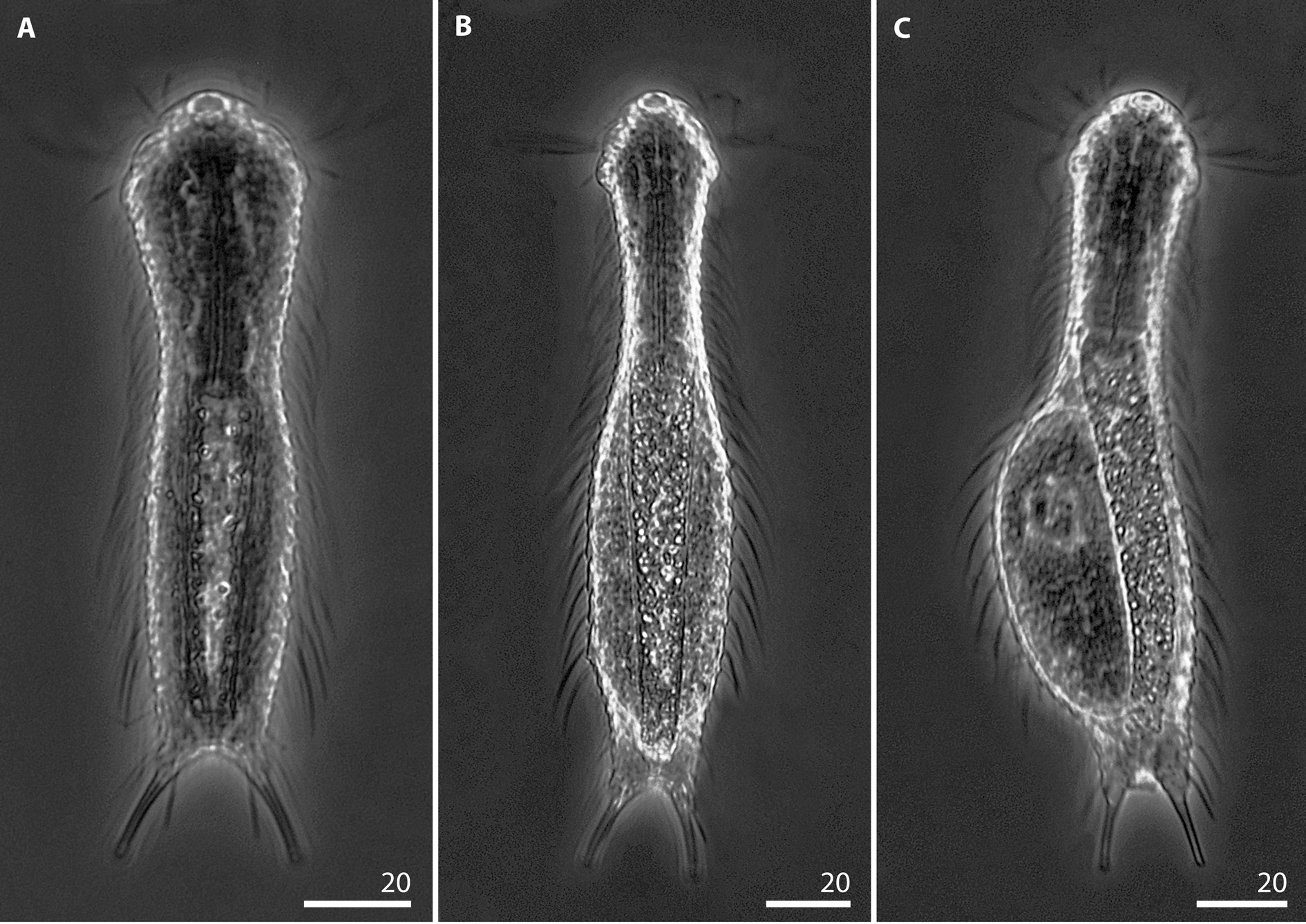
HABITUS. Chaetonotus (Chaetonotus) invitatus sp. nov. has a slender, bottle-shaped body ( Figs 2 View Fig , 5 View Fig ). The head is wide and the neck constriction is strongly marked. The neck gradually tapers to the beginning of the trunk (ca U38) ( Figs 2 View Fig , 5 View Fig ). The trunk is only slightly wider than the head and gradually dilates from ca U39 to ca U59, where it is at its maximum width. Then it gradually tapers slightly towards a weakly demarcated furcal base (from U85) ( Fig. 2 View Fig ). The furcal indentation is parabolic in shape. The furcal branches are set apart and point slightly outwards ( Figs 6 View Fig G–H, 8). The adhesive tubes are long, slightly bent, narrow and very gradually taper slightly towards their ends. The adhesive tube ends are blunt ( Figs 2 View Fig , 6 View Fig G–H, 8).
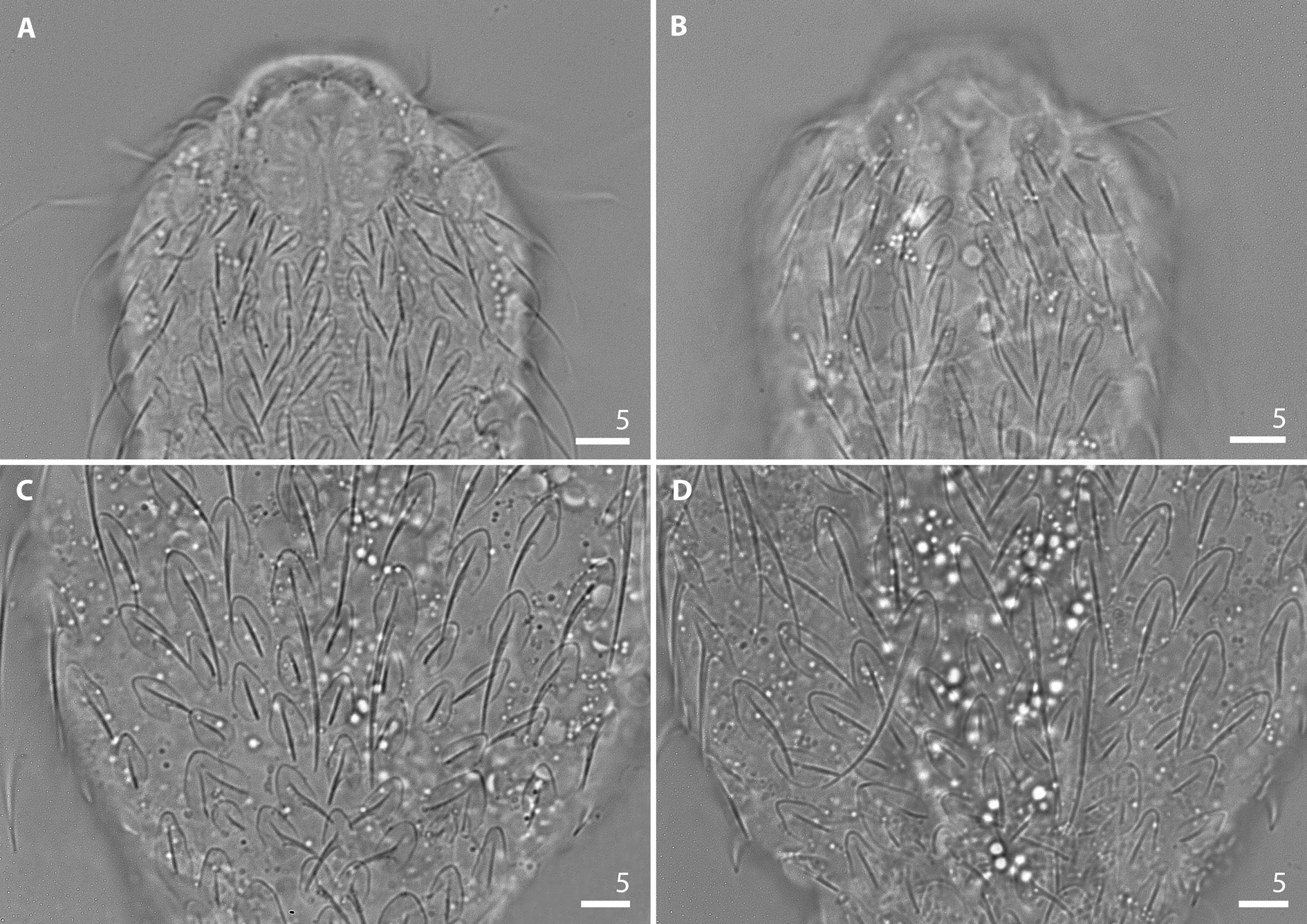
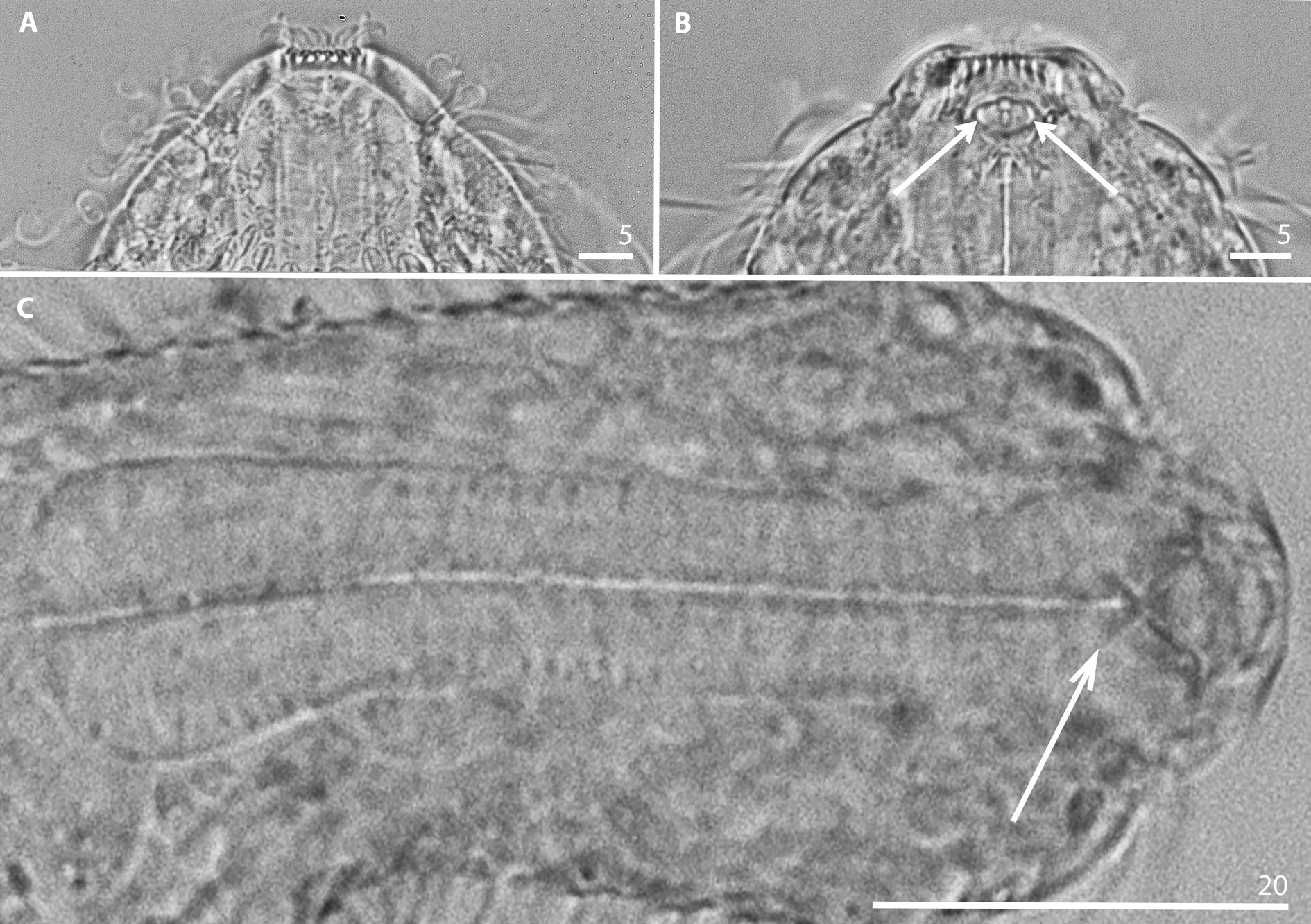
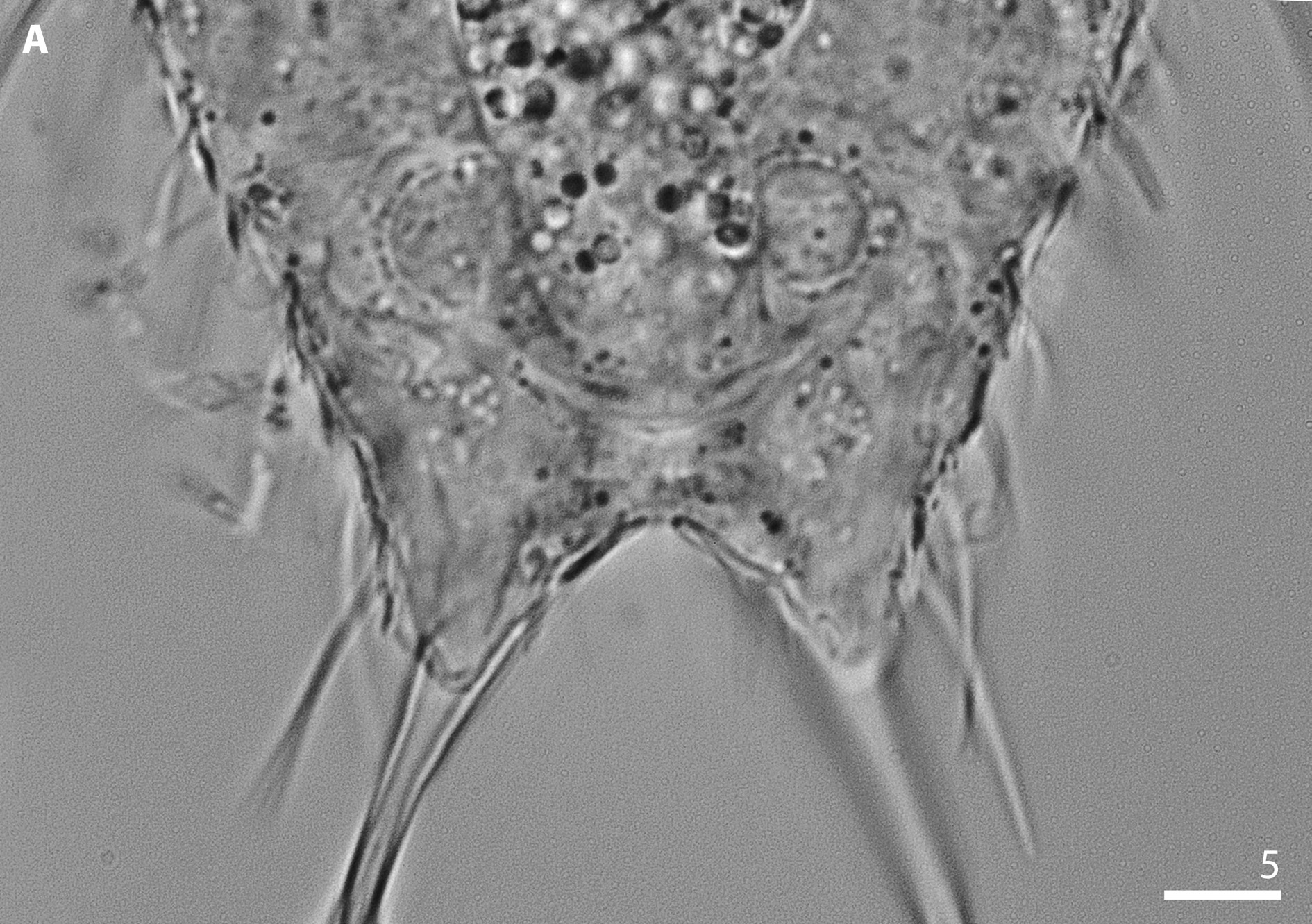
HEAD. The head is five-lobed, long and parabolic. The cephalion (U1–U7) is long and wide, it is visible on the dorsal head surface as an inverted, rounded triangle. It is clearly demarcated in the body outline and its dorsal edge is free and does not adhere to the head surface ( Fig. 7 View Fig A–B). The pleurae are large, convex and clearly demarcated in the head outline, the hypopleurae are only slightly larger than the epipleurae ( Fig. 2 View Fig ). Between the epipleurae and hypopleurae, deep notches are present. The epipleurae (U3–U7) are located on the dorsal, dorsolateral, lateral and ventrolateral surfaces, whereas the hypopleurae (U8– U12) cover the dorsal, dorsolateral, lateral, ventrolateral and ventral surfaces in such a manner that their greatest portion is located on the ventral side. The hypostomium is kidney-shaped and has two horn-like protuberances placed anterolaterally and a pair of strong reinforcements near its anterior edge (U3–U6) ( Figs 2C View Fig , 6B View Fig ). One pair of additional plates is located posterolaterally to the hypostomium, at U6–U7. These plates are wide and short and their lateral edges extend to the edges of the ventral epipleurae ( Fig. 6B View Fig ). Two pairs of cephalic ciliary tufts are present. The anterior tufts emerge laterally between the cephalion edge and the dorsal edge of the epipleurae (at U3). The anterior tufts consist of 5 cilia which are bent and short, and their length increases from the first to the last cilium ( Table A1). The posterior tufts have 5 cilia each and emerge laterally between the epipleurae and the hypopleurae (at U7–U8). Cilia in the posterior tufts are long and nearly straight and their length increases from the first cilium to the last, long cilium ( Table A1). Ocellar granules are absent. The mouth ring is located terminally at U1–U3, is wide and has long, strong finger-like reinforcements as well as very long cuticular inner hairs ( Figs 2 View Fig , 9 View Fig A–B). Inside the mouth ring, two small and weak cuticular teeth arising from the anterior pharynx region are present ( Fig. 9 View Fig B–C). Suboral bristles emerge in three pairs of tufts, laterally to the mouth ring, near the lateral edges of the hypostomium and in lines below the additional hypostomium plates ( Fig. 2C View Fig ).
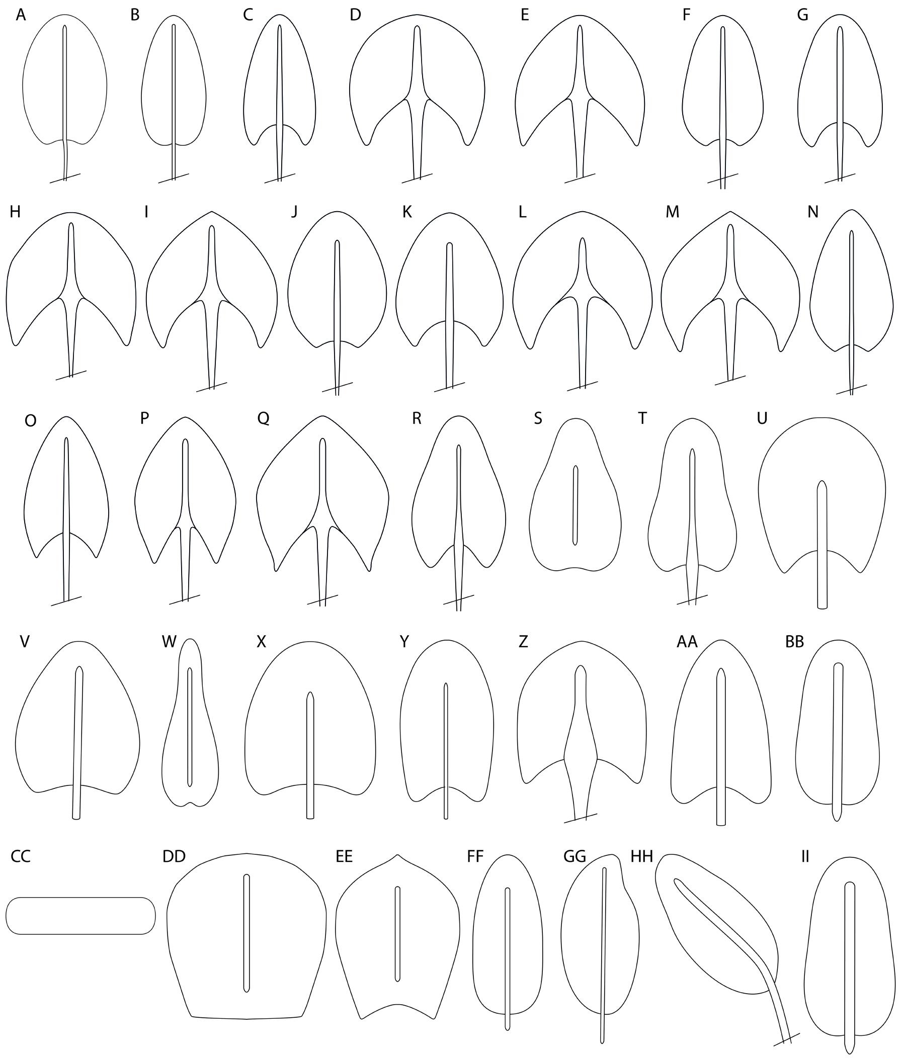
SCALES. The body is covered with one-lobed scales that adhere along their entire surface to the cuticle ( Figs 3 View Fig , 6–7 View Fig View Fig ). The scales have a straight keel and are egg-shaped with very weak posterior notches to very deep posterior notches. The scales are distributed in 23–25 longitudinal alternating rows (5–7D +4DL+6L+4LV+ 4V) with 25–27 scales in the central row. The longitudinal rows of scales run straight and are arranged in parallel to one another from the top of the head to the widest body region. Dorsally and dorsolaterally on the posterior trunk region, furca base and furcal appendages, the scales are arranged in rounded arcs and in a rosette ( Figs 2 View Fig , 7 View Fig C–D, 8A). The scales are located close to one another, but on the head, neck and anterior and central trunk part they do not overlap. The edges of the scales overlap only on the furcal base and furcal appendages ( Figs 2 View Fig , 8A View Fig ). The scales show morphological diversity throughout the particular body regions in terms of shape and size ( Fig. A1 View Fig ). The head anteriormost scales are situated near the posterior edge of the cephalion. These scales are oval with shallow posterior notches (scale 1; Fig. 3A View Fig ). The remaining head dorsal scales (scale 2; Fig. 3B View Fig ) are more elongated and have shallower posterior notches. The head dorsolateral scales (scale 3; Fig. 3C View Fig ) become shorter than the dorsal scales and have deeper posterior notches. The head lateral and ventrolateral scales (scale 4; Fig. 3D View Fig ) become more semi-rounded, shorter and wider than the dorsolateral ones and possess deep posterior notches, whereas the ventral scales are smaller, wide and have more pointed anterior edges (scale 5; Fig. 3E View Fig ). On the neck, the scales are shorter and wider than the scales on the head area (scales 6–9; Fig. 3 View Fig F–I) and their size gradually increases towards the trunk region (scales 10–13; Fig. 3J View Fig ). The neck dorsal scales (scales 6 and 10; Fig. 3F, J View Fig ) are egg-shaped and possess weak posterior notches. The neck dorsolateral scales (scales 7 and 11; Fig. 3G, K View Fig ) become gradually larger than the dorsal scales and have deeper posterior notches. The neck lateral and ventrolateral scales (scales 8 and 12; Fig. 3H, L View Fig ) become more semi-rounded and deeply posteriorly notched. They are wider and gradually larger than the dorsolateral ones, whereas the ventral scales (scales 9 and 13; Fig. 3I, M View Fig ) are smaller, wide and have more pointed anterior edges ( Fig. 2C View Fig ). The trunk dorsal scales (scale 14; Fig. 3N View Fig ) from the anterior to posterior trunk part are egg-shaped, but with a more tapered anterior edge than the neck scales. These scales have weak posterior notches. The trunk dorsolateral scales (scale 15; Fig. 3O View Fig ) become gradually larger than the dorsal scales and have deeper posterior notches. The trunk lateral and ventrolateral scales (scale 16; Fig. 3P View Fig ) become more oval, gradually larger and wider than the dorsolateral ones, have a more pointed anterior edge and possess deep posterior notches. The trunk ventral scales (scale 17; Fig. 3Q View Fig ) are smaller, shorter and wider, have more pointed anterior edges and a very deep posterior notch ( Figs 2A View Fig , 6F View Fig ). The scales in the ventral longitudinal row located closest to the ciliary band are distinctly smaller than the other scales and their anterior edge is oriented towards the bands at an angle of ca 40° ( Figs 2C View Fig , 6B, D, F, H View Fig ). Dorsally on the trunk posterior region (at U73–U77), two or three slightly larger scales with deeper posterior notches and much longer and stronger spines are present (scale 18; Fig. 3R View Fig ). Near the scales with longer and stronger spines and posteriorly to them (at U75–U80), smaller, trifle pear-shaped scales are located with weak posterior notches, straight keels and without spines (scale 19; Fig. 3S View Fig ). Subsequently, on U80–U85, dorsally on the posterior trunk region, 8 scales, that are triangle-shaped with strongly rounded edges, with clear posterior notches and a straight keel and spine, are situated (scale 20; Fig. 3T View Fig ). On the dorsal posterior trunk region, one pair of small, semirounded scales with a straight keel and a short, straight spine is located in the notch of a pair of double keeled scales with trunk sensory bristles (at U82–U83) (scale 21; Fig. 3U View Fig ). Dorsolaterally on the posterior trunk region and furcal base, two pairs of small semi-triangular scales with strongly rounded edges and shallow posterior notches, straight keels and straight rudimental spines are present (at U84– U87) (scale 22; Fig. 3V View Fig ). Laterally on the posterior trunk region and on the furcal base, at U82–U88, two pairs of narrow, elongated pear-shaped scales with very weak posterior notches are present. These scales have long straight keels but are not spined (scale 23; Fig. 3W View Fig ). Dorsally on the furcal base and furcal appendages at U85–U88, three semi-rounded scales with weak posterior notches, straight keels and short spines are present (scale 24; Fig. 3X View Fig ). Laterally to them (to scales 24), on the dorsal furcal base, one pair of large (5.4–9.1 ×3.2–5.3 μm), oval scales with a shallow posterior notch, straight keel and a short, straight spine is situated (at U84–U87) (scale 25; Fig. 3Y View Fig ). Dorsolaterally on the furcal appendages at U87–U89, one pair of large, rounded scales with prominent posterior notches is present (scale 26; Fig. 3Z View Fig ). These scales have straight keels and long, rigid spines. Laterally to this pair of scales (to scales 26) at U88–U89, two pairs of small (2.6–6.1×1.8–4.1 μm), oval-shaped scales with a shallow indentation and straight keels and spines are present (scale 27; Fig. 3 View Fig AA). On the lateral surface of the furcal appendage, two pairs of scales with parafurcal spines are present. The first pair is located at U88–U89 and the second one, i.e. the last pair of furcal lateral scales, is located at U90–U91. Both pairs are similar in shape to the posterior trunk scales ( Figs 2 View Fig , 6G View Fig ). Ventrolaterally and ventrally on the furcal appendages and furcal base, two pairs of small (2.2–5.3 × 1.4–2.8 μm) oval scales without any posterior notches are present (at U85–U88). These scales have straight keels and straight rudimentary spines (scale 28; Fig. 3 View Fig BB).
SPINES. All spines emerging from the posterior scale region have no lateral denticles. The dorsal, dorsolateral ( Fig.4A View Fig ),lateral and ventrolateral ( Fig. 4B View Fig )spines are basally bent and thick,and subsequently strongly and gradually taper towards the slightly bent hair-like ends. The spines arising from the lateral, ventrolateral and ventral scales are much more strongly bent than the dorsal and dorsolateral spines. The ventral scale spines are hair-like along their entire length ( Fig. 4C View Fig ). The length of the spines gradually increases from the head towards the widest body region (from 3.9–24.1 to 10.1–31.1 μm). Then the length of the dorsal spines gradually shortens to the posterior trunk region. Moreover, the length of the spines gradually increases from the dorsal surface, along the dorsolateral, lateral, ventrolateral and ventral surfaces towards the ciliary bands (head: from 1.6–7.4 to 13.3–26.4 μm; neck: from 5.0–10.7 to 15.9–31.9 μm; trunk: from 7.8–16.5 to 19.6–34.3 μm) ( Table A1). On the dorsal posterior trunk region, two or three spines of different types are present (arising from scales 18). These spines are much longer (16.6–20.0 μm) and stronger than the other dorsal spines and taper to blunt ends ( Figs 2A View Fig , 6 View Fig C–D). The spines arising from the dorsal and dorsolateral posterior trunk and furcal base scales (namely from scales 20, 22, 24, 27) are thick and straight, tapering to blunt ends ( Fig. 4D View Fig ). The posteriormost lateral trunk spines (16.1–25.1 μm) do not vary in length from the other lateral spines in the posterior trunk region. From the dorsolateral furcal appendage scales (scale 26) arise very long (14.5–18.4 μm), thick, rigid, basally bent and spike-like spines that extend to the inner furcal indentation ( Figs 2A View Fig , 4E View Fig , 6G View Fig , 8A View Fig ). These spines taper slightly to pointed ends. Two pairs of parafurcal spines, emerging from the lateral scales of the furcal appendages (at U88–U89 and at U90–U91), are thicker and longer (11.3–13.5 μm) than the other lateral body spines ( Fig. 4F View Fig ). These spines are basally bent and taper slightly towards their pointed ends ( Figs 6G View Fig , 8A View Fig ).
DORSAL SENSORY BRISTLES. This species has three pairs of dorsal sensory bristles. The first pair is located on the head, directly between the lateral edge of the cephalion and the epipleurae (at U4), and emerges from small, round papillae. The second pair of sensory bristles is located on the neck (U19) and emerges from small, rounded papillae. The third posterior pair of sensory bristles emerges from the doublekeeled scales located on the posterior trunk region at U81–U83. These scales are shaped like triangles with strongly rounded edges and a deep posterior notch. The keels of these scales are slightly bent, not connected and pass into rudimentary spines ( Figs 2A View Fig , 8A View Fig ).
VENTRAL CILIARY BANDS AND VENTRAL INTERCILIARY FIELD. Longitudinal ventral ciliary bands begin at U8 and run back to U86 ( Figs 2C View Fig , 6B, D, F, H View Fig ). On the head, at ca U8–U10, the ciliary bands are merged and lie close to the hypopleurae. The ventral interciliary field scales are distributed in single (on the pharyngeal region) and 5–7 (on the intestinal region) longitudinal alternating rows, with 29–33 scales in the central row, including 15–18 plate-like scales and 14–17 one-lobed scales. The entire pharyngeal region (from U10 to U31) is covered with short, wide plates (scale 29; Fig. 3 View Fig CC) and their size gradually increases from the anterior to the posterior end (from 1.0–1.7 ×1.9–3.5 μm to 2.6–5.2 ×7.1–11.4 μm). On the intestinal region, one-lobed scales with overlapped edges are present (from U32 to U84). On the anterior part of the intestinal region the scales are semi-rectangular in shape, have weak keels and their posterior edges are straight, without posterior notches (scale 30, Fig. 3 View Fig DD). Towards the furcal base the keels become more pronounced and on the edges of the anterior scales small, horn-like perturbances begin to appear; the posterior edges become notched (scale 31; Fig. 3 View Fig EE). The last alternating row of interciliary field scales is of a different type, i.e., they are oval in shape without posterior notches, have straight keels and present rudimentary spines (scale 32; Fig. 3 View Fig FF). The longitudinal row of ventral field scales closest to the ventral ciliary bands has significantly smaller and narrower scales than in the central rows. The first pair of terminal scales on the ventral interciliary field is large (14.8–17.1 × 6.6–8.1) and located at U84–U87. They are oval, asymmetrical in shape, possess a straight keel and a straight, thick spine (scale 33; Fig. 3 View Fig GG). The second pair of terminal scales on the ventral interciliary field is located on the furcal appendages (at U86–U88). These scales are oval in shape, have long, straight keels and thick, curved spines reaching to the internal furcal indentation (scale 34; Fig. 3 View Fig HH). The first and second pairs slightly overlap. The third (at U87–U88) and fourth (at 88–U89) pairs are definitely small (2.0–4.8× 1.3–3.0 μm), egg-shaped, are located on the furcal appendages, and have straight keels and rudimentary spines (scale 35; Fig. 3 View Fig II).
INTERNAL MORPHOLOGY. The pharynx (from U2 to U31) is narrow and has marked anterior and posterior dilatations. The posterior dilatation is stronger than the anterior one ( Fig. 9C View Fig ). In the anterior dilatation, weak cuticular reinforcements are located in the form of two straight, connected rods (at U4). The pharynx is connected through the pharyngeal–intestinal junction by the straight intestine (running from U32 to U86). The pharyngeal–intestinal junction is clearly demarcated, short and wide (U32) ( Fig. 2B View Fig ). The intestine does not have a separate, different anterior section. The X-organ of this species (observed in one specimen) is located at U84–U86 near the terminal part of the intestine. It is bilobed, built from two extensions enveloped in a thin coat and connected by a thinner band located below the intestine end, at the ventral surface ( Fig. 10 View Fig ). The extensions have a granular appearance. The thin coat and the cellular bridge connecting the extensions have a smooth and homogeneous structure. The sperm packets of this species were not observed.
Remarks
Because of the long pharynx in relation to the length of the body and intestine, the pharynx–intestine ratio (I) has high values in this species. Juvenile and subadult specimens have a pharynx–intestine ratio (I) higher than 65%, rather than higher than 55%, as is usually observed in other species. In juvenile specimens, the first head scales from each dorsal longitudinal row are located under the free dorsal cephalion edges, whereas in adults they are nto located under the cephalion but below their dorsal edges ( Fig. 7 View Fig A–B). Of the 25 reported adults, one individual had a large, developing egg ( Fig. 5C View Fig ) and another possessed a distinct X-organ ( Fig. 10 View Fig ). Four specimens had live, motile euglenids inside their intestine ( Fig. 11 View Fig ) and were listed in an earlier paper as Chaetonotus (Chaetonotus) sp. 1 ( Kisielewska et al. 2015).
Differential diagnosis
Of all the 92 currently known nominal representatives of the subgenus Chaetonotus (Chaetonotus) Ehrenberg, 1830 , C. (C.) invitatus sp. nov. most closely resembles C. (C.) maximus Ehrenberg, 1830 , C. (C.) microchaetus Preobrajenskaja, 1926 , C. (C.) similis Zelinka, 1889 , C. (C.) heterospinosus Balsamo, 1978 , C. (C.) laroides Marcolongo, 1910 and C. (C.) polyspinosus Greuter, 1917 . Of all 32 species belonging to the subgenus Hystricochaetonotus Schwank, 1990 , C. (H.) trispinosus Balsamo, 1990 seems to be the most similar to the newly described taxon, whereas of all the currently known 22 nominal representatives of the subgenus C. (Primochaetus) Kisielewski, 1997b, the new species most closely resembles C. (P.) mutinensis Balsamo, 1978 . Chaetonotus (C.) maximus , C. (C.) microchaetus , C. (C.) similis , C. (C.) heterospinosus , C. (C.) laroides , C. (C.) polyspinosus and C. (P.) mutinensis are the most similar to the newly described species in terms of: body shape similarity, range of body length, range of pharynx length, range of adhesive tube length, presence of a scale of a different type on the dorsal and dorsolateral surfaces on the furcal base, presence of rigid, long spines on the furcal appendages and a similar type of terminal scales on the ventral interciliary field. Chaetonotus (H.) trispinosus was selected for comparison with C. (C.) invitatus sp. nov. due to its similarity in terms of having three larger scales with longer and stronger spines present on the dorsal posterior trunk region, length variation of large spines on different body sides and by the presence of scales of a different type on the dorsal and dorsolateral surfaces of the furcal base. Despite the fact that these eight species have the highest number of common features with the newly described species, they are significantly different from C. (C.) invitatus sp. nov. – most strikingly by scale type and shape variation, spine types and length variation. Comparisons between the new species and the most morphologically similar taxa are summarised in Table A2. Taxa chosen for the differential diagnosis often occur in different regions of the world and are presented very differently by various researchers. Thus, a different description/ specification or taxonomic classification of theoretically the same species may suggest that we are dealing with a very large range of plasticity of these taxa or with a larger number of undiagnosed, separate species. To avoid any doubts in comparisons with C. (C.) invitatus sp. nov., only the original descriptions of these species and three of the most detailed papers with morphological data were used ( Balsamo 1983; Schwank 1990; Kisielewski 1997a). For information regarding C. (P.) mutinensis , only original data are considered in the differential diagnosis.


No known copyright restrictions apply. See Agosti, D., Egloff, W., 2009. Taxonomic information exchange and copyright: the Plazi approach. BMC Research Notes 2009, 2:53 for further explanation.
|
Kingdom |
|
|
Phylum |
|
|
Order |
|
|
Family |
|
|
Genus |