
Chaetonotus ( Hystricochaetonotus ) inaequabilis, 2019
|
publication ID |
https://doi.org/10.5852/ejt.2019.511 |
|
publication LSID |
lsid:zoobank.org:pub:8FDAD45D-1B7D-446F-8B34-026EDF192210 |
|
DOI |
https://doi.org/10.5281/zenodo.5620069 |
|
persistent identifier |
https://treatment.plazi.org/id/82BC94AE-15DD-4A5D-A727-B34013BC6B7A |
|
taxon LSID |
lsid:zoobank.org:act:82BC94AE-15DD-4A5D-A727-B34013BC6B7A |
|
treatment provided by |
Plazi |
|
scientific name |
Chaetonotus ( Hystricochaetonotus ) inaequabilis |
| status |
sp. nov. |
Chaetonotus ( Hystricochaetonotus) inaequabilis View in CoL sp. nov.
urn:lsid:zoobank.org:act:
Figs 26–32 View Fig View Fig View Fig View Fig View Fig View Fig View Fig , A 7 View Fig ; Tables 5, A5–A View Table 5 6 View Table 6
Diagnosis
Stocky body, measuring from 98.2 to 108.5 μm in length. Head five-lobed, cephalion narrow, epipleurae and hypopleurae small. All cephalic plates weakly demarcated in head outline. Hypostomium small, rectangular in shape. Ocellar granules absent. Almost all scales three-lobed. Scales distributed in 15–17 total longitudinal rows (5–7D+ 2DL+2L+ 4LV+ 2V), with 12–13 scales in central row. Scales strongly differ morphologically in various body areas. Nine significantly larger scales on dorsal trunk area with very long, thick spines with a strong lateral denticle. Two pairs of posteriormost trunk lateral scales with long, thick spines with strong lateral denticle. Remaining scales with simple, shorter and thinner spines, or with rudimentary spines, or without spines on central dorsal trunk area and on furcal appendages. Scales with parafurcal spines absent. Spines on ventral surface hair-like, with narrow, delicate lamellae along entire length. Entire ventral interciliary field covered with small scales. Four pairs of ventral interciliary field terminal scales. Pharynx wide, with pronounced anterior and posterior dilatations. Intestine straight without anterior section differing in form and morphology.
Etymology
From the Latin ʻ inaequabilis ʼ = ʻheterogeneousʼ, referring to the very numerous types of scales on the body.
Type material
Holotype POLAND • adult; Kraków, Botanical Garden, Jubilee Greenhouse, site 2 ; 50°03'38" N, 19°57'30" E; 15 Nov. 2013; M. Kolicka leg.; NHC-GCHI-23-1-25/h (photomicrographs, also in the author's collection). GoogleMaps
Paratypes POLAND • 6 adults; same locality as holotype; sites 2–3; 15 Nov. 2013 and 17 Apr. 2014; M. Kolicka leg.; NHC-GCHI-23-26-60/p (photomicrographs, also in the author's collection) GoogleMaps .
Description
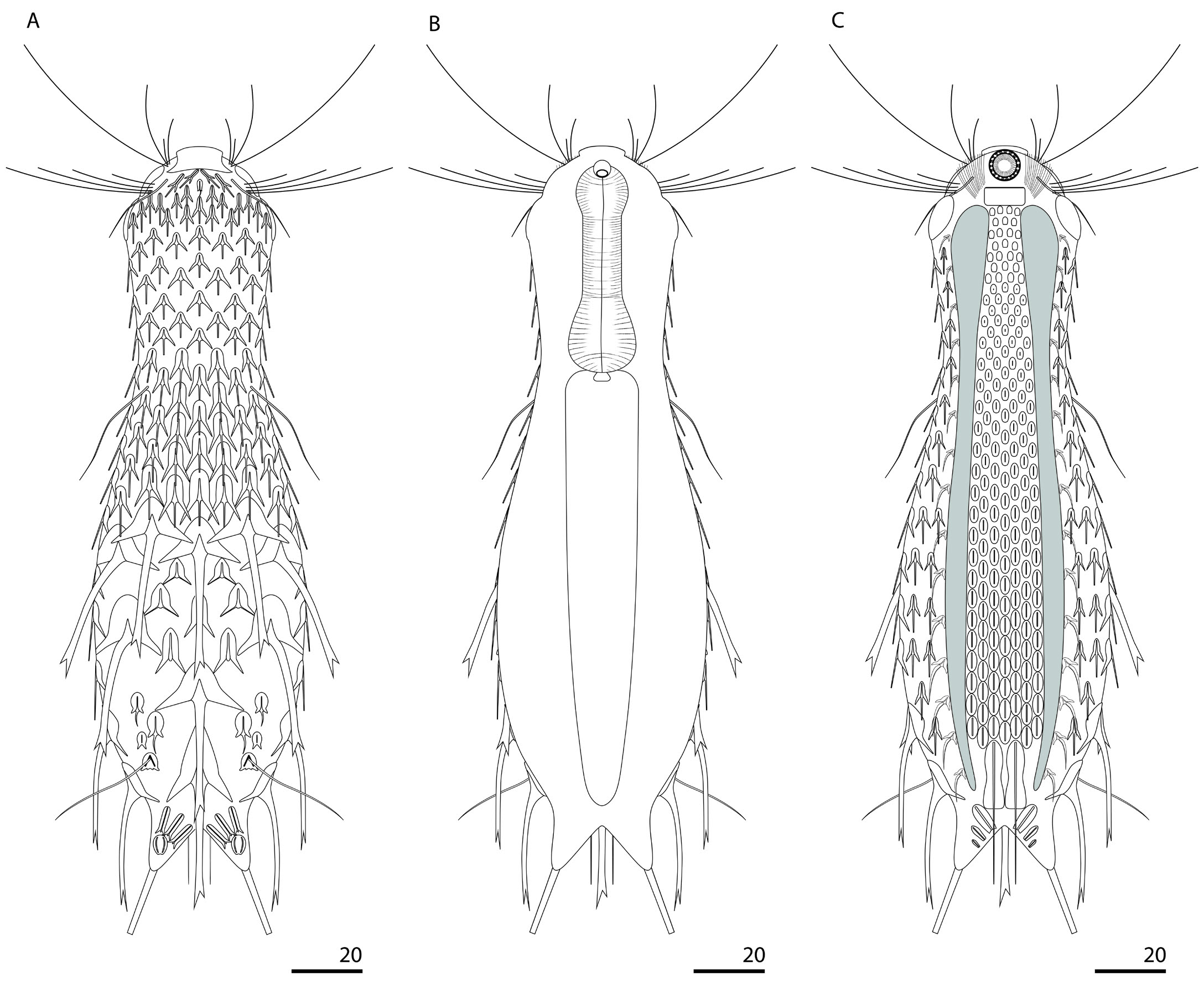
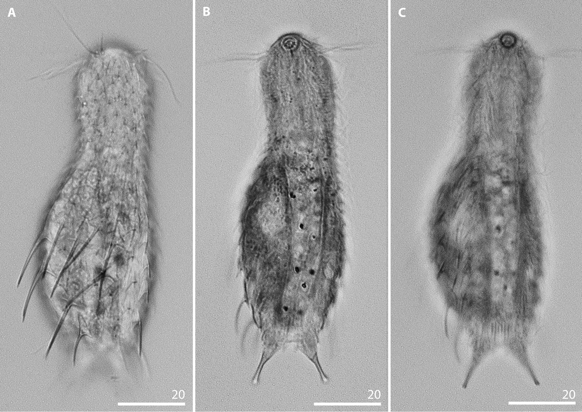
HABITUS. Chaetonotus ( Hystricochaetonotus) inaequabilis sp. nov. has a stocky body ( Figs 26 View Fig , 29 View Fig ). Its head is only slightly wider than the neck, and the neck constriction is weakly demarcated. The neck gradually tapers from the head (from U20) to the beginning of the trunk (ca U30) ( Figs 26 View Fig , 29 View Fig ). The trunk is slightly wider than the head and gradually dilates from ca U31 to ca half of its length (U56), where it is at its maximum width. Then it gradually tapers towards the narrow furcal base (from U84) ( Figs 26 View Fig , 29 View Fig ). The furcal base is clearly demarcated and narrow. The furcal indentation is V-shaped. The furcal branches are set narrowly apart and point slightly outwards ( Figs 26 View Fig , 29 View Fig ). The adhesive tubes are relatively short in comparison to the whole body length ( Table 5 View Table 5 ), straight, fairly thick and not tapered towards the blunt ends ( Figs 26 View Fig , 29 View Fig ).
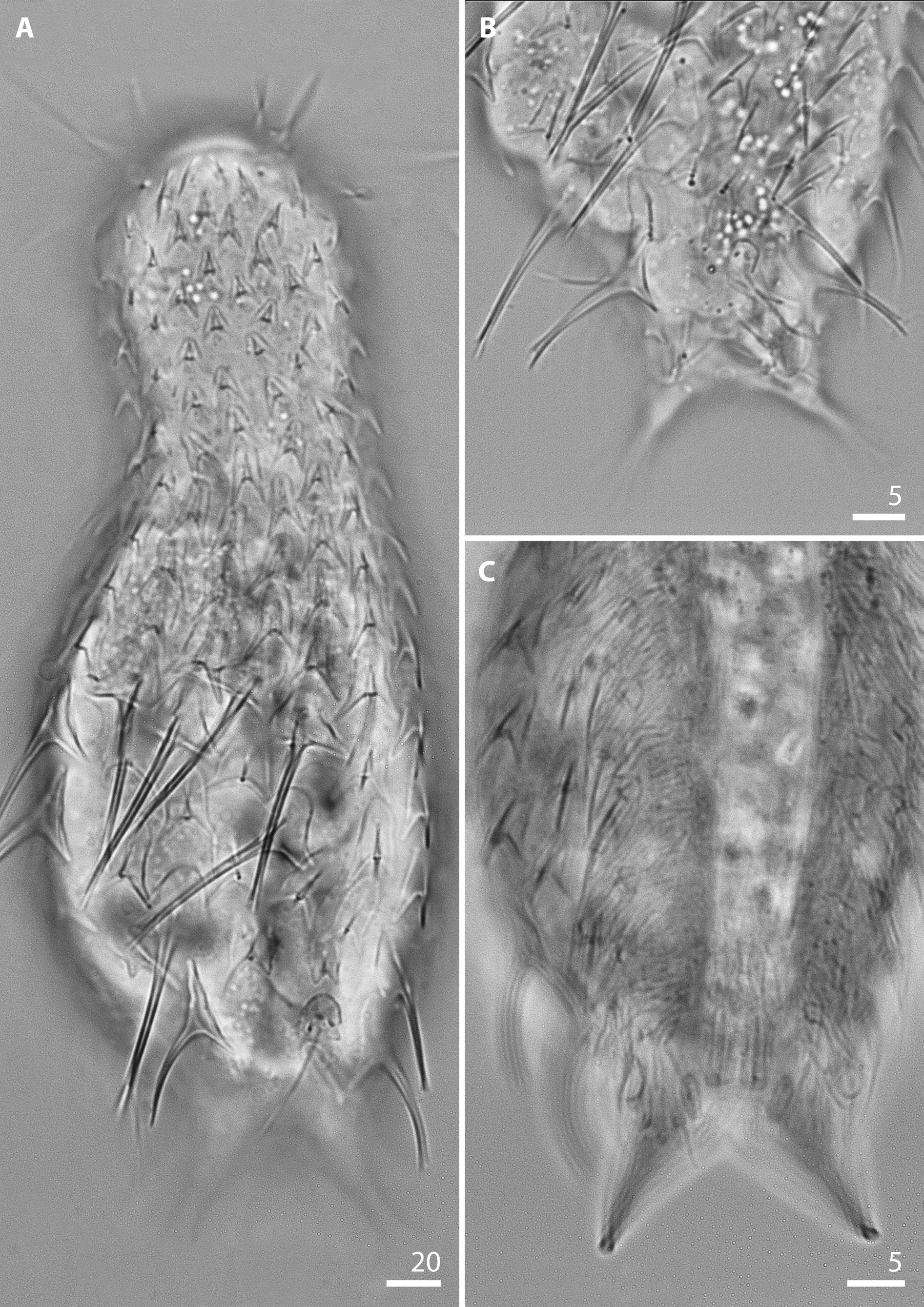
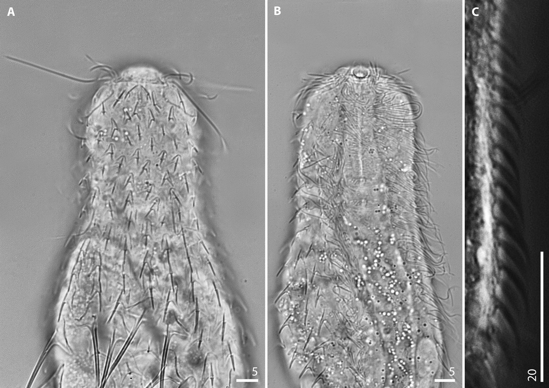
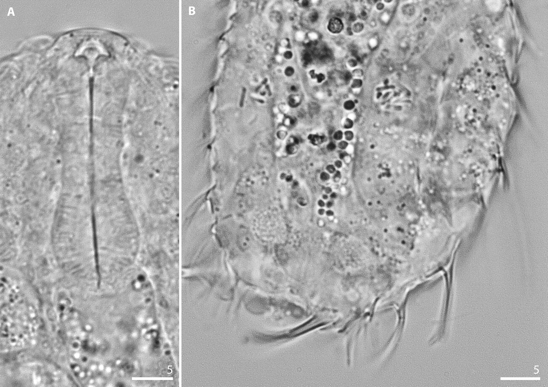
HEAD. The head is five-lobed, short and semicircular in shape. The cephalion (U1–U3) is narrow, short and straight, on the dorsal body side. It adheres to the head along its entire length and rapidly extends laterally near the dorsal edge ( Figs 26A View Fig , 31A View Fig ). The epipleurae (U3–U7) are small, narrow, slightly arched and weakly demarcated in the head outline. They are visible on the dorsal, dorsolateral and lateral head sides. The hypopleurae (U7–U12) are similar in size to the epipleurae and located entirely on the ventrolateral and ventral head sides ( Fig. 26 View Fig ). On the dorsal head surface, between the lateral edges of the cephalion and epipleurae, is a prominent space; their edges meet only in the cephalion extension place. Deep notches are present between the cephalion and epipleurae and between the epipleurae and hypopleurae. The hypostomium (U6–U8) is small, rectangular, with a slightly reinforced anterior edge ( Figs 26C View Fig , 31B View Fig ). Two pairs of cephalic ciliary tufts are present. The anterior tufts emerge in the area between the cephalion and epipleurae on the dorso-terminal head surface (at U2–U3) and consist of four cilia. The anteriormost cilium in both anterior tufts is fairly short. The second cilium is short, shorter than the anteriormost one. The third cilium is longer than either of the preceding cilia. The fourth, last cilium in the anterior tufts is very long, the longest in both pairs of tufts. The posterior tufts (at U6–U7) have four straight cilia each and emerge on the ventral head surface, above the anterior edge of the hypopleurae. The length of the cilia in the posterior tuft gradually increases from the anteriormost to the fourth cilium. Ocellar granules are not present. The mouth ring is large and subterminal at U2–U5. It has very strong, granular cuticular reinforcements and short inner hairs ( Figs 26C View Fig , 29C View Fig ). Suboral bristles are located in two tufts located beneath and laterally to the mouth ring and reaching half the length of the lateral hypostomium edges (between U5 and U7).
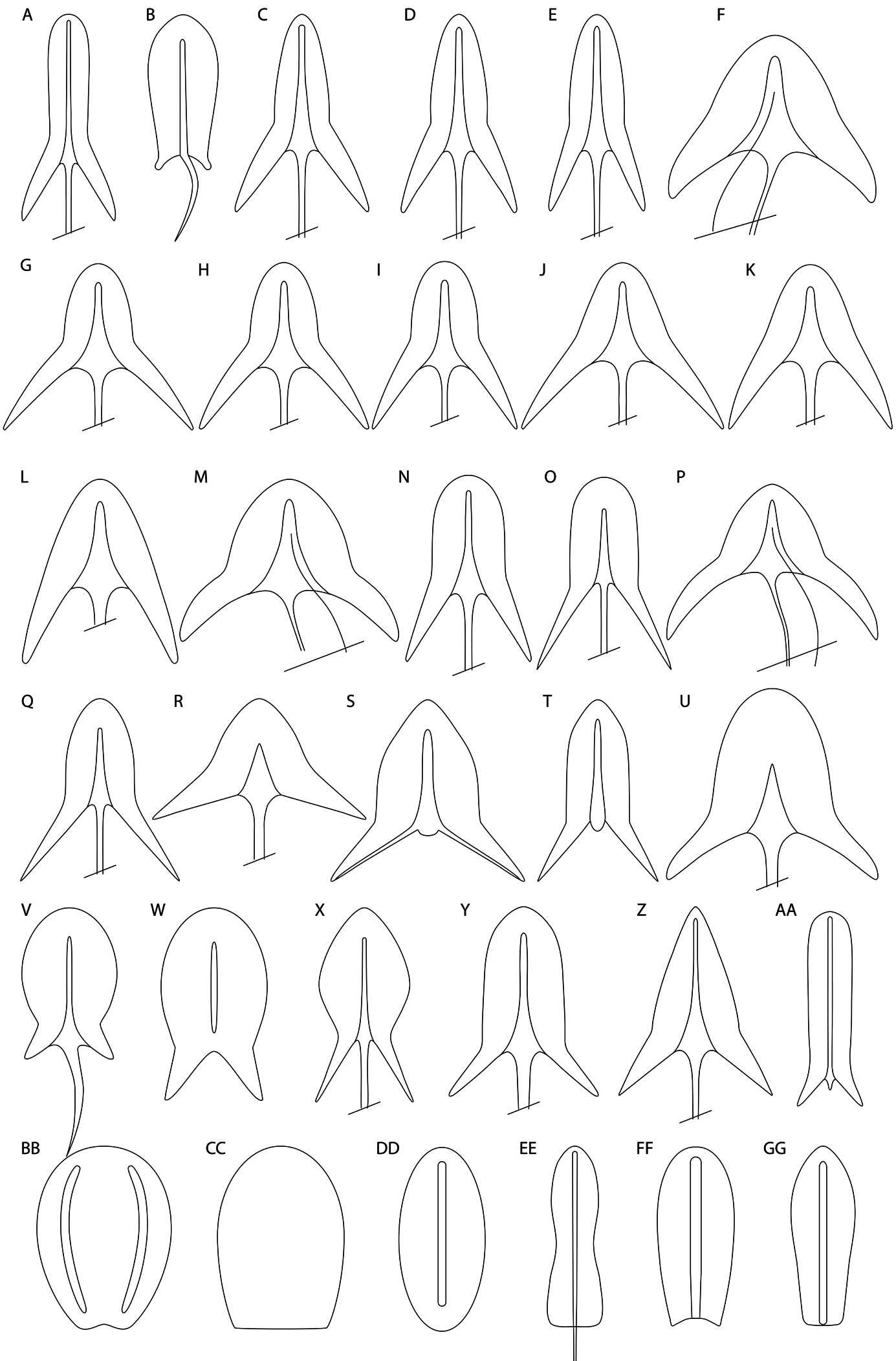
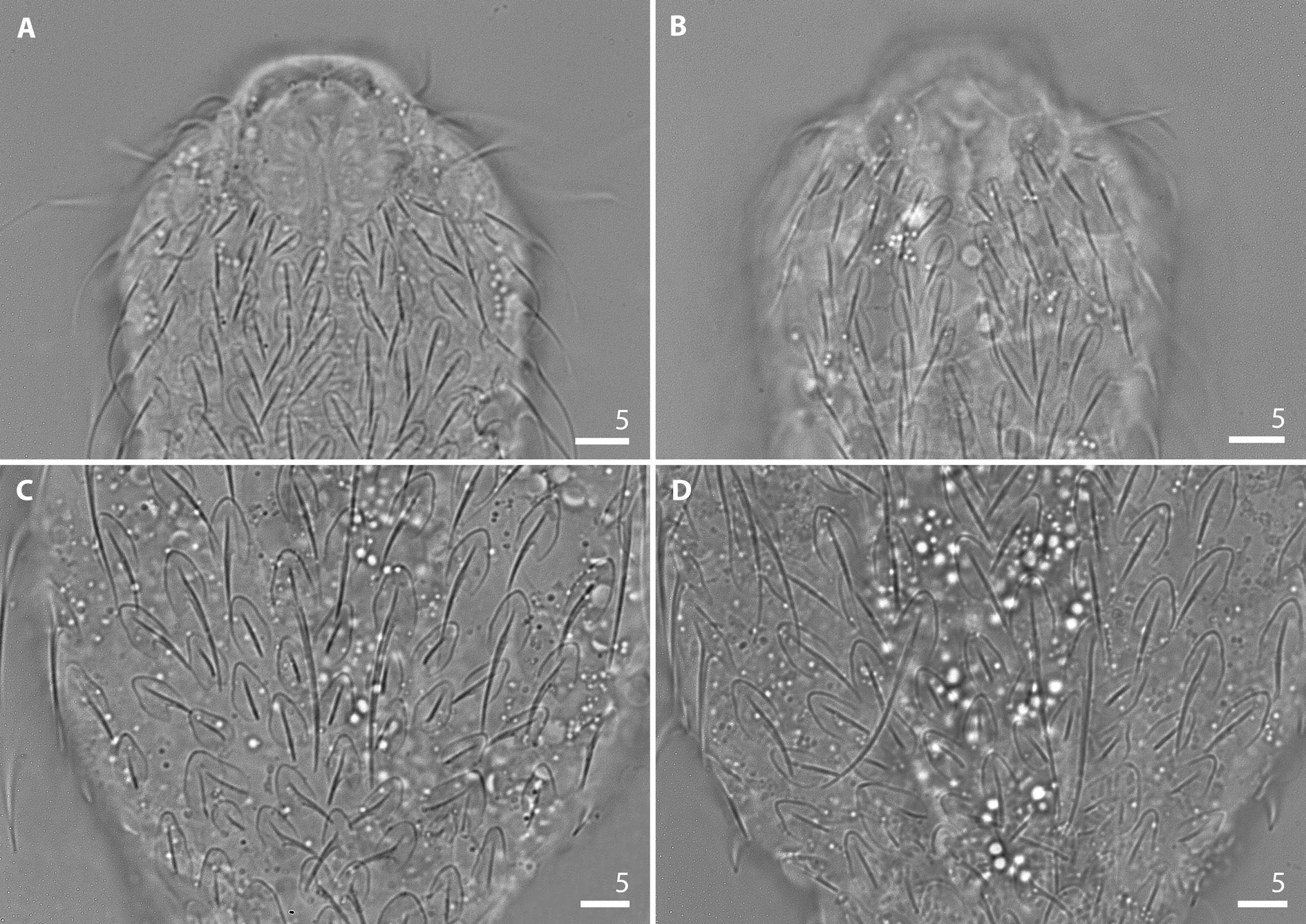
SCALES. The entire body, except for one pair of scales on the furcal appendages, is covered with threelobed scales that adhere to the cuticle along their entire surface ( Fig. A7 View Fig ). All scales have a strong, triangular keel and are shaped like triangles with a deep posterior notch. The scales are distributed in 15–17 total longitudinal alternating rows (5–7D +2DL+2L+4LV+ 2V), with 12–13 scales in the central row. The longitudinal rows of scales begin on the head directly beneath the posterior edge of the cephalion, epipleurae and hypopleurae. The scales show a strong diversity in size and shape between the various parts of the body. On the head and anterior neck region the scales are located far from each other, while on the posterior neck and anterior trunk region the scales become gradually juxtaposed but do not overlap. The dorsolateral, lateral, ventrolateral and ventral trunk scales are juxtaposed, whereas the dorsal trunk scales and scales on the furcal appendages have overlapping posterolateral edges ( Fig. 30 View Fig ). The head anteriormost scales are situated near the posterior edge of the cephalion and epipleurae. The dorsal anterior scales are located aslant, whereas the dorsolateral scales are arranged parallel to the longitudinal body axis. These scales are elongated, their central lobes are long and wide, and they have very long keels. The posterolateral lobes are narrow, slanted downward and create a narrow, deep V-shaped posterior scale notch (scale 1; Fig. 27A View Fig ). The central anteriormost head scales are located at U5–U6, beneath the aslant arranged scales, are smaller, more rounded and have weaker separate posterolateral lobes (scale 2; Fig. 27B View Fig ). The remaining scales of the head are wider. The central lobes of these scales become shorter, and simultaneously the posterolateral lobes become slightly longer, whereas their keels are shorter. The posterolateral lobes are directed diagonally downward and laterally and create a deep and wide posterior scale notch (scales 3 and 7; Fig. 27C, G View Fig ). The anterior neck scales are shorter than the head scales. Their central lobes are wide and short. Their posterolateral lobes are weaker and separate from the central lobes (scale 10; Fig. 27J View Fig ). The subsequent neck scales become larger but relatively narrower than the preceding neck scales ( Table A5 View Table 5 ). Their central lobes are wide, with strongly rounded anterior edges, whereas their postero-lateral lobes are more separated and directed more downward and create a deep V-shaped posterior scale notch (scale 14; Fig. 27N View Fig ). The posterolateral lobes of the dorsolateral, lateral and ventrolateral scales of the head and neck become gradually more directed downward towards the ciliary bands as compared to the dorsal scales (scales 3–14; Fig. 27 View Fig C–N). From the neck towards the trunk, the size of the scales gradually increases (from 2.1– 2.5× 2.4–2.9 to 4.2–5.4× 5.1–6.5 μm). The anterior trunk scales have a wide and rounded anterior central lobe and clearly separate, long, narrow and sharp posterolateral lobes. The posterolateral lobes are directed diagonally downward and slightly laterally, and they create a deep, narrow V-shaped posterior scale notch (scale 15; Fig. 27O View Fig ). The dorsolateral, lateral and ventrolateral trunk scales, arranged from the central to posterior trunk region (U50 to U83), become gradually larger than the anterior trunk scales (from 2.3–7.1 ×2.7–5.1 to 3.5–8.8× 4.1–7.1 μm) and they have a more pointed anterior edge of the central lobe and longer, more sharp posterolateral lobes directed more apart and diagonally downward (scale 17; Fig. 27Q View Fig ). Dorsally, on the central trunk part (at U47–U57), five different scales with very long and strong spines are located in one transverse row. These scales are very large (6.4–13.0 ×5.2– 8.3 μm) and very thick. They are triangular, their central lobes are short, wide and pass seamlessly into wide, posterolateral lobes directed very far apart. The posterior edges of these scales are almost straight and only very narrowly notched. Their keels start from the half length of the central lobes and are very pronounced, strongly triangular in shape (scale 18; Fig. 27R View Fig ). Directly beneath the row of large scales, two pairs of smaller scales with long keels and spiny processes are located (at ca U55–U61). These scales are triangular, have longer central lobes with pointed anterior edges and narrower posterolateral lobes directed slightly more downward (scale 19; Fig. 27S View Fig ). Posteriorly to these scales, at the centre of the dorsal trunk region, three scales with long keels and spiny processes are located (at U60–U68). These scales are narrower and have longer central lobes than the preceding scales. Their posterolateral lobes are directed clearly downward, creating a V-shaped posterior scale notch (scale 20; Fig. 27T View Fig ). Laterally to these three scales and posteriorly to the transverse row with wide, large scales, another pair of large scales with very long and strong spines is located (at U60–U66). These scales have longer central lobes, narrower posterolateral lobes directed more downward (7.3–9.2 ×7.2–9.5 μm) and more rounded edges than the preceding large scales (scale 21; Fig. 27U View Fig ). In the central dorsal longitudinal row, at U67–U74, a single scale of the same type as the large scales from the transverse row at U55–U59 is present (scale 18; Fig. 27R View Fig ). Laterally to it, at U70–U74, two pairs of smaller scales are located, with one pair after another in the longitudinal row. These scales have a long and strongly rounded central lobe and clearly separated, short and wide posterolateral lobes ( Fig. 30B View Fig ). Of these two scale pairs, the pair situated below (U72–U74) is larger than the upper pair (U70–U72) (scale 22; Fig. 27V View Fig ). On the posterior trunk region laterally to these rounded scales, directed above the double-keeled scales with trunk dorsal sensory bristles, one pair of small (3.3–4.9×2.2–3.3 μm) scales is arranged at U74–U75 (scale 23; Fig. 27W View Fig ). These scales have a strongly rounded central lobe and posterolateral lobes that are weakly separated from the central lobes, wide, short and directed downward, creating a V-shaped posterior notch. Their keels are strong but short and pass into short spines which do not extend to the posterior edge of the scales. On the lateral surface of the posterior trunk region, one pair of second to last trunk scales is located (at U71–U78). These scales are large (7.4–11.4 ×4.1–6.1 μm) and have a long, wide, strongly rounded and clearly pointed anterior central lobe. Their postero-lateral lobes are strongly separated from the central lobe and have long, narrow and sharp edges that are directed diagonally downward. These scales have strong keels and strong, long spines (scale 24; Fig. 27X View Fig ). Posteriorly to them, the last pair of trunk lateral scales is situated at the furcal base (at U78–U84). This pair of scales is large (6.4–10.8 ×4.9–8.3 μm); these scales have a long, wide and strongly anteriorly rounded central lobe with a very strong and high keel and strong, long spines and short, narrow posterolateral lobes. The posterolateral lobes of these scales are directed diagonally wide apart, downward and laterally, creating a wide and shallow V-shaped posterior scale notch (scale 25; Fig. 27Y View Fig ). On the dorsal posterior trunk region and furcal base, at U76–U83, one median large scale that is different in type is located. This scale is triangular, has a long and pointed central lobe with a long keel and a long, strong spine, and clearly separate, relatively short postero-lateral lobes directed diagonally downwards (scale 26; Fig. 27Z View Fig ). On the dorsal surface of the furcal appendages (at U84–U89), three pairs of three-lobed, elongated and narrow scales with long central and short posterolateral lobes are present. These scales have a long keel with a spiny process (scale 27; Fig. 27 View Fig AA). Dorsolaterally and laterally to the furcal appendages (at U88–U90), one pair of scales with two long and slightly bent keels is present. These scales are oval in shape and have very shallow posterior notches (scale 28; Fig. 27 View Fig BB). The edges of the scales on the furcal appendage are slightly overlapping ( Fig. 30B View Fig ). The size of the scales gradually decreases from the dorsal, dorsolateral and lateral surfaces towards the ventrolateral surface. The scales in the ventral longitudinal row located closest to the ciliary bands are short and wide, their central lobes are short and their posterolateral lobes are weakly separated. From the head to the widest body region the length of the posterolateral lobes of these scales, as well as their separation from the central lobes, gradually increase. The notches of the posterior edges of these scales are semicircular in shape. The ventral scales are much smaller than the scales of the other rows and their anterior edge is oriented towards the bands at an angle of ca 20°.
SPINES. In this species, five main types of spines may be distinguished ( Fig. 28 View Fig ). The first type are basally bent spines tapering towards the end, without any lateral denticle ( Fig. 28A View Fig ). These spines emerge from single, central anteriormost head scales located at U5–U6 ( Figs 27A View Fig , 31A View Fig ) and from two pairs of dorsal posterior trunk region scales located at U70–U74 (scale 12) ( Fig. 27A View Fig ). The second type are simple spines that do not taper towards the blunt ends and are without any lateral denticle ( Fig. 28B View Fig ). These spines emerge from the dorsal, dorsolateral, most of the lateral and ventrolateral head, neck and anterior trunk region scales and dorsolaterally, laterally and ventrolaterally on the remaining trunk part scales. The length and thickness of the spines gradually increase from the head top towards the widest body region (from 0.3–12.0 to 3.1–15.2 μm) (ca U57). Moreover, their length gradually decreases towards the ventrolateral surface (head: from 0.5–3.1 to 0.3–2.4 μm; neck: from 2.1–5.7 to 1.6–4.4 μm; trunk: from 3.9–8.2 to 3.1–7.1 μm) ( Table A5 View Table 5 ). The third type are very long (11.4–21.6 μm) and thick, straight spines with a prominent lateral denticle ( Fig. 28C View Fig ). Together, nine dorsal spines and four (two pairs) lateral spines of this type are present. Five of them arise from large dorsal trunk scales located in one transverse row at U47–U57 (scale 18) ( Figs 29A View Fig , 31 View Fig A–B). The next two spines arise from one pair of dorsal trunk scales located at U60–66 (scale 21) ( Figs 29A View Fig , 31 View Fig A–B). The eighth of these long spines arises from the central dorsal scale located at U67–U74 (scale 18) ( Figs 29A View Fig , 31 View Fig A–B). This spine is the longest (11.4–21.6 μm) and the thickest body spine and it has the most prominent lateral denticle. The ninth and last of these long dorsal spines arises from the central dorsal scale located at the posterior trunk region and on the furcal base (at U84–U89) (scale 26) ( Fig. 31 View Fig A–B). This spine is slightly shorter than the preceding long spine (11.4–14.2 μm). The two pairs of long lateral spines arise from the trunk second to posteriormost pair of lateral scales (at U71–U78) (scale 24) and from the trunk posteriormost pair of lateral scales (at U78–U84) (scale 25; Figs 28D View Fig , 31 View Fig A–B). The posteriormost pair is longer (11.4– 14.0 μm) and thicker than the second to posteriormost pair of lateral spines (10.1–12.7 μm long). The fourth type are short, simple spines ( Fig. 28E View Fig ) or spiny processes without any lateral denticle ( Fig. 28F View Fig ). These spines arise from the central dorsal trunk scales located at U55–U59 and U60–U68 ( Fig. 31A View Fig ), from one pair of trunk dorsal scales located at U74–U75, above the double-keeled scales with the dorsal sensory bristle, and from three pairs of elongated scales located on the dorsal and dorsolateral surface of the furcal appendages at U84–U89 ( Fig. 31A View Fig ). The fifth type are long (7.3–15.2 μm), thin and hair-like, with narrow, delicate lamellae along their entire length ( Fig. 28G View Fig ). These spines arise on the ventral surface, from scales arranged in one pair of a longitudinal row located closest to the ciliary bands. The lamellae on these spines are widest near the spine base and gradually taper towards the end of the spine ( Fig. 31C View Fig ).
DORSAL SENSORY BRISTLES. This species has three pairs of dorsal sensory bristles ( Fig. 26A View Fig ). The first, anterior pair is located on the dorsal surface of the head at U5, beyond the dorsolateral cephalion edges, where the second pair of sensory bristles is located on the dorsal surface of the posterior neck at U31. The first and the second pairs emerge from small, spherical papillae. The third, posterior pair of sensory bristles is located dorsolaterally on the posterior trunk region and furcal base and emerges from the small three-lobed scales with two strong keels situated at U77–U79. These scales have keels which are connected in the centre of the scales and have a strongly rounded, wide central lobe and short, weakly separated posterolateral lobes. These scales have a double, shallow posterior notch ( Fig. 31A View Fig ).
VENTRAL CILIARY BANDS AND VENTRAL INTERCILIARY FIELD. On the ventral surface, the longitudinal ciliary bands begin at U9 and run back to U82 ( Fig. 29C View Fig ). The ciliary bands are wide, and wider on the head region than on other parts of the body. The entire ventral interciliary field is covered with thin, small scales. They are distributed in seven longitudinal rows, with 21–25 scales in the central row ( Figs 30C View Fig , 31B View Fig ). The ventral interciliary field anterior scales are partially recessed on the cuticle and isolation from the cuticle increases towards the posterior body part. On the anterior region the scales have an oval shape with a straight posterior edge and are located far from each other (scale 29; Fig. 27 View Fig CC). Towards the posterior body region the scales become gradually larger (from 1.4–1.9× 0.6–1.1 to 3.8–5.2 ×2.7– 4.1 μm), more elongated and with a rounded posterior edge (scale 30; Fig. 27 View Fig DD). The anterior scales are keelless and spineless, but towards the trunk posterior region the keels on the scales become more distinct and the distance between the scales decreases ( Figs 26C View Fig ). Four pairs of ventral interciliary field terminal scales are present. The first pair (at U76–U85) is elongated and has a narrowing halfway along the length of the scale ( Fig. 30C View Fig ). They have a long, narrow keel and a long, thin and straight spine extending beyond the internal furcal indentation (scale 31; Fig. 27 View Fig EE). The second pair (at U84–U87) is elongated, has a pointed anterior edge and gradually tapers to the posterior end. This pair has long, narrow keels and is spineless (scale 32; Fig. 27 View Fig FF). The third (at U87–U88) and fourth (at U88–U89) pairs are small, elongated and oval-shaped, with a shallow posterior notch (scale 33; Fig. 27 View Fig GG). They have a long, straight keel and are spineless. The second, third and fourth pairs of ventral interciliary field terminal scales are situated at the furcal appendages.
INTERNAL MORPHOLOGY. The pharynx (from U3 to U29) is wide and has distinct, marked, rounded anterior and posterior dilatations. The posterior dilatation is wider than the anterior one ( Fig. 32A View Fig ). The pharynx is connected through the pharyngeal–intestinal junction to a straight intestine, running from U29 to U84. The pharyngeal–intestinal junction is clearly demarcated, short and narrow (U29–U30). The X-organ of this species (observed in one specimen) is located at U80–U83 near the terminal part of the intestine. It is bilobed, built from two extensions enveloped in a thin coat and connected by a thinner band located behind the intestine at the ventral side. The extensions, the thin coat and the cellular bridge connecting the extensions have have a grain-like structure. The pair of sperm packets of this species are circular in shape and contain spermatozoids (12 per packet) in the form of a short rod. They are located at U62–U64 on both sides, juxtaposed to the intestine ( Fig. 32B View Fig ).
Remarks
Juvenile specimens of Chaetonotus ( Hystricochaetonotus) inaequabilis sp. nov. were not observed; furthermore, specimens with large, developing eggs were not present. Out of seven adults, one had an X-organ and two had sperm packets.
Differential diagnosis
Within the subgenus Hystricochaetonotus , Chaetonotus ( H.) inaequabilis sp. nov. most closely resembles C. ( H.) enormis Stokes, 1887 , C. ( H.) schlitzensis Schwank, 1990 , C. ( H.) octonarius Stokes, 1887 , C. ( H.) novenarius Greuter, 1917 , C. ( H.) aemilianus Balsamo, 1978 , C. ( H.) ferrarius Schwank, 1990 , C. ( H.) spinulosus Stokes, 1887 and C. ( H.) longispinosus Stokes, 1887 . All of these species were chosen for comparison because they possess a set of similar features such as: range of body length; body shape; presence of very long spines on dorsal and/or dorsolateral trunk surfaces; presence of lateral denticle only on the long spines (for C. ( H.) ferrarius see Table A6 View Table 6 ); and absence of parafurcal spines. Moreover, C. ( H.) enormis is similar to the new species in having two pairs of furcal base lateral spines which are longer and stronger than the remaining lateral spines. Chaetonotus ( H.) schlitzensis , C. ( H.) octonarius , C. ( H.) novenarius , C. ( H.) aemilianus , C. ( H.) ferrarius and C. ( H.) spinulosus share with C. ( H.) inaequabilis sp. nov. the presence of scales without spines or with spiny processes on the central dorsal trunk region. Chaetonotus ( H.) novenarius and C. ( H.) aemilianus also possess the same number of scales with long spines as the newly described species. Despite the fact that among the hitherto known species in this subgenus, those listed above have the highest number of common features with the newly described species, they are significantly different from C. ( H.) inaequabilis sp. nov. – most strikingly by size, type, and shape of the scale coverage as well as the arrangement of scales with long spines. In order to avoid any doubts in comparisons with the new species, only the original descriptions of these species and three of the most detailed papers with morphological data were used ( Balsamo 1983; Schwank 1990; Kisielewski 1997a). Comparisons between the new species and the morphologically most similar taxa are summarised in Table A6 View Table 6 .

No known copyright restrictions apply. See Agosti, D., Egloff, W., 2009. Taxonomic information exchange and copyright: the Plazi approach. BMC Research Notes 2009, 2:53 for further explanation.
|
Kingdom |
|
|
Phylum |
|
|
Order |
|
|
SubOrder |
Paucitubulatina |
|
Family |
|
|
SubFamily |
Chaetonotinae |
|
Genus |