
Podisus ventralis ( Dallas, 1851 )
|
publication ID |
https://doi.org/ 10.11646/zootaxa.4958.1.33 |
|
publication LSID |
lsid:zoobank.org:pub:D586C16A-7D57-4BE6-A1DA-2CF7703E60BC |
|
DOI |
https://doi.org/10.5281/zenodo.4711391 |
|
persistent identifier |
https://treatment.plazi.org/id/894E3E5C-D562-FF90-F9AE-929AB762FEE9 |
|
treatment provided by |
Plazi |
|
scientific name |
Podisus ventralis ( Dallas, 1851 ) |
| status |
|
Podisus ventralis ( Dallas, 1851)
( Figs. 1–9 View FIGURES 1–15 , 16–18 View FIGURES 16–24 , 25–27 View FIGURES 25–33 , 34–35 View FIGURES 34–39 , 40–42 View FIGURES 40–48 , 49–50 View FIGURES 49–54 )
Arma ventralis Dallas, 1851: 100 ( Figs 1–3 View FIGURES 1–15 ).
Arma pallipes Dallas, 1851: 101 ( Figs 4–6 View FIGURES 1–15 ) syn. nov.
Podisus neniator Breddin, 1904: 154 ( Figs 7–9 View FIGURES 1–15 ) (syn. Thomas 1992: 99).
Podisus (Podisus) ventralis (Dallas) : Stål 1870: 51.
Podisus pallipes (Dallas) : Walker 1867: 144; Stål 1870: 54.
Apateticus (Eupodisus) ventralis (Dallas) : Schouteden 1907: 72.
Apateticus (Eupodisus) neniator (Breddin) : Schouteden 1907: 72.
Apateticus (Eupodisus) pallipes (Dallas) : Schouteden 1907: 72.
Apateticus (Podisus) neniator (Breddin) : Kirkaldy 1909: 20.
Apateticus (Podisus) pallipes (Dallas) : Kirkaldy 1909: 20.
Apateticus (Podisus) ventralis (Dallas) : Kirkaldy 1909: 21.
Podisus ventralis (Dallas) : Thomas 1992: 99.
Types examined. Arma ventralis Dallas , syntype ♀ at NHMUK, labels: “ Venezuela / 47 26”; “21. ARMA VENTRALIS”; “a”; “ Type ”; “NHMUK 010592337”. ( Figs 1–3 View FIGURES 1–15 )
Arma pallipes Dallas , syntype ♂ at NHMUK, labels: “ Venezuela / 47 26”; “29. ARMA CRASSIMARGO. ”; “a”; “BRIT.MUS”; “ Podisus sp. 1-198”; “NHMUK 010938912”. ( Figs 4–6 View FIGURES 1–15 )
Podisus neniator Breddin , lectotype ♀ at SDEI, labels: “Balzapamba (Ecuad.) R. Haensch S.”; “ Podisus neniator Bredd.”; “coll. Breddin”; “Holo-typus”; “DEI EBRESWALDE [sic!]”; “DEI Hemimetabola #100183”. ( Figs 7–9 View FIGURES 1–15 )
Non-types examined. COLOMBIA: Cundinamarca, Reserva Chicaque, Refugio , 04.6148667°N, 74.3112833°W, 2221m, 24-28-II-2014, D. Forero col., 2♀ 1♂ ( MPUJ) GoogleMaps ; Choachi, La Victoria, N 4° 31, W 73° 55, 1927 m, 18-VI-2011, J. Pérez col., ♂ ( UNAB) GoogleMaps ; Boyacá, Buenavista, Patiño, Las Lomas , N 5° 3, W 73° 24, 1989 m, 15-IV-2004, W. Ávila col., ♀ ( UNAB) GoogleMaps ; Risaralda, La Suiza, 1900m, VIII-1992, Yaces col., ♀ ( MPUJ) .
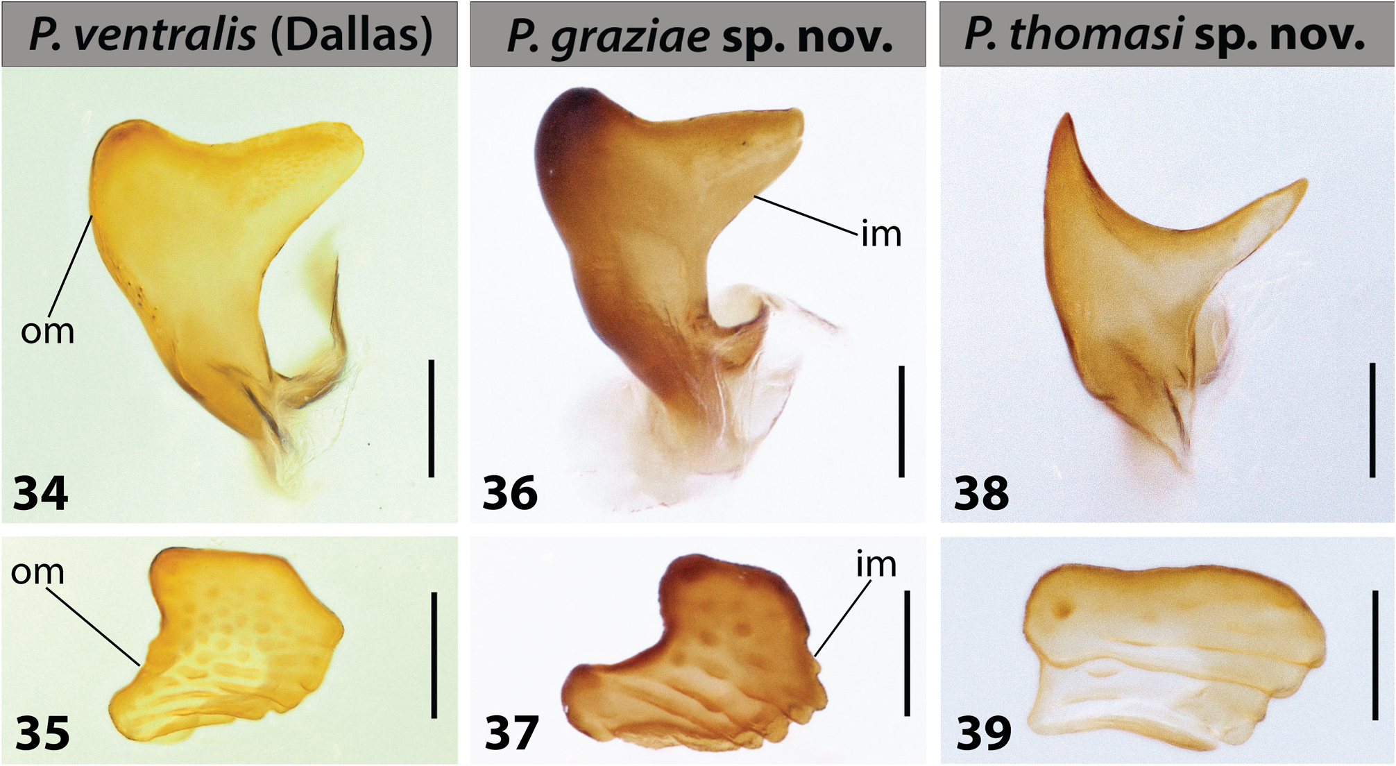
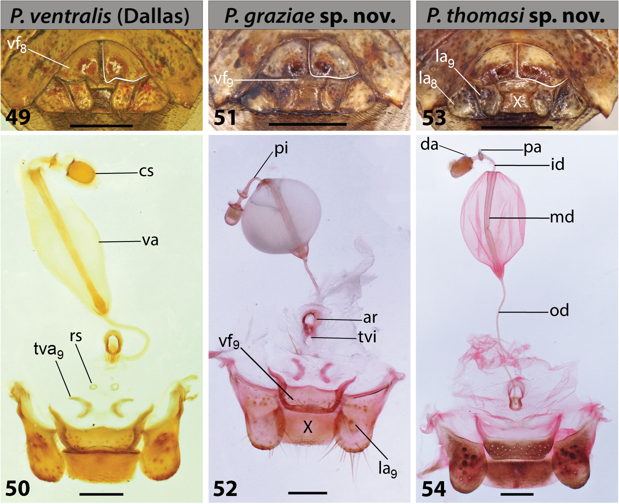
Diagnosis. Pronotum without spots, anterolateral pronotal margins calloused, crenulated, paler than pronotal disc; humeral angles angular, without a posterior tooth, laterally directed. Apex of scutellum concolourous with disc ( Figs 1, 4, 7 View FIGURES 1–15 , 16 View FIGURES 16–24 ). Apices of parameres obtuse ( Figs 25–27 View FIGURES 25–33 , 34 View FIGURES 34–39 , par). Posterior margins of valvifers VIII strongly sinuous, forming a V-projection on posterior angles over valvifers IX ( Fig. 49 View FIGURES 49–54 , vf 8).
Redescription. Body length 9.86–11.29 mm (♀) and 10 mm (♂).
Body castaneous to brown or rubrous, anterior half of lateral margins of pronotum forming a marginal wide carina, lighter than rest of pronotum. A thin central line mostly without punctures or lighter than rest of dorsum extends from anterior margin of pronotum to posterior margin of scutellum ( Figs 1, 4, 7 View FIGURES 1–15 , 16, 18 View FIGURES 16–24 ).
Head subrectangular, mandibular plates uniformly punctured, clypeus as long as or slightly surpassing apex of mandibular plates; ocelli lying behind an imaginary line through posterior margins of compound eyes; antennal tubercles partially visible from above, proportion of antennomeres: I<II>III<IV>V ( Figs 1, 4, 7 View FIGURES 1–15 , 16, 18 View FIGURES 16–24 ); bucculae evanescent posteriorly, apex rounded; labium extending to posterior margin of metasternum, proportion of labiomeres: I<II>III>IV ( Figs 2, 5, 8 View FIGURES 1–15 , 17 View FIGURES 16–24 ).
Pronotum hexagonal, uniformly punctured; cicatrices flat, almost indistinguishable; anterior and lateral margins concave in dorsal view, lateral margins slightly crenulated and lighter than disc on anterior half, anterior half of lateral margins preceded by a marginal sulcus; humeral angles angular, slightly produced, without a posterior tooth. Scutellum longer than wide, reaching an imaginary line connecting fifth connexives medially; frenal margins longer than post frenal margins. Coria longer than scutellum, each attaining abdominal segment IV, uniformly punctured; hemelytral membranes well surpassing apex of abdomen, with apex spotted in brown ( Figs 1, 4, 7 View FIGURES 1–15 , 16 View FIGURES 16–24 ). Pro-, meso- and metasterna covered by small thin setae; prosternum with a weak median carina; mesosternum carinated longitudinally; limit between mesosternum and metasternum tumid; metasternum flat. Pro-, meso-, and metapleura sparsely punctured, evaporatoria on posterior margin of each mesopleura; metapleural evaporatoria surrounding each ostiolar peritreme; each ostiolar peritreme discal-type, elongated, with a darker spot on apical angle; each ostiolar opening directed laterally ( Figs 2, 5, 8 View FIGURES 1–15 , 17 View FIGURES 16–24 ).
Abdomen sparsely punctured, posterolateral angles of each connexivum projected as small spines, longer on seventh segment. Mesial tubercle projecting from sternite III not surpassing metacoxae ( Figs 2, 5, 8 View FIGURES 1–15 , 17 View FIGURES 16–24 ).
Male. Without glandular patches on ventral surface of abdomen.
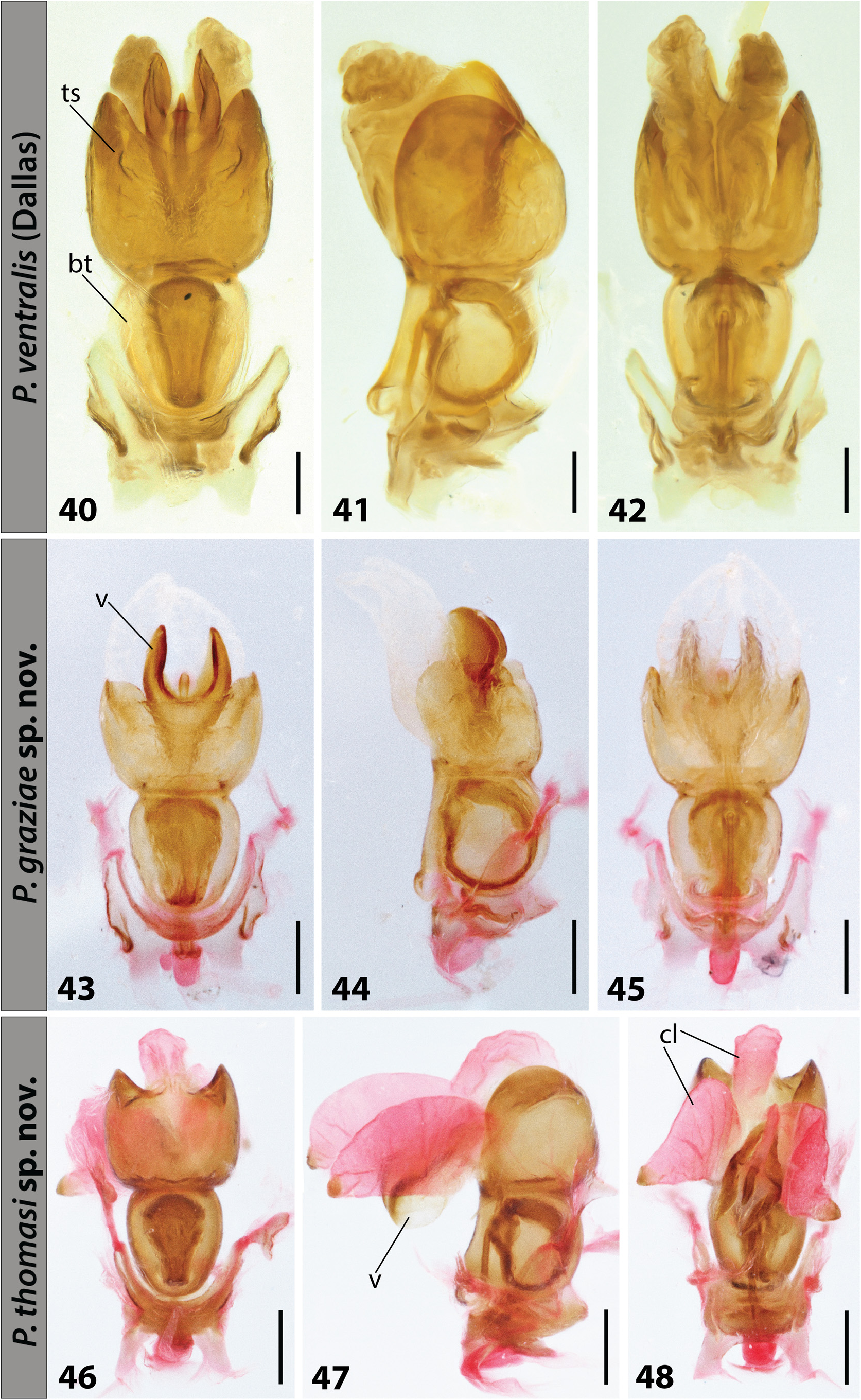
Genitalia. Pygophore bowl-shaped, opened posteriorly, setose, setae denser on ventral rim ( Figs 25–27 View FIGURES 25–33 ); dorsal rim concave with 1+1 dorsal projections near segment X ( Figs 25–27 View FIGURES 25–33 , dr, dp); ventral rim concave, with 1+1 ventral projections near posterolateral angles ( Fig. 27 View FIGURES 25–33 , vr, vp, pa); surface between inferior and superior layers of ventral rim excavated ( Figs 26–27 View FIGURES 25–33 , vr). Posterolateral angles acute, slightly surpassing seventh tergite ( Fig. 5 View FIGURES 1–15 ). Segment X tubular, ventrally directed. Head of parameres divided into two arms, each apex obtuse, well visible posteriorly ( Figs 25–27 View FIGURES 25–33 , 34 View FIGURES 34–39 , par). Pseudoclasper subquadrate, outer margins sinuous, placed dorsolaterally to parameres, with globose projections and elevations ( Figs 25–27 View FIGURES 25–33 , 35 View FIGURES 34–39 , pc, om). Phallus. Basal theca and thecal shield subequal in length, thecal shield slightly wider than basal theca ( Figs 40–42 View FIGURES 40–48 , bt, ts), vesica golf club-shaped in lateral view ( Fig. 41 View FIGURES 40–48 , v), apices of conjunctival lobes slightly sculptured ( Fig. 40–42 View FIGURES 40–48 , cl).
Female. Genitalia. Genital plates positioned ventroposteriorly ( Fig. 17 View FIGURES 16–24 ); valvifers VIII subtriangular, posterior margins setose, strongly sinuous, forming a V-projection on posterior angles over valvifers IX, sutural margins straight, juxtaposed ( Fig. 49 View FIGURES 49–54 , vf 8). Laterotergites VIII triangular with spiracles on proximal angle ( Fig. 49 View FIGURES 49–54 , la 8). Exposed portion of valvifers IX rectangular, wider than long ( Fig. 49 View FIGURES 49–54 , vf 9). Exposed portion of laterotergites IX tongue-like, setose apically ( Fig. 49 View FIGURES 49–54 , la 9), surpassing segment X and almost attaining mediotergite VIII. Segment X subquadrate ( Fig. 49 View FIGURES 49–54 , X). Inner portion of valvifers IX projected in 1+1 straight elongated arms with acute apices ( Fig. 50 View FIGURES 49–54 , vf 9). Valvulae IX with 1+1 small lunate secondary thickenings ( Fig. 50 View FIGURES 49–54 , tva 9). Ring sclerites small, circular, placed posteriorly to thickening of vaginal intima ( Fig. 50 View FIGURES 49–54 , rs, tvi); thickening of vaginal intima short, obtuse ( Fig. 50 View FIGURES 49–54 , tvi); arcus placed right anteriorly of thickening of vaginal intima ( Fig. 50 View FIGURES 49–54 , ar). Vesicular area almost four times length of pars intermedialis plus capsula seminalis ( Fig. 50 View FIGURES 49–54 , va, pi, cs). Inner and median ducts of vesicular areas of uniform diameter, median duct slightly enlarged basally ( Fig. 50 View FIGURES 49–54 , id, md). Pars intermedialis tubular, shorter and narrower than capsula seminalis; capsula seminalis oval, elongated, smooth ( Fig. 50 View FIGURES 49–54 , pi, cs). Proximal and distal annular crests of similar width ( Fig. 50 View FIGURES 49–54 , pa, da).
Distribution: Venezuela, Colombia, Ecuador.
Remarks: Dallas (1851) described A. ventralis and A. pallipes in the same work. He examined one or more females of A. ventralis and one or more males of A. pallipes . It is unclear how many specimens he had at the time of the description of each species as the lettering system he used is different from the one that had been agreed for the catalogues of zoological specimens in the collection of the British Museum, the one that, for instance, Francis Walker dutifully followed ( Kondorosy et al. 2006; Roell et al. in prep.). After a morphological analysis of the typespecimens found by Roell et al. (in prep.), we can confirm our original hypothesis: Dallas’s syntype of A. pallipes is the male of Podisus ventralis .
Arma submarginata Walker, 1867 , synonymized under P. ventralis by Thomas (1992), was recently synonymized under Podisus distinctus (Stål, 1860) by Brugnera et al. (2020).
Breddin (1904) described Podisus neniator from an undetermined number of female specimens (syntype (s)). Gaedike (1971), finding a unique female specimen in the collection of SDEI, labelled and listed it as the holotype. As syntypes from Breddin’s collection are still found scattered in many depositories and even still discovered in SDEI by experts of different hemipteran families ( Kondorosy & Rédei 2015; Simões & Campos 2015; Rédei et al. 2016), we have accepted that Gaedike (1971) has designated the female specimen deposited at SDEI as lectotype by inference of holotype ( ICZN 1999, Art. 74.6). Should further syntypes be found, they should be considered paralectotypes.


| UNAB |
Universidad Nacional, Facultad de Agronomia |
No known copyright restrictions apply. See Agosti, D., Egloff, W., 2009. Taxonomic information exchange and copyright: the Plazi approach. BMC Research Notes 2009, 2:53 for further explanation.
|
Kingdom |
|
|
Phylum |
|
|
Class |
|
|
Order |
|
|
Family |
|
|
Genus |
Podisus ventralis ( Dallas, 1851 )
| Roell, Talita, Brugnera, Ricardo & Lemaître, Valérie A. 2021 |
Podisus ventralis (Dallas)
| Thomas, D. B. 1992: 99 |
Apateticus (Podisus) neniator (Breddin)
| Kirkaldy, G. W. 1909: 20 |
Apateticus (Podisus) pallipes (Dallas)
| Kirkaldy, G. W. 1909: 20 |
Apateticus (Podisus) ventralis (Dallas)
| Kirkaldy, G. W. 1909: 21 |
Apateticus (Eupodisus) ventralis (Dallas)
| Schouteden, H. 1907: 72 |
Apateticus (Eupodisus) neniator (Breddin)
| Schouteden, H. 1907: 72 |
Apateticus (Eupodisus) pallipes (Dallas)
| Schouteden, H. 1907: 72 |
Podisus neniator
| Thomas, D. B. 1992: 99 |
| Breddin, G. 1904: 154 |
Podisus (Podisus) ventralis (Dallas)
| Stal, C. 1870: 51 |
Podisus pallipes (Dallas)
| Stal, C. 1870: 54 |
| Walker, F. 1867: 144 |
Arma ventralis
| Dallas, W. S. 1851: 100 |
Arma pallipes
| Dallas, W. S. 1851: 101 |