
Persica qeshmensis, Maghsoudlou & Bulnes & Rahimian, 2015
|
publication ID |
https://doi.org/ 10.1080/00222933.2015.1006278 |
|
publication LSID |
lsid:zoobank.org:pub:736C2B3B-41A6-4FAD-92A4-1159027830FD |
|
DOI |
https://doi.org/10.5281/zenodo.4327869 |
|
persistent identifier |
https://treatment.plazi.org/id/8A374B5A-8729-6D46-36F0-2840FC9A0532 |
|
treatment provided by |
Carolina |
|
scientific name |
Persica qeshmensis |
| status |
sp. nov. |
Persica qeshmensis sp. nov.
( Figures 2–6 View Figure 2 View Figure 3 , 7A–C)
Diagnosis
Background colour of the dorsal surface sandy beige to light brown, with pale brown microdots; ventrally light grey; coiled ejaculatory duct immersed in a parenchymatous cell mass; elongate sigmoid stylet, and well developed penis sheath situated in a small male atrium; a non-muscular blind chamber bulging anteriorly from the distal vagina; well-developed genital sucker between male and female gonopore.
Material examined
Holotype: Seven slides of posterior end as sagittal serial sections, ZUTC Platy.1255.
Paratypes: Posterior ends of three specimens in serial sagittal sections, two specimens mounted whole, and eight specimens preserved in 70% ethanol. For further details, see Table 1.
Etymology:
The specific epithet alludes to the type locality, Qeshm Island, in the Persian Gulf .
Description
External morphology
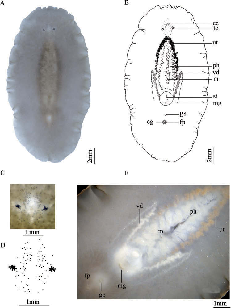
Body broadly oval, soft and translucent. Dorsal ground colour sandy beige to light brown, darker around the pharynx, with pale brown microdots, except over the pharynx and towards the body margin ( Figure 2A View Figure 2 ). Ventrally light grey to whitish. Fixed specimen 22 mm long, 12 mm wide ( Figure 2A–B View Figure 2 ). Small nuchal tentacles present. Tentacular eyes more crowded at the base of tentacles, each cluster with about 20 eye-spots. Cerebral eyes smaller, in two clusters behind the brain, each with about 39 eye-spots ( Figure 2C View Figure 2 ). Marginal eyes absent ( Figure 2B–D View Figure 2 ).
Digestive system
Ruffled pharynx located centrally, with about 13 lateral folds, 7 mm in length (one third of the body length), mouth in posterior third of the pharyngeal cavity, opening to main intestine anteriorly to the male apparatus ( Figure 2E View Figure 2 ).
Body wall
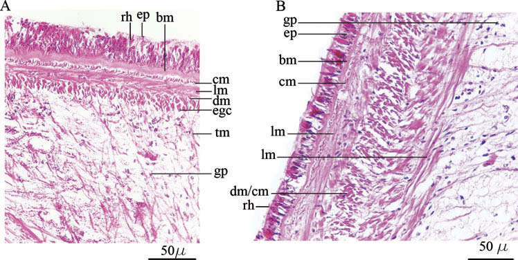
Dorsally 72 µm high. Cellular ciliated epidermis with columnar cells, rhabdites apparent, basement membrane 11 µm high. Three distinct subepidermal muscle layers: an outer circular, middle longitudinal and inner diagonal. Transversal muscle fibres well developed. Eosinophilic gland cells present beneath the muscular layers ( Figure 3A View Figure 3 ).
Ventral body wall 85 µm high, thicker between gonopores, where the genital sucker is located (130–140 µm high). Cellular epidermis sparsely provided with rhabdites, basement membrane 6 µm high. Subepidermal outer circular muscle layer is followed inwards by a thinner layer of longitudinal muscles and a welldeveloped diagonal muscle layer interspersed with circular fibres ( Figure 3B View Figure 3 ). Large eosinophilic glandular cells between male gonopore and genital sucker. Smaller basophilic cells extend from this point posteriorly towards the female system, merging with cement glands. Scattered granular pigment cells between ventral and dorsal muscle fibres as well as in the parenchyma ( Figure 3A–B View Figure 3 ).
Male copulatory complex
Male apparatus directed backwards, with true seminal vesicle, interpolated tubular prostatic vesicle and elongated stylet ( Figure 4A–B). Testes ventral and located toward the margin along the whole body. Vasa deferentia extend forwards up to the mouth and then recurve, forming ( Figure 2E View Figure 2 ) spermiducal bulbs, shortly before entering the seminal vesicle from the posterior side ( Figure 4B). Seminal vesicle located immediately behind the pharynx, spherical, 520 µm in diameter and occupying more than half of body width. Its muscular wall is highly developed ( Figure 4A–B). The rounded interpolated prostatic vesicle measures about 650 × 640 µm and is surrounded by a strongly developed muscular wall ( Figure 4A–C). Glandular epithelium of the prostatic vesicle tubular and divided into four chambers, while numerous extra-vesicular glands discharge their secretion into the lumen ( Figure 4B, D). The ejaculatory duct projects into the prostatic vesicle and is not immediately attached to the glandular lining. The ejaculatory duct possesses a well-defined muscular wall. Between the prostatic vesicle and the stylet, there is a mass of specialised parenchymatous tissue, (440 × 280 µm), where the highly muscularised ejaculatory duct bends before entering the stylet. This parenchymatous tissue consists of large, loosely arranged vacuolated cells, with thin cell walls and a scattered intercellular matrix, limited by scattered muscle fibres ( Figures 4A, 5A–D). The elongated, sigmoid stylet (910 µm long) is housed in an elongated penis sheath and a small male atrium ( Figures 2B View Figure 2 , 5D). The seminal vesicle, prostatic vesicle, specialised parenchymatous tissue, ejaculatory duct and penis sheath are surrounded by concentric longitudinal muscle fibres, albeit these do not form a distinct bulb ( Figure 4B). In resting position, when the stylet lies in the male atrium, the male copulatory complex is not aligned with the main longitudinal body axis, but is coiled ( Figure 4B).
Female system
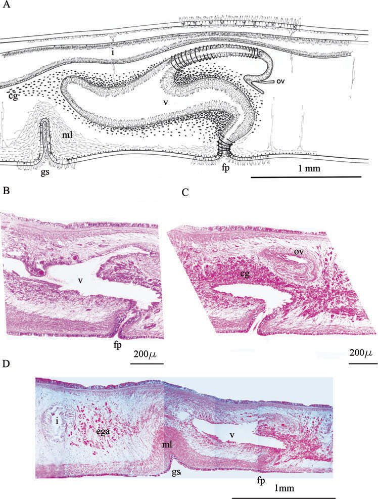
Ovaries dorsal. Uteri well developed anterior to mouth ( Figure 2B, E View Figure 2 ), giving rise to narrow paired oviducts, which open into the vagina at its ventral side. From this point, the vagina turns dorsally and runs anteriorly, then turns ventrally to the gonopore ( Figure 6A View Figure 6 ). Proximal part of the female genital canal slender, with cuboidal ciliated epithelium, surrounded by a well-developed muscular wall. Distally, the vaginal tract is surrounded by an adjacent mass of glandular cells. The vagina is lined with cylindrical secretory and ciliated cells. A blind vaginal chamber extends forwards. The wall of this chamber possesses the same structure as the distal tract of the vagina ( Figure 6A–D View Figure 6 ). Gonopores separated (2.3 mm), located in the posterior third at last third of the body ( Figure 2B, E View Figure 2 ). Well-developed genital sucker between gonopores, 0.2 mm deep ( Figure 6A, D View Figure 6 ).
The size of the collected specimens ranged between 12.1 × 6.7 mm and 22 × 12 mm ( Table 1). In concordance with this variability, bigger animals develop more eye-spots and some variability in the body outline and size ( Figure 7A–C). Nevertheless, the anatomy of all examined specimens reveals a great uniformity. The position of the mouth, the presence of apparent spermiducal bulb, the presence of a genital sucker, the highly muscularised male copulatory complex, the long and slender stylet and the characteristic morphology of the female system leave no doubt as to species identity. For these reasons, we found Faubel’ s system more suitable for the determination of this new species.
No known copyright restrictions apply. See Agosti, D., Egloff, W., 2009. Taxonomic information exchange and copyright: the Plazi approach. BMC Research Notes 2009, 2:53 for further explanation.