
Sphaerocetum, Ek, Martin Fiká Č, 2010
|
publication ID |
https://doi.org/ 10.5281/zenodo.197617 |
|
DOI |
https://doi.org/10.5281/zenodo.6197658 |
|
persistent identifier |
https://treatment.plazi.org/id/8B1C87A8-1272-FFF9-FF7E-BC7A1B5DFDA3 |
|
treatment provided by |
Plazi |
|
scientific name |
Sphaerocetum |
| status |
gen. nov. |
Sphaerocetum View in CoL gen. n.
Type species. Sphaerocetum malayanum sp. n. (hereby designated).
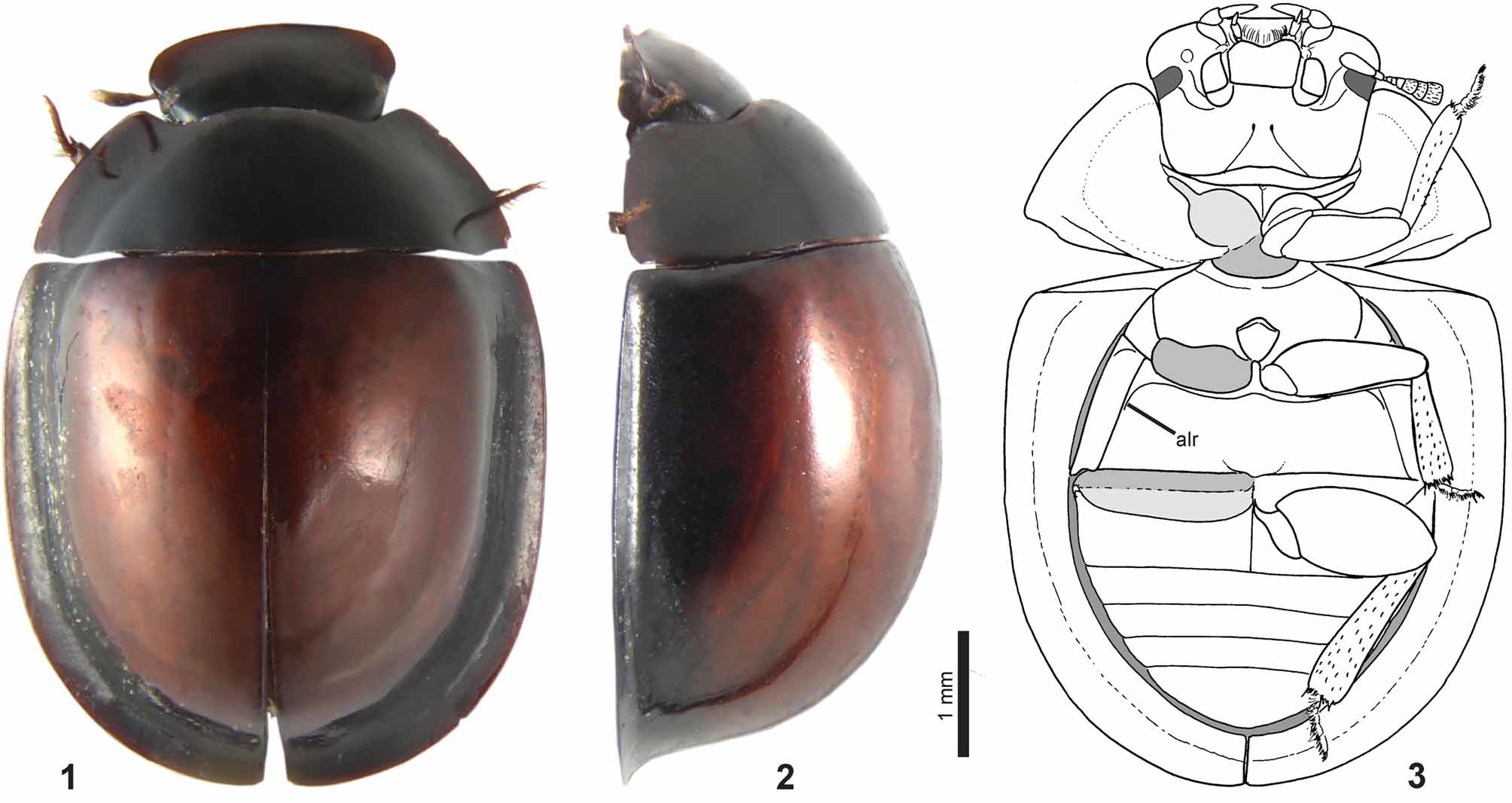
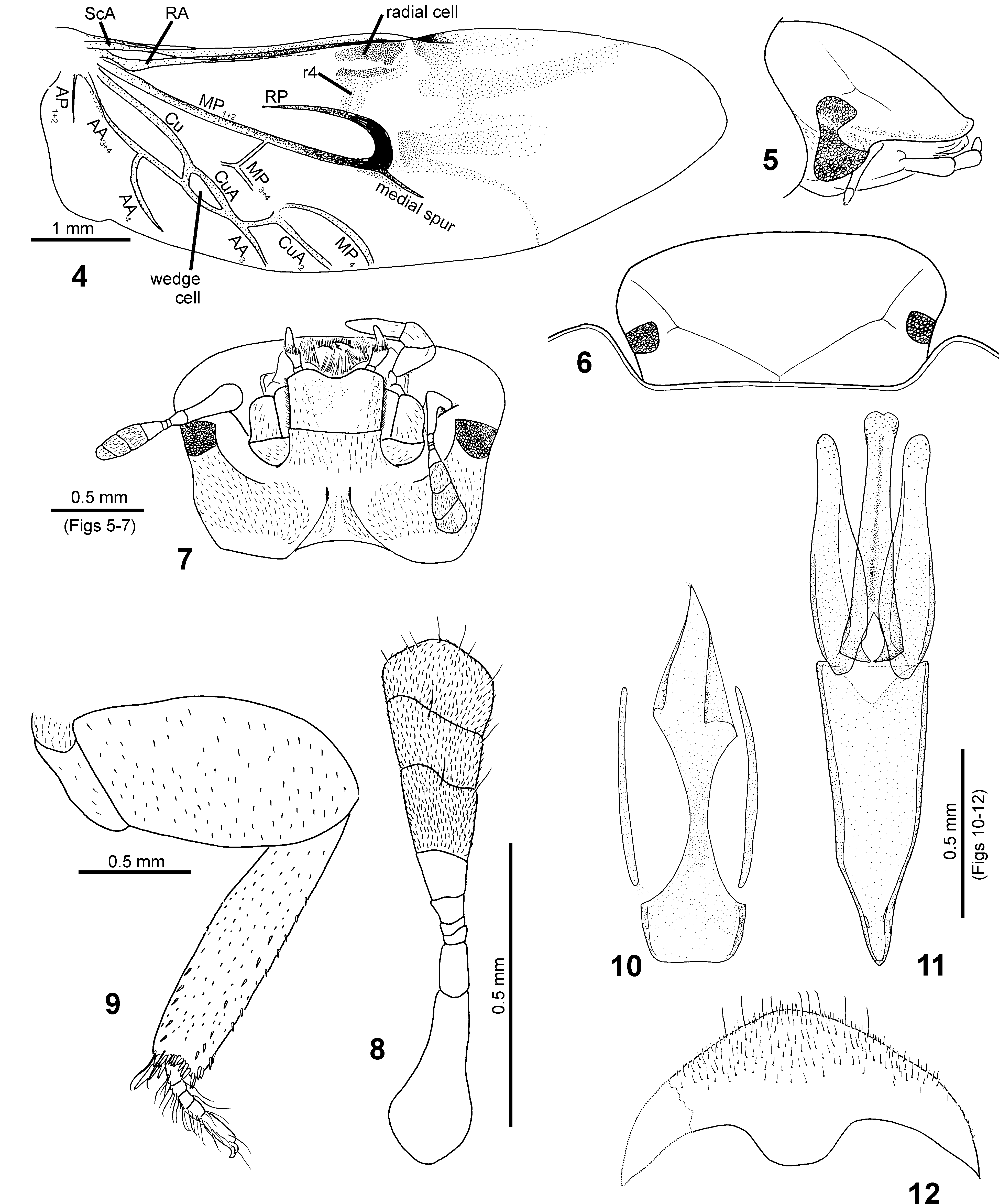
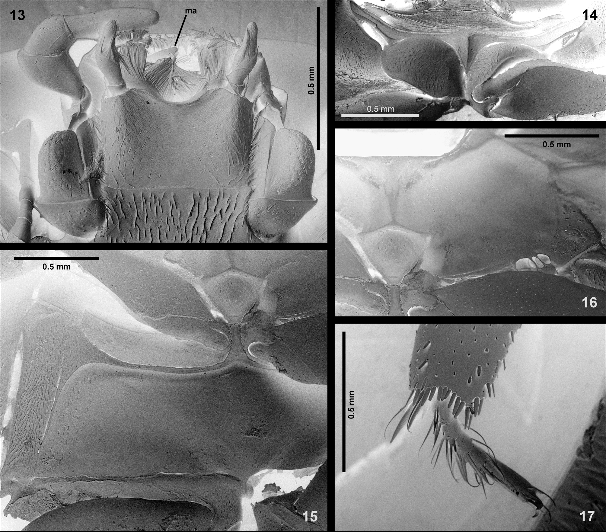
Diagnosis. Body highly convex in lateral view ( Fig. 2 View FIGURES 1 – 3 ); lateral margins of elytra widely explanate ( Figs 1– 2 View FIGURES 1 – 3 ); head widened anterior of eyes ( Fig. 6 View FIGURES 4 – 12 ); antenna with nine antennomeres; antennal club compact ( Fig. 8 View FIGURES 4 – 12 ); prosternum carinate medially; prosternum very narrow anterior of procoxae ( Fig. 14 View FIGURES 13 – 17 ); transverse fold below posterior margin of pronotum well developed, almost reaching lateral margin; mesoventrite and mesanepisternum fused, anapleural suture absent; posteromedian portion of mesoventrite elevated into subpentagonal preepisternal plate; preepisternal plate without median carina ( Fig. 15 View FIGURES 13 – 17 ); mesothoracic grooves for reception of procoxae absent ( Fig. 16 View FIGURES 13 – 17 ); elytral series inconspicuous; epipleura wide throughout, horizontal in posterior portion of elytra ( Fig. 3 View FIGURES 1 – 3 ); metaventrite with very distinctly developed anterolateral ridges ( Figs. 3 View FIGURES 1 – 3 , 15 View FIGURES 13 – 17 ); anteromedian metaventral process narrow and long; wedge cell of hind wing present, closed ( Fig. 4 View FIGURES 4 – 12 ); vein AA4 long, nearly reaching posterior margin of hind wing; AP1 +2 present but short; femoral lines of metaventrite absent; femora with very deep tibial grooves; tarsi very short, much shorter than tibiae ( Fig. 9 View FIGURES 4 – 12 ); first metatarsomere short, ca. twice as long as metatarsomere 2; abdominal ventrite 1 carinate medially; median lobe of aedeagus not extending into phallobase; phallobase nearly symmetrical; male sternite 9 with tongue-shaped median projection; sternite 8 with broad median basal process ( Fig. 12 View FIGURES 4 – 12 ).
Description. Body widely oval, highly convex in lateral view; pronotal and elytral outline continuous in dorsal and lateral views.
Head ( Figs 5–7 View FIGURES 4 – 12 , 13 View FIGURES 13 – 17 ). Clypeus widened anteriorly of eyes, anteromedian margin of clypeus straight. Frontoclypeal suture present but rather inconspicuous. Eyes small, subrectangular in dorsal view, deeply emarginate anteriorly in lateral view, interocular distance>7× the width of one eye in dorsal view. Labrum small, membranous, nearly totally concealed under clypeus, ca. 0.25× as wide as maximum width of head. Mandibular apex bifid. Cardo and basistipes strongly protruding laterad, lateral margin of basistipes strongly arcuate, mediostipes very distinctly divided from basistipes. Maxillary palpus with four palpomeres, palpomere 1 minute, palpomeres 2–4 long and wide; palpomere 2 strongly widened distally, palpomere 3 slightly narrower than distal portion of palpomere 2; palpomere 4 longest and narrowest. Mentum slightly wider than long, lateral margin parallel-sided, each bearing dense row of setae; anterior margin of mentum bisinuate; labial palpus with three palpomeres, palpomere 2 widened distally, bearing group of densely arranged long setae on distal portion, palpomere 3 ca. as long as palpomere 2 but slightly narrower; submentum sparsely pubescent. Antenna with nine antennomeres; scapus rather long and thick, its basal portion bent dorsad; pedicel ca. as long as antennomeres 3–5 combined, only indistinctly narrowing distally; antennomeres 3–4 minute, slightly widened distally, antennomere 5 ca. as long as antennomeres 3–4 combined; cupula (= antennomere 6) rather long, widened distally, bare; antennal club compact, depressed dorsoventrally, slightly widened distally, blunt at apex; entire club densely pubescent with few longer and thicker setae on sides and ventrally in distal margins of antennomeres. Ridge arising from posterolateral ventral margin of eyes long, strongly developed; genae sparsely pubescent. Posterior tentorial pits narrow, elongate; gular sutures strongly divergent posteriad of tentorial pits. Gula sparsely pubescent laterally, bearing blunt, low median longitudinal carina.
Prothorax ( Figs 1–3 View FIGURES 1 – 3 , 14 View FIGURES 13 – 17 ). Pronotum weakly explanate along lateral margins. Anterior margin of pronotum deeply emarginate, posterior margin weakly arcuate except for posterolateral corners which are angulate and slightly emarginate ( Fig. 1 View FIGURES 1 – 3 ); lateral, anterior and lateral portions of posterior margins with sharp but narrow rim. Prosternum carinate medially, prosternal process without posterior emargination; prosternal portion anterior of procoxae very narrow. Procoxal cavities large, open posteriorly, anterolateral aperture of procoxal cavity open. Notopleural suture very short. Hypomeron with large pubescent inner portion and moderately wide marginal bare portion. Transverse fold below posterior margin of pronotum present, long, nearly reaching lateral margin.
Mesothorax ( Figs 3–4 View FIGURES 1 – 3 View FIGURES 4 – 12 , 15 View FIGURES 13 – 17 ). Mesoventrite completely fused with anepisternum 2; epimeron 2 welldelimited, divided from anepisternum 2 by a suture; anterior collar of mesothorax narrow. Posteromedian portion of mesoventrite elevated into preepisternal plate, the plate subrhomboid in shape, lacking median longitudinal carina throughout. Grooves for reception of procoxae not defined. Elytron highly convex, lateral margin widely explanate anteriorly as well as at apex; elytron bearing 10 very indistinct elytral series, series 1–7 consisting of punctures only slightly larger than interval punctation and only very indistinctly impressed, series 8–10 with coarser but still only indistinctly impressed punctures; all series reduced subapically. Epipleuron very wide and horizontal throughout, with outer bare portion (= ‘pseudepipleuron’) ca. as wide as inner pubescent portion (=‘epipleuron’). Mesocoxal cavities transverse, very narrowly divided medially by an anterior projection of metaventrite.
Metathorax ( Figs 3–4 View FIGURES 1 – 3 View FIGURES 4 – 12 , 15 View FIGURES 13 – 17 ). Metaventrite flat, without median elevation, bare on whole surface, anteromedially projecting between mesocoxae by long and narrow metaventral process. Anterolateral ridges very distinctly developed, joint mesally, arcuatelly bent posteriad sublaterally, continuing along lateral margin of metaventrite laterally. Anepisternum 3 elongate, parallel-sided, bearing a transverse ridge anteriorly. Hind wings with closed wedge cell, vein AA4 nearly reaching posterior margin of the wing and short but clearly developed AP1+2.
Legs ( Figs 3 View FIGURES 1 – 3 , 9 View FIGURES 4 – 12 , 17 View FIGURES 13 – 17 ). Procoxa globular, sparsely pubescent; profemur with deep tibial groove delimited by high ventral and dorsal ridges, ventral surface sparsely pubescent except for small bare distal area; protibia cylindrical. Mesotrochanter slightly sinuate on posterior margin; ventral surface of mesotibia sparsely covered by short and stout spines, tibial groove deep, developed throughout, delimited by high ventral and low dorsal ridges; mesotibia flattened, with scattered short spines and three rows of stouter spines on ventral surface, distal apex with series of short spines and several longer spurs. Metatrochanter large, strongly bisinuate on posterior margin; metafemur large and wide, ventral surface covered by scattered short spines, tibial groove well developed, deep. Metatibia flattened, bearing scattered short spines and three series of larger spines on ventral surface, distal portion with series of moderately long spines and two long spurs. Tarsi much shorter than tibiae, each tarsomere bearing a brush of long setae ventrally and single to few long setae dorsally; metatarsomere 1 as wide as, but ca. twice as long as metatarsomere 2; claws small, simply arcuate.
Abdomen ( Fig. 3 View FIGURES 1 – 3 ) with five ventrites, ventrite 1 carinate medially, ventrite 5 without apical notch.
Male genitalia ( Figs 10–12 View FIGURES 4 – 12 ). Aedeagus simple, trilobate, rather weakly sclerotized. Base of median lobe attached to the bases of parameres, not extending into the phallobase; apical portion of median lobe without distinct gonopore. Parameres gradually narrowing towards widely rounded apex. Phallobase widest at junction with parameres, gradually narrowing posteriad, nearly symmetrical, without distinctly detached manubrium. Sternite 8 with broad median anterior process. Sternite 9 with long, well-sclerotized median tongue-shaped portion, its posterior part slightly coiled.
Etymology. Consisting of the prefix Sphaero-, derived from the Latin noun sphaera (ball) referring the highly convex and semiglobular body of the genus, and of the ending – cetum derived from the name of the protosternine genus Mucetum d’Orchymont, 1926 . Gender: neuter.
Distribution. Oriental Region, so far known from the peninsular Malaysia only.
No known copyright restrictions apply. See Agosti, D., Egloff, W., 2009. Taxonomic information exchange and copyright: the Plazi approach. BMC Research Notes 2009, 2:53 for further explanation.