
Enchodelus hopedorus ( Thorne, 1929 ) Thorne, 1939
|
publication ID |
https://doi.org/ 10.1163/156854107779969646 |
|
DOI |
https://doi.org/10.5281/zenodo.8111780 |
|
persistent identifier |
https://treatment.plazi.org/id/8C029544-A917-FFCA-FCF2-FE30FB58CF48 |
|
treatment provided by |
Carolina |
|
scientific name |
Enchodelus hopedorus ( Thorne, 1929 ) Thorne, 1939 |
| status |
|
Enchodelus hopedorus ( Thorne, 1929) Thorne, 1939 = Dorylaimellus hopedorus Thorne, 1929
( Figs 8 View Fig , 9 View Fig )
MATERIAL EXAMINED
Nineteen females, mounted on slide labelled Enchodelus hopedorus 5c, and collected from Longs Peak, Colorado, USA, in summit soil, ont 19 July 1924; in acceptable condition although the specimens have become somewhat flattened.
MEASUREMENTS
See Table 4. View Table 4
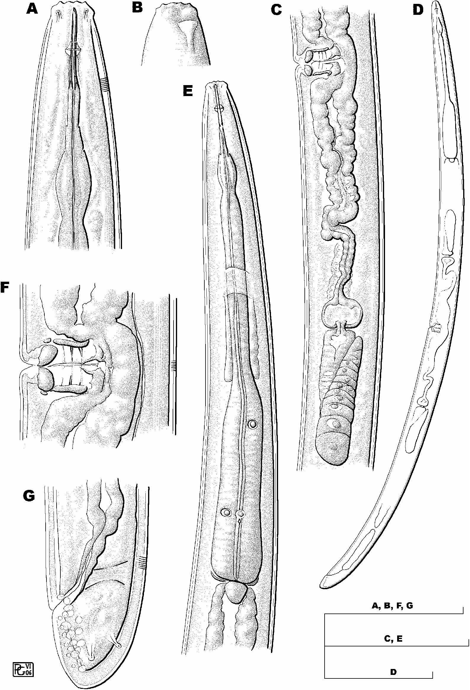
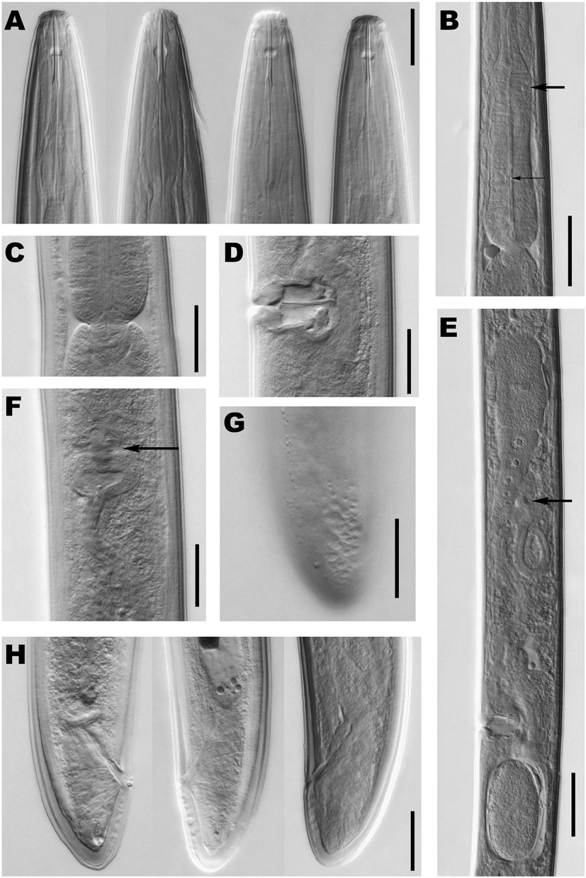
DESCRIPTION
Female
Moderately stout nematodes of medium size, 1.4- 1.8 mm long. Habitus after fixation slightly ventrad curved. Body cylindrical, tapering towards both ends but more so towards anterior. Cuticle 2.5-3.0 µ m thick at anterior region, 2.5-3.5 µ m at midbody and 4.5-6.5 µ m at tail; its outer layer practically smooth or bearing very fine transverse striations, and much thinner than inner, especially at level of tail. Lateral chord relatively narrow, occupying 13-18% of midbody diam., lacking any particular differentiation. Lateral pores obscure. Lip region practically continuous or barely offset by a shallow depression, 2.4-3.1 times as broad as high and ca one-third of body diam. at neck base. Lips amalgamated, with their inner portions visibly projecting; labial and cephalic papillae distinct. Amphid fovea cup-shaped, opening at level of cephalic depression and occupying one-half to three-fifths of corresponding body diam. Cheilostom a truncate cone, with thickened walls. Odontostyle long and slen- der, with distinct walls and narrow lumen, slightly curved; 11-16 times as long as wide or 1.9-2.2 times as long as lip region diam.; aperture small, one-twelfth to one-tenth of total length. Odontophore lacking basal flanges but with basal portion slightly thickened, clearly separated from pharyngeal lining; 1.3-1.6 times longer than odontostyle. Guiding ring double, at 17-21 µ m or 1.1-1.3 times lip region diam. from anterior end. Pharynx consisting of a slender but well muscular anterior portion expanding gradually into basal expansion at 59-64% of total neck length, reaching full diam. at 65-70%; pharyngeal expansion occupying 33-38% of total neck length, and one-half to four-fifths of the corresponding body diam. Pharyngeal gland nuclei located as follows: DN = 66-72; S 1 N 1 =?; S 1 N 2 =?; S 2 N 1 = 52-63; S 2 N 2 = 53-64. Weak membrane-like structure surrounding base of pharyngeal expansion. Cardia very small, rounded conoid, as wide as long. In some specimens, intestine containing greenish material. Genital system didelphic-amphidelphic, with both branches equally and well developed. Ovaries relatively large, 69-187 µ m long, but usually not reaching sphincter level; oocytes first in two or more rows, then in one row. Oviduct 79-156 µ m long and consisting of a slender portion with prismatic cells and a well developed pars dilatata bearing distinct lumen. Sphincter distinct, located between oviduct and uterus. Uterus long, 189-270 µ m or 2.9-4.2 times corresponding body diam.; tripartite with wider proximal portion with distinct lumen followed by narrower and often twisted intermediate length region with narrow lumen joining a well developed spheroid pars dilatata distalis. Sperm not observed in genital tract. Vagina extending inwards 39-55% of body diam.; pars proximalis 12-17 × 16-25 µ m, with almost straight or somewhat sigmoid walls, enveloped by weak circular musculature; pars refringens with (in lateral view) two close together drop-shaped sclerotisations which have a combined width of 13-19 µ m; pars distalis short, 3.0-6.0 µ m long. Vulva a transverse slit. Prerectum 1.5-4.6, rectum 0.8-1.2 anal body diam. long. Tail rounded conoid, slightly more straight ventrally; numerous saccate bodies distinct in tail, most in ventral side. Two pairs of caudal pores, both subdorsal, one at middle of tail, other subterminal.
Male
Unknown.
DIAGNOSIS
Enchodelus hopedorus is distinguished by its body 1.4- 1.8 mm long, lip region practically continuous and 14.5- 16 µ m diam., odontostyle 29-33 µ m long or 1.9-2.2 times the lip region diam., odontophore lacking distinct basal flanges and longer (1.3-1.6 times) than the odontostyle, neck length 305-374 µ m long, pharyngeal expansion 106- 136 µ m and occupying 33-38% of total neck length, female genital system amphidelphic, uterus long and tripartite, pars refringens vaginae with two drop-shaped sclerotisations, vulva in form of a transverse slit and equatorial (V = 48-55), tail rounded conoid (27-39 µ m, c = 41-53, cļ = 0.7-1.0) with numerous saccate bodies, and males unknown.
REMARKS
There is no doubt concerning the identity of these specimens with those Thorne (1939) refers to in his description. The re-examination of this material has provided some remarkable differences from Thorne’s original description. The lip region is practically continuous, not “set off by slight constriction”; in fact, Thorne’s Figure 70 shows a constriction, but the specimens are viewed in dorsal or ventral position, not laterally. The presence of basal flanges in the odontophore is not confirmed, because the nature of odontophore base resembles that observed in E. macrodoroides (see under E. geraldi) rather than the corresponding to E. macrodorus, as can be seen if we compare his Figures 70a, 68c and 76b, respectively.
After its original description, E. hopedorus was reported from Poland ( Brzeski, 1963; Winiszewska-Slipinska, 1987) and Nepal ( Zullini, 1973), but some doubts persist concerning the identity of non-American material. Brzeski (1963) studied two females having shorter odontostyle (20-23 µ m) and neck (b = 5.9-7.1), and slightly anterior vulva (V = 46); he also described two males. Zullini (1973) recorded one female, comparatively smaller (L = 1.15), and having a shorter odontostyle (21 µ m) and more anterior vulva (V = 43). Also, Winiszewska-Slipinska (1987) studied three females having a shorter neck (b = 5.0-5.7), relatively longer tail (cļ = 1.1-1.2) and more anterior vulva (V = 45-46).
No known copyright restrictions apply. See Agosti, D., Egloff, W., 2009. Taxonomic information exchange and copyright: the Plazi approach. BMC Research Notes 2009, 2:53 for further explanation.