
Enchodelus arcuatus Thorne, 1939
|
publication ID |
https://doi.org/ 10.1163/156854107779969646 |
|
DOI |
https://doi.org/10.5281/zenodo.8111774 |
|
persistent identifier |
https://treatment.plazi.org/id/8C029544-A919-FFDE-FCE9-FA04FE4FC838 |
|
treatment provided by |
Carolina |
|
scientific name |
Enchodelus arcuatus Thorne, 1939 |
| status |
|
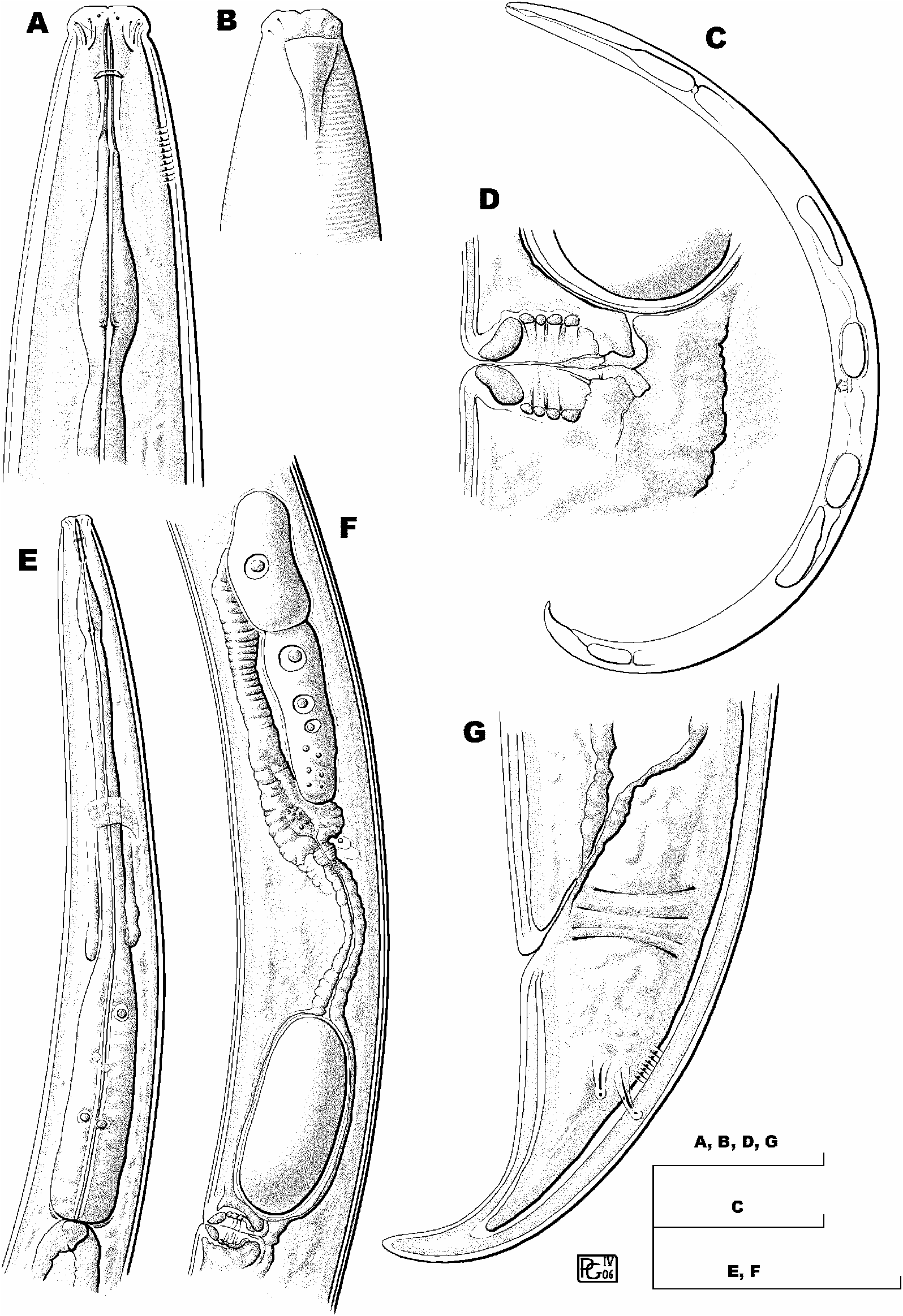
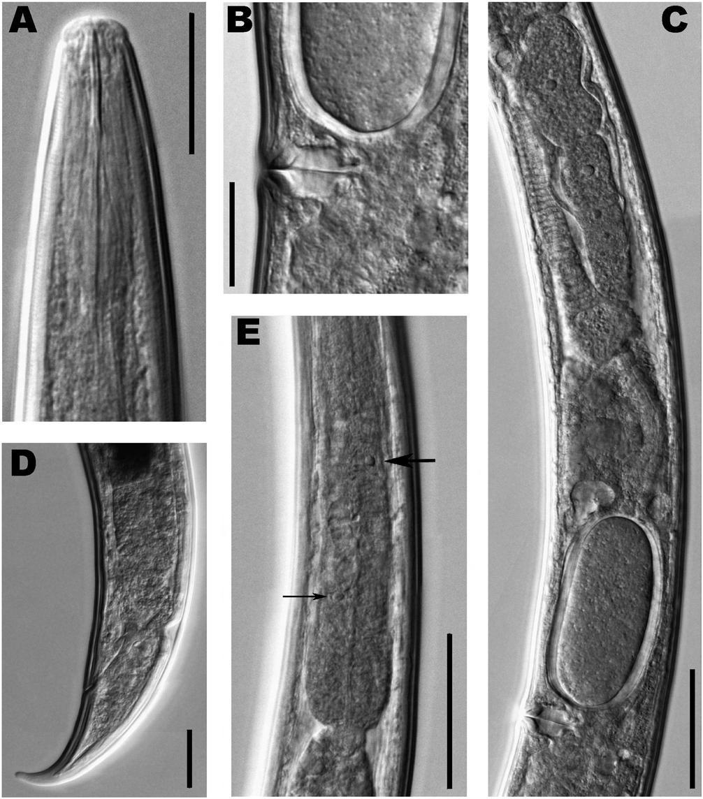
Enchodelus arcuatus Thorne, 1939
( Figs 2 View Fig , 3 View Fig )
MATERIAL EXAMINED
Two females, one mounted onslide labelled Enchodelus arcus, and collected from Arlington Farm, Virginia ( USA) on April 8, 1931, in quite good condition. Another female mounted on slide labelled Enchodelus arcus, and collected from Granddaddy Lake, Utah ( USA) in August, 1924, in poor condition.
MEASUREMENTS
See Table 2 View Table 2 .
DESCRIPTION
Female
Moderately slender nematodes of medium size, 1.5,
2.0 mm long. Body cylindrical, tapering towards both ends but more so towards posterior. Cuticle 2.5, 3.5 µ m thick at anterior region, 3.0, 4.0 µ m at mid-body and 4.0, 6.0 µ m at tail; its outer layer thinner than inner, with very fine transverse striations. Lateral chord 13 µ m wide or ca one-fifth of mid-body diam., lacking any particular differentiation. Lateral pores obscure. Lip region rounded, offset by shallow constriction, 2.8, 3.0 times as broad as high and ca one-third of body diam. at neck base; lips practically amalgamated; perioral area weakly differentiated; labial and cephalic papillae hardly perceptible. Amphid fovea cup-shaped, opening at level of cephalic constriction and occupying almost two-thirds of corresponding body diam. Cheilostom a truncate cone, with no particular differentiation. Odontostyle slender, slightly dorsad curved; its length 10.9, 12.9 times as long as wide or 1.5, 1.6 times as long as lip region diam.; aperture small, ca one-tenth of total length. Odontophore rod-like, lacking any specialisation, 1.3, 1.4 times the odontostyle length long. Guiding ring apparently double, at 10.5-12.0 µ m, or 0.9 times lip region diam., from anterior end. Pharynx consisting of a slender, but well muscled portion expanding gradually into basal expansion at 54, 58% of total neck length, reaching full diam. at 63, 67%; pharyngeal expansion occupying ca two-fifths (38, 42%) of total neck length, and ca three-fifths of corresponding body diam. Pharyngeal gland nuclei located as follows: DN = 70, 65; S 1 N 1 = 23; S 1 N 2 = 27; S 2 N 1 = 51, 48; S 2 N 2 = 53, 49. Base of pharyngeal expansion surrounded by a weakly refractive membrane-like structure. Cardia rounded conoid, practically as long as wide (8 µ m). Intestine containing abundant green-coloured material, mainly in posterior portion. Genital system didelphic-amphidelphic, with both branches equally and well developed. Ovaries relatively large, 87-139 µ m long, but not reaching sphincter level; oocytes first in two or more rows, then in one row. Oviduct 108-147 µ m long or 1.6-2.4 times corresponding body diam., consisting of a slender portion with prismatic cells and a well developed pars dilatata with distinct lumen. Sphincter clearly visible between oviduct and uterus; a group of hyaline cells surrounding sphincter and distal part of uterus. Uterus bipartite, i.e., consisting of a wider proximal portion with distinct lumen, and a narrower distal part with narrow lumen and refractive inner lining, portion close to sphincter bearing very small refractive granules; uterus length 133-161 µ m or 2.0-2.7 times body diam. at its level. Sperm not observed in genital tract. Uterine egg measuring 35, 38 × 80, 81 µ m (n = 2). Vagina extending inwards to ca two-fifths of body diam.; pars proximalis vaginae 11.5, 13.5 × 16.5, 15.0 µ m, with almost straight walls and enveloped by moderately developed circular musculature; pars refringens vaginae with (in lateral view) two, rather close, trapezoidal sclerotisations that have a combined width of 12.0, 18.0 µ m; pars distalis vaginae short, 2.5, 5.0 µ m long. Vulva a transverse slit. Prerectum 2.1, 2.9; rectum 1.1, 1.2 anal body diam. long. Tail conical, regularly ventrad curved, with rounded tip; hyaline terminal portion 16, 23 µ m or 26, 29% of total length. Two pairs of caudal pores at the middle of tail: one subdorsal, another practically lateral.
Male
Unknown.
DIAGNOSIS
Enchodelus arcuatus is distinguished by its body 1.5- 2.0 mm long, lip region 12-14 µ m diam., odontostyle 18.5-22 µ m long or 1.5-1.6 times the lip region diam., odontophore rod-like and 1.3-1.4 times the odontostyle, neck length 287-340 µ m long, pharyngeal expansion 108- 142 µ m or occupying ca two-fifths (38-42%) of total neck length, female genital system amphidelphic, uterus bipartite, pars refringens vaginae with two well developed trapezoid sclerotisations, vulva in form of a transverse slit and slightly posterior (V = 52-54), tail conical and regularly ventrad curved (62-80 µ m, c = 24-25, cļ = 2.0- 2.2), and males unknown.
REMARKS
Thorne (1939) provided measurements, description and illustrations of the female collected from Arlington Farm, and mentioned that he had studied another two females from Kailab Forest, Arizona, and three from Tryal Lake, Uinta Mountains, Utah. One of the two females examined here is, without doubt, that from Arlington Farm; another might be one of the three collected from Uinta Mountains.
After its original description, E. arcuatus was later reported from several habitats and localities. Andrássy (1958) studied one Bulgarian female having longer prerectum (4-5 ABD) and shorter tail (c = 35.9). Also Andrássy (1959) described one female from Romania having longer body (L = 2.2), more anterior vulva (V = 47) and shorter tail (c = 32.7, cļ = 1.8). Zullini (1970) reported fourteen females from Italy having a more anterior vulva (V = 49-52, n = 3) and shorter tail (c = 27-35, n = 3). Taking into account that the differences observed are morphometric and with no large gap among them, and that only a few specimens have hitherto been studied, it is tentatively assumed that these four records belong to the same species, although European material can be distinguished from that from America by a shorter tail.
On the other hand, Vinciguerra and De Francisci (1973) and Vinciguerra and La Fauci (1978) described two Italian populations characterised by a smaller body (L = 1.1-1.5, n = 18), shorter odontostyle (13-16 µ m), pars refringens vaginae apparently absent (“la vagina... e la sua estremità prossimale è cuticolarizzata, ma non sclerificata”), and male as frequent as the female. This material is certainly not conspecific with those referred above.
No known copyright restrictions apply. See Agosti, D., Egloff, W., 2009. Taxonomic information exchange and copyright: the Plazi approach. BMC Research Notes 2009, 2:53 for further explanation.