
Enchodelus brevidentatus Thorne, 1939
|
publication ID |
https://doi.org/ 10.1163/156854107779969646 |
|
DOI |
https://doi.org/10.5281/zenodo.8111776 |
|
persistent identifier |
https://treatment.plazi.org/id/8C029544-A91F-FFD3-FFA9-FDD0FE17CE2A |
|
treatment provided by |
Carolina |
|
scientific name |
Enchodelus brevidentatus Thorne, 1939 |
| status |
|
Enchodelus brevidentatus Thorne, 1939
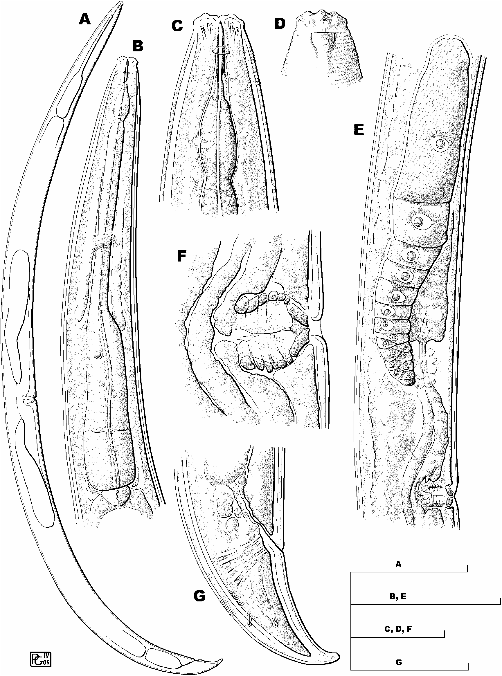
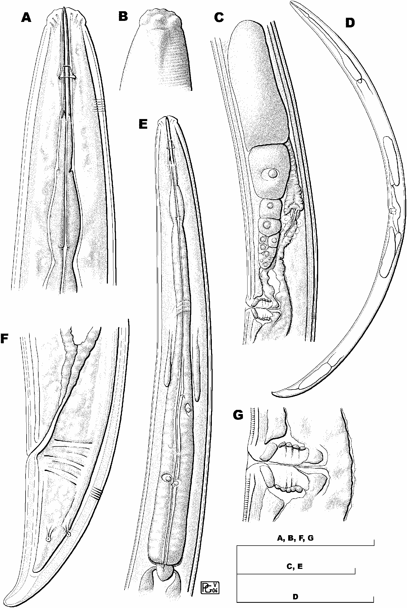
( Figs 4 View Fig , 5 View Fig )
MATERIAL EXAMINED
Nineteen females, 18 of them mounted on one slide labelled Enchodelus brevidens, and collected from foothill soil in Lehi , Utah ( USA) on March 8, 1926; a few specimens in acceptable condition, but most of them becoming flattened. The remaining female, mounted on slide labelled Enchodelus macrodoroides 2a, and collected from Wellsville Hill, Utah ( USA) on March 28, 1925, is in quite bad condition.
MEASUREMENTS
See Table 2. View Table 2
DESCRIPTION
Female
Moderately slender nematodes of medium size, 1.6- 1.9 mm long. Body cylindrical, tapering towards both ends but more so towards posterior. Cuticle 2.0-3.0 µ m thick at anterior region, 2.0-4.0 µ m at midbody and 3.5- 6.0 µ m at tail; its outer layer thinner than inner, bearing fine but distinct transverse striations that are more visible at level of anterior body region and caudal region. Lateral chord occupying 18-30% of midbody diam, with granular appearance. Lateral pores obscure. Lip region rather angular, offset by distinct depression, 2.4-2.9 times as broad as high and ca one-third (some specimens ca one-fifth, but obviously flattened) of body diam. at neck base; lips mostly amalgamated; labial and cephalic papillae protruding, in particular the inner labial ones. Amphid fovea cup-shaped, opening at level of cephalic depression and occupying one-third to one-half of corresponding body diam. Cheilostom a truncate cone to practically cylindrical, with no particular differentiation. Odontostyle relatively slen- der, practically straight; 7.7-11.3 times as long as wide or 1.1-1.4 times as long as lip region diam.; aperture small, one-tenth to one-fifth of total length. Odontophore rod-like, lacking any specialisation, 1.2-1.5 times odontostyle length long. Guiding ring simple, at 8.5-10.5 µ m or 0.7- 0.8 lip region diam. from anterior end. Pharynx consisting of a slender but well muscular portion expanding gradually into basal expansion at 54-65% of total neck length, reaching full diam. at 62-70%; pharyngeal expansion occupying 33-42% of total neck length, and ca three-fifths of corresponding body diam. Pharyngeal gland nucleilocatedas follows: DN = 67-72 (n = 10); S 1 N 1 = 22; S 1 N 2 = 31 (n = 1); S 2 N 1 = 49-55; S 2 N 2 = 50- 57 (n = 7). Base of pharyngeal expansion surrounded by faint membrane-like structure. Cardia rounded conoid, wider than long, 16 × 11 µ m. Genital system didelphic-amphidelphic, with both branches equally and well developed. Ovaries relatively large, 109-320 µ m long, usually reaching and surpassing sphincter level; oocytes first in two or more rows, then in one row. Oviduct 120-273 µ m long or 2-3 times corresponding body diam., consisting of a slender portion with prismatic cells and a well developed pars dilatata with distinct lumen. Sphincter present between oviduct and uterus. Uterus bipartite, i.e., with a wider proximal portion with distinct lumen, and a narrower distal part with narrow lumen and refractive inner lining; length 65-126 µ m or 0.9-2.0 times body diam. at its level; a group of hyaline cells observed surrounding distal part of uterus. Sperm not observed in genital tract. Only one uterine egg observed in female from Wellsville, measuring 91 × 35 µ m. Vagina extending inwards one-third (flattened specimens) to two-fifths of body diam.; pars proximalis vaginae 11-16 × 13-25 µ m, with almost straight or slightly convergent walls and enveloped by moderately developed circular musculature; pars refringens vaginae with (in lateral view) two trapezoidal sclerotisations which have a combined width of 9.5-15.5 µ m and are somewhat separated by a weakly sclerotised intermediate area; pars distalis vaginae short, 2.5-5.0 µ m long. Vulva a transverse slit. Prerectum 1.8-3.2, rectum 0.8-1.2 anal body diam. long. Tail conical, regularly ventrad curved, with rounded tip; hyaline terminal portion 10-15 µ m or 15-21% of total length. Two pairs of caudal pores at the middle of the tail: one subdorsal, another practically lateral.
Male Unknown.
Enchodelus brevidentatus is distinguished by its body 1.6-1.9 mm long, lip region 12-13.5 µ m diam., odontostyle 14.5-17 µ m long or 1.1-1.4 times the lip region diam., odontophore rod-like, neck length 264-308 µ m long, pharyngeal expansion 91-125 µ m long or occupying 33-42% of total neck length, female genital system amphidelphic, uterus bipartite, pars refringens vaginae with two trapezoidal sclerotisations, vulva in form of a transverse slit (V = 47-53), tail conical and regularly ventrad curved (55-71 µ m, c = 24-33, cļ = 1.3-2.1), and males unknown.
REMARKS
The material examined is certainly the original described by Thorne (1939) and, as a consequence, the above description fits that by the American author, although ratio a is lower (vs a = 30-35) due to flattening of the specimens.
Since its original description, E. brevidentatus has been recorded from Italy ( Vinciguerra, 1972), Hungary ( Andrássy, 1996) and Spain ( Liébanas et al., 2004). The material described by Vinciguerra differs from type population in several relevant features: shorter body (L = 1.2-1.5) and odontostyle (10-12 µ m) and males as frequent as females. Some doubts therefore exist as to the true identity of the Italian material.
No known copyright restrictions apply. See Agosti, D., Egloff, W., 2009. Taxonomic information exchange and copyright: the Plazi approach. BMC Research Notes 2009, 2:53 for further explanation.