
Koreagna obtecta, Mikhaljova, Elena V. & Lim, Kil-Young, 2008
|
publication ID |
https://doi.org/10.5281/zenodo.184776 |
|
DOI |
https://doi.org/10.5281/zenodo.6229641 |
|
persistent identifier |
https://treatment.plazi.org/id/8C058934-C42B-FF91-FF41-FF44FC3D972F |
|
treatment provided by |
Plazi |
|
scientific name |
Koreagna obtecta |
| status |
sp. nov. |
Koreagna obtecta sp. n.
Figs 1–6 View FIGURES 1 – 6
Material examined: Holotype: 1 male (ChNU), Hwaseong, Gyeonggi–do, South Korea, 21 April 1991, leg. K.Y. Lim; Paratypes: 1 male (ChNU), Dodong, Ulreungdo, Gyeongsangbuk–do, South Korea, 4 April 1991, leg. K.Y. Lim; 1 male (ChNU), 1 male, 1 female ( ZMUM), 1 male, 1 female ( IBSS), same locality as for holotype, 21 April 1991, leg. K.Y. Lim; 3 males, 3 females (ChNU), Ganghwa, Gyeonggi–do, South Korea, 22 April 1991, leg. K.Y. Lim; 1 male, 2 females (ChNU), Goyang, Gyeonggi–do, South Korea, 17 May 1991, leg. K.Y. Lim.
Description: Male. Length 11–12 mm, width 0.9–1.0 mm. Coloration in alcohol light tan or yellowish– white with transverse (distinct or not) light brown strip on metazona. Legs light tan, with marbled dark brown and brown distal part. Eyes black. Antennae dark brown. Anterior portion of head marbled brown, vertex with brown spot.
Body with 29 segments. Head covered with long and short setae, vertigial suture hardly visible. Eye patches subtriangular, each composed of 21–23 ocelli. Collum semicircular. Body width gradually increasing until somite 7, body parallel–sided on somites 8–18(19), gradually tapering posteriorly. Beginning from somite 2, somites with poorly developed dorsolateral bulges which gradually grow less distinct toward hind part of body. Metazonital macrochaetae in transverse row on somites 24–27(28), like elongate triangle on preceding somites. Macrochaetae short, pointed apically, but not very sharply so.
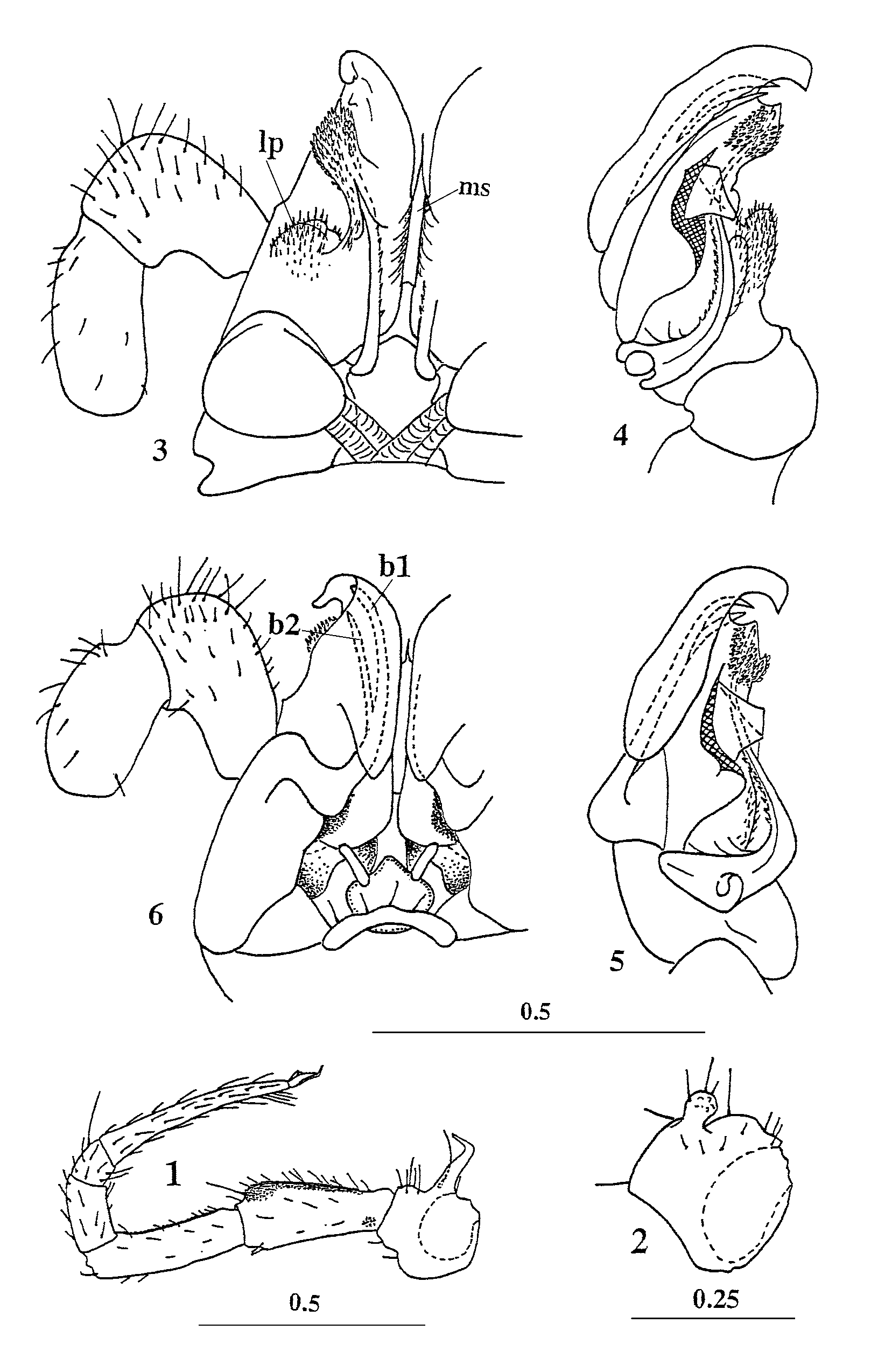
Legs long and slender. Each claw of legs 1–2 at base with two small additional claws dorsally and long setiform outgrowth ventrally. Leg pairs 3–7 not larger than other walking legs. Leg pairs 3–7 with a group of funnel–shaped tarsal papillae apically near claw and a shagreen sole part of the praefemur and femur; size of papillar tarsal field being equal for each leg. Each claw of leg pairs 3–7 with a long setiform outgrowth ventrally only i.e. without dorsal additional small claws. Postgonopodal legs (including leg pairs 10 and 11) without tarsal papillae but with a shagreen sole part of the praefemur and femur; each claw at base with a long setiform outgrowth ventrally and two small additional claws dorsally. Legs 10 and 11 with coxal glands. Coxa 10 with a long process curved forward ( Fig. 1 View FIGURES 1 – 6 ). Coxa 11 with small knob caudally ( Fig. 2 View FIGURES 1 – 6 ). One of male paratypes with deformed stout claws of leg pair 10.
Coxosternum of anterior gonopods with several setae in middle part. Flagelliform anterior gonopod telopodite 1–segmented, its distal part positioned inside elevated sheaths covered with tiny knobs and short thick bacilli, its frontal portion set with either short setae or cuticular spinules (that is very difficult to discern) ( Figs. 3–5 View FIGURES 1 – 6 ). Apices of the anterior gonopods telopodites unmodified. Mesal sheath processes of posterior gonopod colpocoxites fused medially into single low, long structure (ms) drawn out ventrally and being the place of colpocoxite fusion. Lateral sheath processes of colpocoxites (lp) low, knob–shaped, densely set with either setae or thin, setiform, cuticular spinules (that is very difficult to discern). Apex of colpocoxite curved posterad. Posterior gonopod angiocoxite with a globule and without process in posterior view, depressed centrally in anterior view. Single anterior angiocoxal process long, divided into two branches distally. Distal portion of anterior angiocoxal process placed inside front fold of colpocoxite ( Fig. 6 View FIGURES 1 – 6 ). Each branch of anterior angiocoxal process (b1, b2) somewhat protruding outside, apices pointed. Anterior angiocoxal mesodistal angle extended frontally in order to support long anterior angiocoxal process.
Female. Length 12–13 mm, width 1.0–1.1. Body with 29 segments. Ocelli 21–23. Vulvae not dissected.
Etymology: The specific epithet refers to the certain details of the posterior gonopods caudally and the anterior gonopod telopodites frontally covered with setae and spinules.
| ZMUM |
Zoological Museum, University of Amoy |
No known copyright restrictions apply. See Agosti, D., Egloff, W., 2009. Taxonomic information exchange and copyright: the Plazi approach. BMC Research Notes 2009, 2:53 for further explanation.
|
Kingdom |
|
|
Phylum |
|
|
Class |
|
|
Order |
|
|
Family |
|
|
Genus |