
Lichomolgus papuensis, Kim & Boxshall, 2021
|
publication ID |
https://doi.org/ 10.11646/zootaxa.5013.1.1 |
|
publication LSID |
lsid:zoobank.org:pub:BBB1CB11-1AEA-4678-8F6C-B43B7F35E453 |
|
persistent identifier |
https://treatment.plazi.org/id/8D4A87BF-FF9A-FFAA-FF19-FDC19DDCFBC4 |
|
treatment provided by |
Plazi |
|
scientific name |
Lichomolgus papuensis |
| status |
sp. nov. |
Lichomolgus papuensis sp. nov.
( Figs. 21 View FIGURE 21 , 22 View FIGURE 22 )
Type material. Holotype ♀ (MNHN-IU-2009-5188) and paratype ♀ (MNHN-IU-2014-21599). Dissected paratype ♀ (MNHN-IU-2014-21489) from Οhopalaea cẚrcula Monniot F. & Monniot C., 2001: Madang Resort, Madang, Papua New Guinea, PAPUA NIUGINI Expedition, Stn PR 72 (05°12.5'S, 145°48.5'E), MNHN coll., 21 November 2012. GoogleMaps
Etymology. The name of the new species is taken from its type locality, Papua New Guinea.
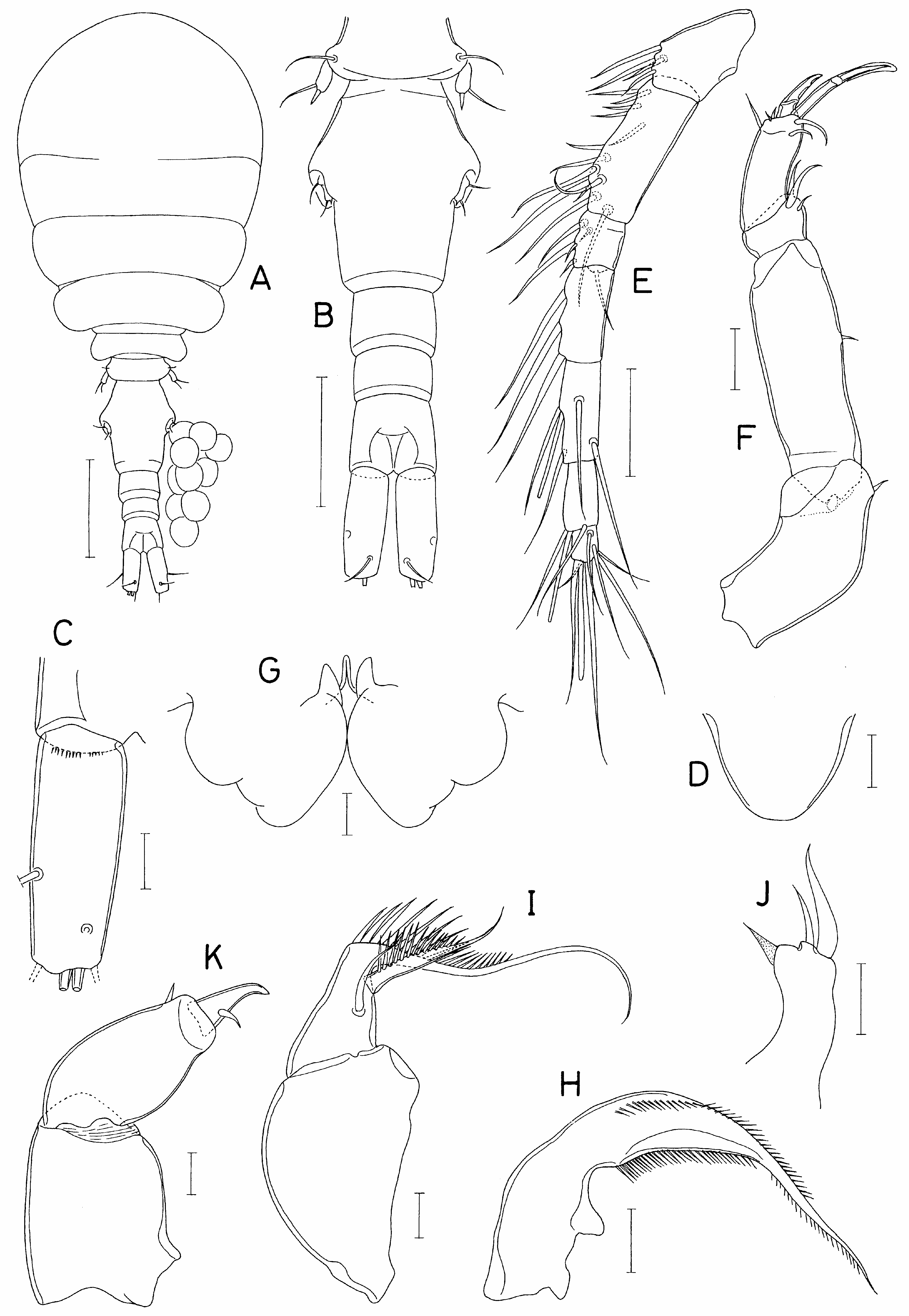
Description of female. Body ( Fig. 21A View FIGURE 21 ) consisting of broad prosome and moderately narrow urosome; body length 1.08 mm in dissected paratype; prosome 704×500 μm. Cephalothorax expanded, sub-globular, with faint suture line dorsally between cephalosome and first pedigerous somite. Urosome ( Fig. 21B View FIGURE 21 ) 5-segmented; fifth pedigerous somite 112 μm wide. Genital double-somite 163×135 μm, widest at about 45% of double-somite length; genital apertures located dorsolaterally just posterior to widest region of double-somite; posterior region gradually narrowing posteriorly. Three free abdominal somites 48×67, 35×63, and 56×67 μm, respectively; anal somite with minute spinules along posteroventral border. Caudal ramus ( Fig. 21C View FIGURE 21 ) broad, about 2.6 times longer than wide (87×34 μm), with 6 caudal setae (or insertion scars of setae); lateral seta located at about 63% of ramus length.
Rostrum ( Fig. 21D View FIGURE 21 ) broadly rounded, with sclerotized lateral margins and faint posterior margin. Antennule ( Fig. 21E View FIGURE 21 ) 270 μm long, 7-segmented; terminal segment shortest; armature formula 4, 13, 6, 3, 4+aesthetasc, 2+aes- thetasc, and 7+aesthetasc; all setae naked. Antenna ( Fig. 21F View FIGURE 21 ) 4-segmented; armature formula 1, 1, 3, and 5+2 claws; setae small, setule-like; terminal segment about 2.3 times longer than wide (35×15 μm): 2 terminal claws very unequal in length, inner claw as long as terminal segment and twice as long as outer claw, with membranous tip.
Labrum ( Fig. 21G View FIGURE 21 ) with swollen posterior lobes and deep median incision; each lobe strongly tapering, with 2 convex swellings on outer margin. Mandible ( Fig. 21H View FIGURE 21 ) slender, with elongate lash and 1 row of spinules along both margins of blade and lash. Maxillule ( Fig. 21J View FIGURE 21 ) as lobe, slightly broadening distally, armed with 3 setae (2 apical and 1 inner distal); inner distal seta transparent. Maxilla ( Fig. 21I View FIGURE 21 ) 2-segmented; proximal segment (syncoxa) unarmed; distal segment with elongate distal lash, armed with spiniform inner and slender anterior setae; inner spiniform seta ornamented with spinules along outer margin; distal lash whip-like, directed at right angle from basis, ornamented with row of spinules proximally on outer margin. Maxilliped ( Fig. 21K View FIGURE 21 ) 3-segmented; first segment unarmed; second segment with single small seta subdistally; small terminal segment tapering and claw-like, with 1 small seta proximally.
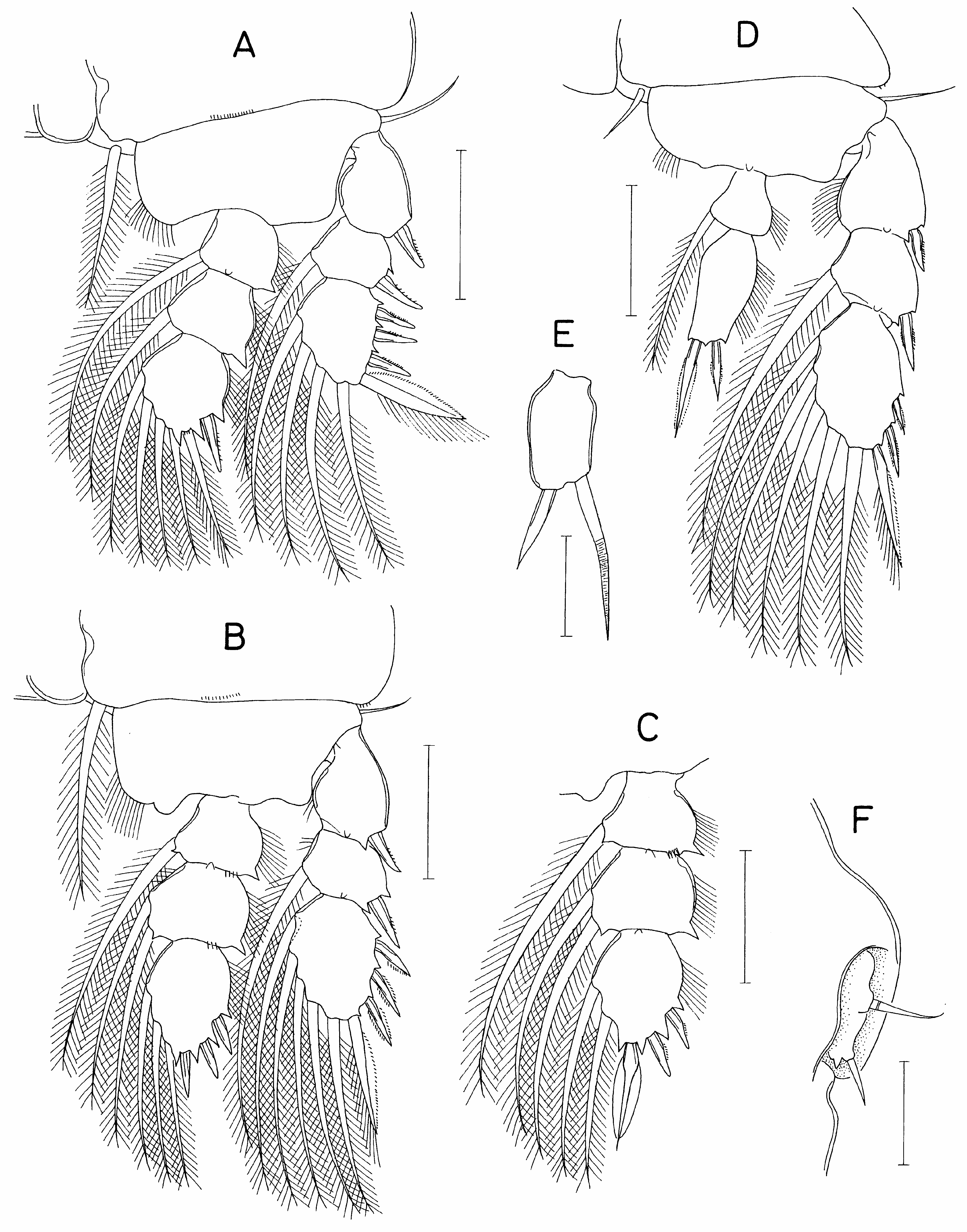
Legs 1-3 with 3-segmented rami ( Fig. 22 View FIGURE 22 A-C). Basis of legs 1-4 with naked outer seta. Basis of legs 2 and 3 with semicircular lobe on inner distal margin. Third endopodal segment of leg 3 armed with 3 spines and 2 setae ( Fig. 22C View FIGURE 22 ), otherwise leg 3 same as leg 2. Leg 4 ( Fig. 22D View FIGURE 22 ) with 3-segmented exopod and 2-segmented endopod; distal endopodal segment 2.05 times longer than wide (45×22 μm); 2 terminal spines 23 μm (outer) and 40 μm (in- ner). Outer spines on rami of legs 1-4 small, naked or weakly serrate. Armature formula for legs 1–4 as usual for the genus.
Coxa Basis Exopod Endopod
Leg 1: 0-1 1-0 I-0; I-1; III, I, 4 0-1; 0-1; I, 1, 4
Leg 2: 0-1 1-0 I-0; I-1; III, I, 5 0-1; 0-2; I, II, 3
Leg 3: 0-1 1-0 I-0; I-1; III, I, 5 0-1; 0-2; I, II, 2
Leg 4: 0-1 1-0 I-0; I-1; II, I, 5 0-1; 0, II, 0
Leg 5 consisting of dorsolateral seta on fifth pedigerous somite and free exopod; exopodal segment ( Fig. 22E View FIGURE 22 ) roughly rectangular, 1.9 times longer than wide (19×10 μm), armed distally with 1 spine (12 μm long) and 1 seta (25 μm long). Leg 6 ( Fig. 22F View FIGURE 22 ) represented by 2 setae and 1 denticle on genital operculum.
Male. Unknown.
Remarks. The terminal segment of the antenna of species of iẚchomolgus is armed with 1 to 4 claws distally, in addition to setae, and various combinations of claw shapes are expressed, according to species, as follows: a single strong claw, or 2 setiform claws, or 2 strong claws, or 1 strong + 2 setiform claws, or 2 strong + 1 setiform claws, or 1 strong + 3 setiform claws, or 2 strong and 2 setiform claws. Two strong terminal claws on the antenna, as found in the new species, are present in four congeners, i. dẚazonae Gotto, 1961, i. forfẚcula, i. ẚeversẚ Thompson & Scott, 1903, and iK ẚndẚcus Ummerkutty, 1962. The length/width ratios of the caudal rami vary in these species, and this is therefore useful to distinguish between them. The ratio is less than 1:1 (wider than long) in i. ẚndẚcus but more than 5: 1 in other three species. Therefore, these four species are easily distinguished from i. papuensẚs sp. nov. in which the ratio is 2.64:1.
iẚchomolgus papuensẚs sp. nov. can also be compared with congeners that share a similar length/width ratio of the caudal ramus. The ratio in i. papuensẚs sp. nov. is 2.64:1. Similar ratios, (i.e. in the range of 2:1 to 3:1), are found in three species: i. furcẚllatus in which the ratio is about 3:1 measured from the illustration of Sars (1917), i. hẚppopẚ in which the ratio is 2.44:1 ( Humes, 1976), and i. hoẚ Stock, 1995, in which the ratio is about 2.1:1 (60×29 μm, according to Stock, 1995). These three congeners can readily be distinguished from the new species. In i. furcẚllatus, which is associated with ascidians in the North East Atlantic and the Mediterranean Sea ( Humes & Stock, 1973), the third exopodal segment of leg 3 is armed with 3 spines and 5 setae (rather than 4 spines and 5 setae, as in the new species) and the distal endopodal segment of leg 4 is elongate ( Sars, 1917). In i. hẚppopẚ, which is associated with bivalve molluscs in the Moluccas, the antenna is 3-segmented and armed with a single large terminal claw, the maxillule is armed with 2 setae, the third exopodal segment of leg 4 is armed with 4 spines and 5 setae (rather than 3 spines and 5 setae, as in the new species), and the distal endopodal segment of leg 4 has a cusp on the outer margin ( Humes, 1976). Finally, in i. hoẚ, which is associated with bivalve molluscs in New Guinea, the genital apertures of the female are positioned posterior to the middle of the genital double-somite, the distal endopodal segment of leg 4 has a cusp on the outer margin, and the exopodal segment of leg 5 is inflated with a convex inner margin ( Stock, 1995).
| MNHN |
Museum National d'Histoire Naturelle |
No known copyright restrictions apply. See Agosti, D., Egloff, W., 2009. Taxonomic information exchange and copyright: the Plazi approach. BMC Research Notes 2009, 2:53 for further explanation.
|
Kingdom |
|
|
Phylum |
|
|
Class |
|
|
Order |
|
|
Family |
|
|
Genus |