
Lichomolgus canui Sars, 1917
|
publication ID |
https://doi.org/ 10.11646/zootaxa.5013.1.1 |
|
publication LSID |
lsid:zoobank.org:pub:BBB1CB11-1AEA-4678-8F6C-B43B7F35E453 |
|
persistent identifier |
https://treatment.plazi.org/id/8D4A87BF-FFA0-FF90-FF19-FCB79E58FDA0 |
|
treatment provided by |
Plazi |
|
scientific name |
Lichomolgus canui Sars, 1917 |
| status |
|
( Figs. 17 View FIGURE 17 , 18 View FIGURE 18 )
Material examined. 1 ♀ (MNHN-IU-2014-21487) in Molgula hellerẚ Drasche, 1884 (MNHN-IT-2008-5542 = MNHN S3 View Materials /MOL.A/163), Mar Grande , Tarento, Italy , 1978.
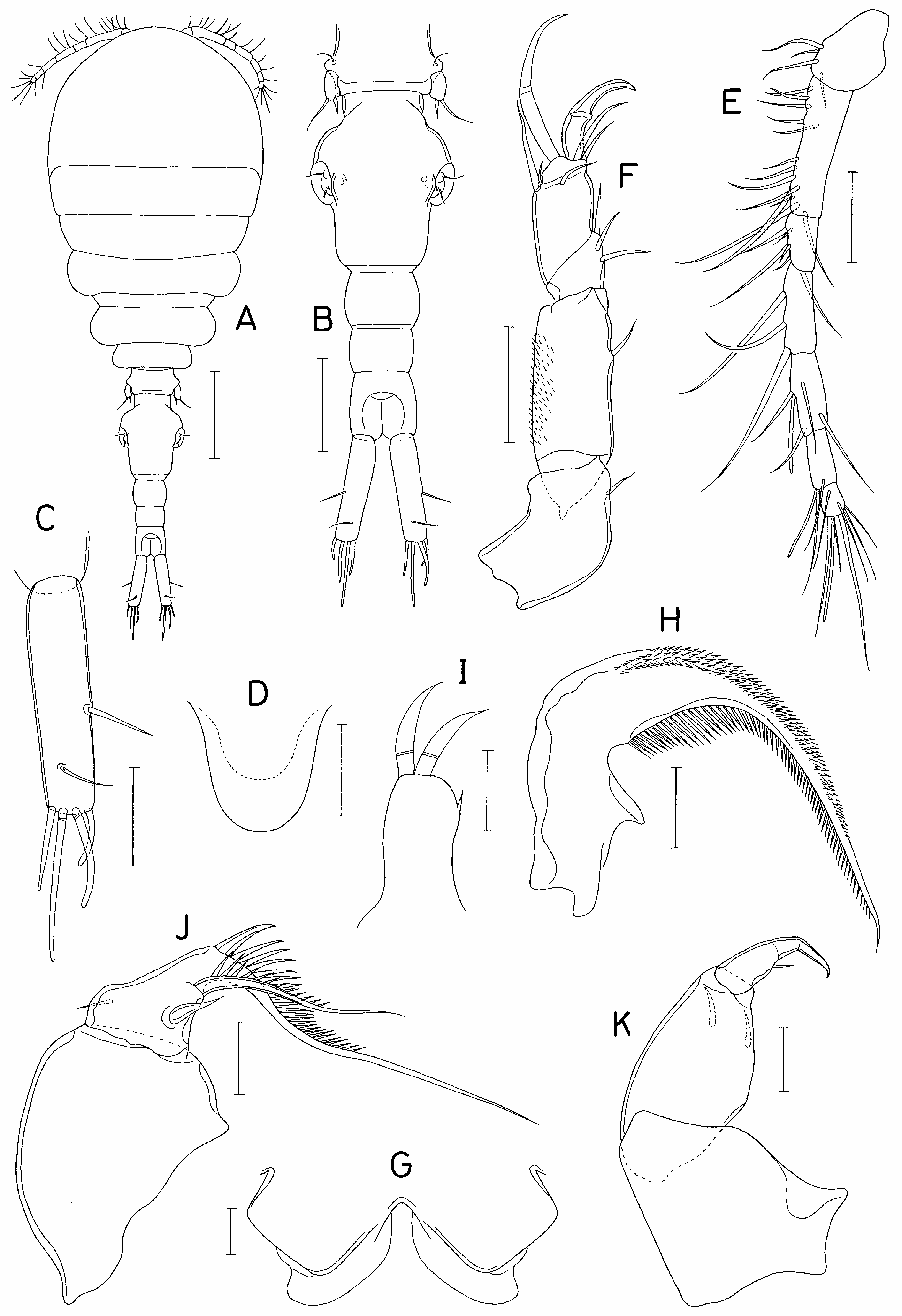
Redescription of female. Body ( Fig. 17A View FIGURE 17 ) with slightly inflated prosome and slender urosome; body length 1.35 mm; prosome 782×505 μm. Cephalothorax sub-globular, with faint dorsal suture line between cephalosome and first pedigerous somite. Urosome ( Fig. 17B View FIGURE 17 ) 5-segmented; fifth pedigerous somite 127 μm wide, distinctly nar- rower than genital double-somite. Genital double-somite longer than wide (193×155 μm); with expanded anterior half; genital apertures positioned dorsolaterally at mid-length of double-somite. Three free abdominal somites unornamented, 60×82, 47×70, and 68×73 μm, respectively. Caudal rami slightly divergent; each ramus ( Fig. 17C View FIGURE 17 ) about 3.0 times longer than wide (120×40 μm): armed with 6 setae; all caudal setae naked and shorter than ramus; 4 distal setae bluntly tipped; lateral seta (seta II) positioned at 57% of ramus length.
Rostrum ( Fig. 17D View FIGURE 17 ) well-developed, nearly spatulate. Antennule ( Fig. 17E View FIGURE 17 ) slender, 280 μm long, 7-segmented; armature formula 4, 13, 6, 3, 4+aesthetasc, 2+aesthetasc, and 7+aesthetasc; all setae naked. Antenna ( Fig. 17F View FIGURE 17 ) 4- segmented; armature formula 1, 1, 3, and 5+2 claws; first endopodal segment (second segment) ornamented with numerous fine spinules on outer surface; third endopodal segment about 1.6 times longer than wide (42×26 μm); 2 terminal claws unequal (inner broader and shorter than outer).
Labrum ( Fig. 17G View FIGURE 17 ) with subquadrate posterior lobes; each lobe with broad membranous fringe along inner margin and angularly projecting lateral margin. Mandible ( Fig. 17H View FIGURE 17 ) slender, with elongate, proximally curved distal lash, ornamented with 4 rows of short spinules along convex margin and 1 row of longer spinules along concave margin. Maxillule ( Fig. 17I View FIGURE 17 ) with 1 small setiform process subdistally and 2 broad, annulated setae distally. Maxilla ( Fig. 17J View FIGURE 17 ) consisting of syncoxa and basis; syncoxa unarmed; basis produced into extremely elongate terminal lash and bearing 3 setae; distal lash ornamented with row of spinules proximally along outer margin (proximalmost spinule thick, claw-like); inner seta (seta I) slightly longer than half length of distal lash, spinulose along outer margin. Maxilliped ( Fig. 17K View FIGURE 17 ) 3-segmented; first segment unarmed; second segment broadened near middle and armed with 2 equal setae; small third segment terminating in spiniform, abruptly bent process, with 1 small seta subdistally.
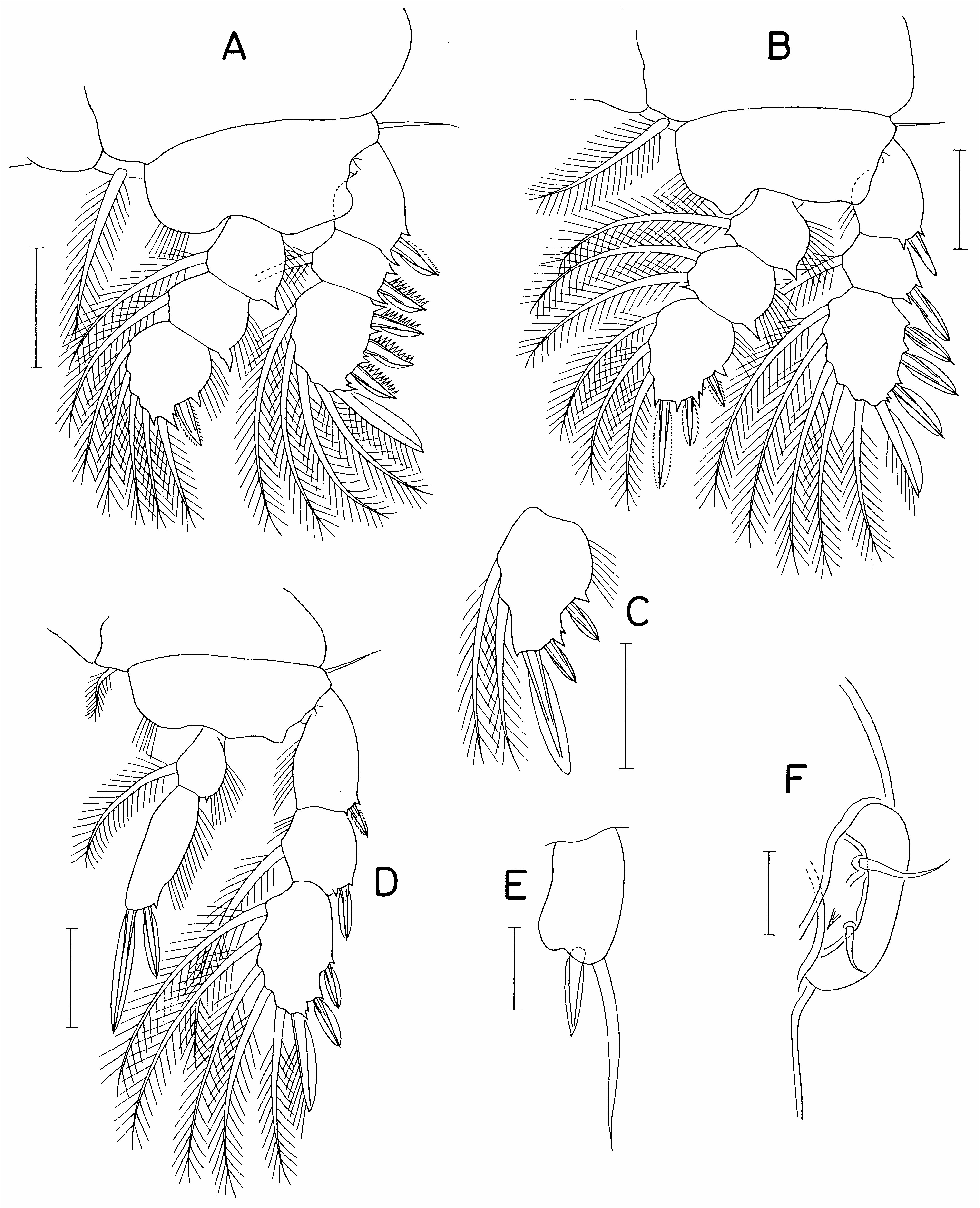
Legs 1–3 with 3-segmented rami ( Fig. 18 View FIGURE 18 A-C). Leg 4 ( Fig. 18D View FIGURE 18 ) with 3-segmented exopod and 2-segmented endopod. Outer spines on second and third exopodal segments of leg 1 ( Fig. 18A View FIGURE 18 ) with serrate proximal margin and fringed with membrane along distal margin. Leg 3 same as leg 2, except third endopodal segment armed with 3 spines and 2 setae ( Fig. 18C View FIGURE 18 ). Second endopodal segment of fourth leg about 2.8 times longer than wide (65×23 μm); lengths of terminal spines, 35 μm (outer) and 64 μm (inner). Armature formula for legs 1–4 as follows:
Coxa Basis Exopod Endopod
Leg 1: 0-1 1-0 I-0; I-1; III, I, 4 0-1; 0-1; I, 1, 4
Leg 2: 0-1 1-0 I-0; I-1; III, I, 5 0-1; 0-2; I, II, 3
Leg 3: 0-1 1-0 I-0; I-1; III, I, 5 0-1; 0-2; I, II, 2
Leg 4: 0-1 1-0 I-0; I-1; II, I, 5 0-1; 0, II, 0
Leg 5 consisting of dorsolateral seta on fifth pedigerous somite and free exopod: exopodal segment ( Fig. 18E View FIGURE 18 ) about 1.8 times longer than wide (32×18 μm), with slightly inflated inner distal margin; terminal spine 21 μm and terminal seta 46 μm. Leg 6 ( Fig. 18F View FIGURE 18 ) represented by 2 naked setae and 1 denticle on genital operculum.
Male. Not found.
Remarks. In the original description of iK canuẚ, Sars (1917) correctly illustrated the shape of the female maxilliped showing the terminal segment as abruptly bent. This form of female maxilliped seems to be a specific feature of i. canuẚ, which is also exhibited by our specimen. Other features of this species, such as the slightly divergent caudal rami which are about twice as long as the anal somite, and the terminal segment of the antenna which is about 1.6 times longer than wide and armed with 1 strong inner claw and 1 elongate outer claw, are also shared by Sars’ type specimens and our Mediterranean female. The ascidian Molgula heller ẚ from which our copepod was extracted is a new host record for i. canuẚ.
| MNHN |
Museum National d'Histoire Naturelle |
No known copyright restrictions apply. See Agosti, D., Egloff, W., 2009. Taxonomic information exchange and copyright: the Plazi approach. BMC Research Notes 2009, 2:53 for further explanation.
|
Kingdom |
|
|
Phylum |
|
|
Class |
|
|
Order |
|
|
Family |
|
|
Genus |