
Intramolgus atlantis, Kim & Boxshall, 2021
|
publication ID |
https://doi.org/ 10.11646/zootaxa.5013.1.1 |
|
publication LSID |
lsid:zoobank.org:pub:BBB1CB11-1AEA-4678-8F6C-B43B7F35E453 |
|
persistent identifier |
https://treatment.plazi.org/id/8D4A87BF-FFB3-FF82-FF19-FA709DC7FE4C |
|
treatment provided by |
Plazi |
|
scientific name |
Intramolgus atlantis |
| status |
sp. nov. |
Intramolgus atlantis sp. nov.
( Figs. 4–6 View FIGURE 4 View FIGURE 5 View FIGURE 6 )
Type material. Holotype ♀ (MNHN-IU-2014-21594, dissected and mounted on a slide) from Styela cha ẚnẚ Monniot C. & Monniot F., 1970 (MNHN-IT-2008-8188); North America Basin (39°46.5´N, 70°43.3´W), R. V. “Atlantis” II, Stn 73, depth 1330-1470 m; WHOI coll., 25 August 1964. GoogleMaps
Etymology. The name of new species alludes to its collection during the Atlantis II cruise.
Description of female. Body ( Fig. 4A, B View FIGURE 4 ) cyclopiform; body length 910 μm; prosome slightly expanded, 618 μm long, 309 μm wide, and 350 μm in dorsoventral depth, with ring of mucus-like material covering lateral margins of first and second pedigerous somites and dorsal borders between cephalosome and first pedigerous somite and between second and third pedigerous somites. Urosome ( Fig. 4C View FIGURE 4 ) much shorter than prosome, 5-segmented: fifth pedigerous somite 46×99 μm, with angular posterolateral corners. Genital double-somite 1.1 times longer than wide (121×109 μm); anterior half slightly broader, with convex lateral margins; genital apertures located dorsolaterally just anterior to middle of double-somite; ventral surface ( Fig. 4D View FIGURE 4 ) ornamented with numerous transverse rows of minute spinules. Three free abdominal somites 39×76, 25×72, and 38×68 μm, respectively; first free abdominal somite ornamented with 1 transverse row of minute spinules along posteroventral border; anal somite also with ventral rows of minute spinules basal to caudal rami ( Fig. 4E View FIGURE 4 ). Caudal rami slightly convergent; each ramus ( Fig. 4E View FIGURE 4 ) about 1.58 times longer than wide (38×24 μm), armed with 6 simple setae (setae II to VII) and row of spinules along posteroventral margin; seta I absent.
Rostrum ( Fig. 4F View FIGURE 4 ) tapering towards beak-like process at apex, with membranous fringe on mid-lateral margins. Antennule ( Fig. 4G View FIGURE 4 ) 7-segmented; first segment with elongate, claw-like process anterodistally and 2 denticles on margin near base of process; armature formula 3, 13, 3, 4, 4+aesthetasc, 2+aesthetasc, and 7+aesthetasc; all setae naked; aesthetasc on fifth segment constricted at proximal third and narrow in distal quarter. Antenna ( Fig. 4H View FIGURE 4 ) 4- segmented, consisting of large coxobasis with 1 seta distally and 3-segmented endopod: first endopodal segment unarmed; second endopodal segment armed with 1 claw and 2 setae (including vestigial proximal seta), and ornamented with 4 spinules on inner margin; third endopodal segment about 2.4 times longer than wide (36×15 μm), armed distally with 3 small claws and 4 small setae.
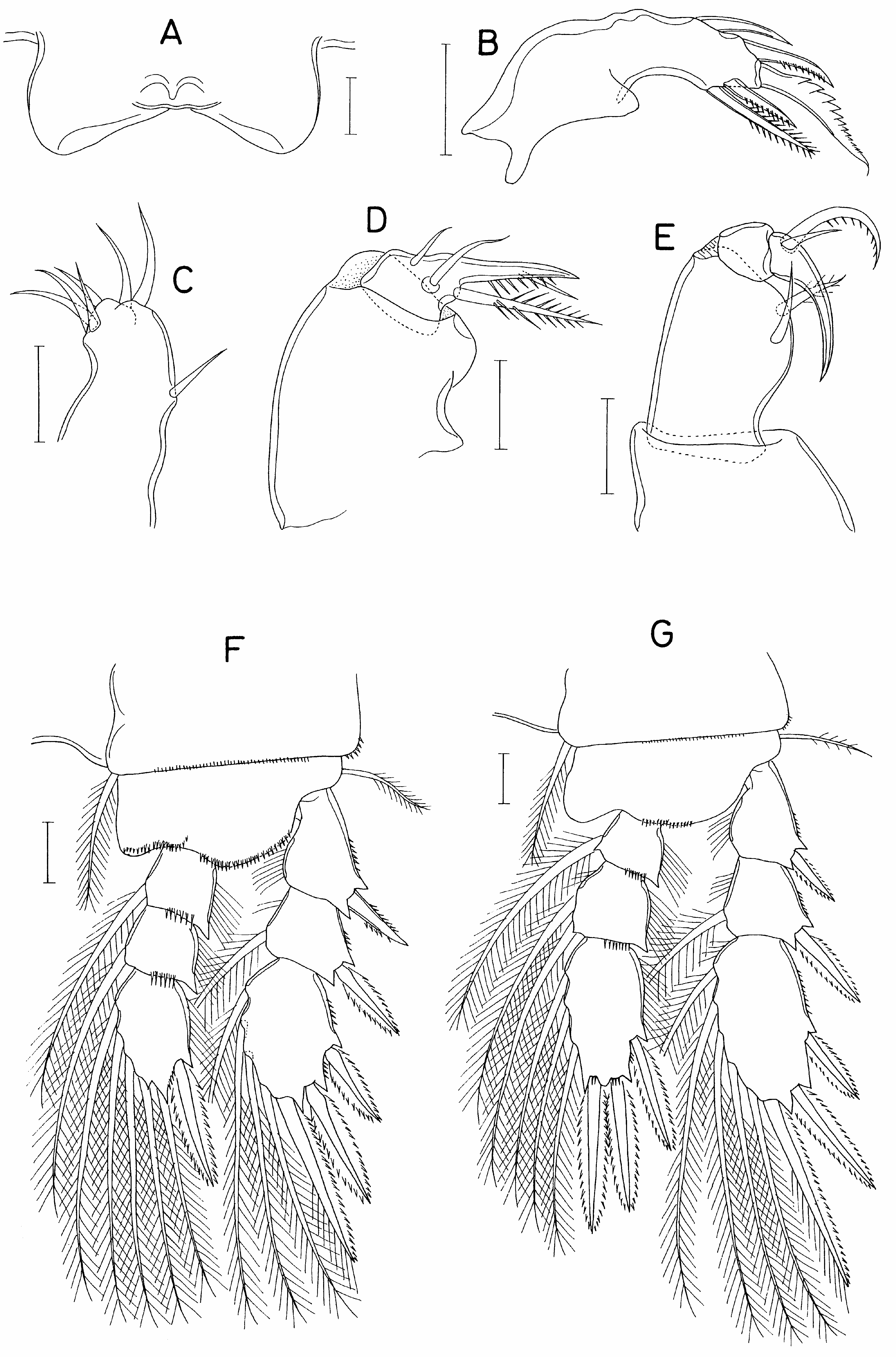
Labrum ( Fig. 5A View FIGURE 5 ) short, broad, medially incised. Mandible ( Fig. 5B View FIGURE 5 ) armed with 5 elements (4 spines and 1 lash-like, serrate terminal element); all elements articulated at base. Maxillule ( Fig. 5C View FIGURE 5 ) armed with 7 setae, weakly bilobed distally. Maxilla ( Fig. 5D View FIGURE 5 ) with large, unarmed syncoxa; basis terminating in spiniform process bearing several spinules, one of spinules much longer than others; armed with 3 setae; largest inner seta spinulose. Maxilliped ( Fig. 5E View FIGURE 5 ) 4-segmented, with fourth segment produced into terminal claw; first segment very broad and unarmed; second segment with 2 setae on expanded inner margin (distal seta weakly pinnate); small third segment unarmed; terminal segment armed proximally with 1 small seta and 1 large unilaterally spinulose seta.
Legs 1-4 ( Figs. 5F, G View FIGURE 5 , 6A, B View FIGURE 6 ) with 3-segmented rami. Armature formula as in preceding species, but all spines on both rami with serrate margins.
Leg 5 ( Fig. 4D View FIGURE 4 ) represented by 2 naked setae. Leg 6 ( Fig. 6C View FIGURE 6 ) represented by 2 setae on genital operculum.
Male. Unknown.
Remarks. The new species can be readily differentiated from its two congeners by the following features: (1) the prosome of the female bears a ring of unusual mucus-like material; (2) the caudal rami of the female are shorter than those of its congeners (1.58 times longer than wide, compared to more than twice as long as wide in both congeners); (3) all caudal setae are naked, compared to the pinnate ornamentation and flagellate tips of some caudal setae of the congeners; (4) the first segment of the antennule is produced anterodistally into an elongate claw-like process, compared to the short and stout process of congeners; (5) the first endopodal segment of the antenna is unarmed, in contrast to armed with 1 seta; and (6) the second endopodal segment of the antenna is armed with 1 claw and 2 setae, compared to 1 claw and 3 setae in both congeners. Collectively these differences support the establishment of a new species.


| R |
Departamento de Geologia, Universidad de Chile |
| V |
Royal British Columbia Museum - Herbarium |
| WHOI |
Woods Hole Oceanographic Institution |
No known copyright restrictions apply. See Agosti, D., Egloff, W., 2009. Taxonomic information exchange and copyright: the Plazi approach. BMC Research Notes 2009, 2:53 for further explanation.
|
Kingdom |
|
|
Phylum |
|
|
Class |
|
|
Order |
|
|
Family |
|
|
Genus |