
Metapontonia scorpio, Ďuriš, Zdeněk & Lin, Chia-Wei, 2016
|
publication ID |
https://doi.org/ 10.11646/zootaxa.4138.3.3 |
|
publication LSID |
lsid:zoobank.org:pub:63A9F8AA-3DB3-4127-A810-B646BBA384DE |
|
DOI |
https://doi.org/10.5281/zenodo.6063578 |
|
persistent identifier |
https://treatment.plazi.org/id/8D7E3756-FFDE-4D63-FF1E-41BCFBD1F8CC |
|
treatment provided by |
Plazi |
|
scientific name |
Metapontonia scorpio |
| status |
sp. nov. |
Metapontonia scorpio View in CoL n. sp.
( Figs 1–7 View FIGURE 1 View FIGURE 2 View FIGURE 3 View FIGURE 4 View FIGURE 5 View FIGURE 6 View FIGURE 7 )
Type material. Taiwan, Pingtung County, Hojie, 21º 57’ 20.4" N, 120º 42’ 41.4" E, from coral Diploastrea heliopora , coll. C.-W. Lin:—2 spms: 1 female ovig. holotype PoCL 2.3 mm (NMMBCD 4047), and 1 female ovig. paratype PoCL 2.2 mm ( RMNH. CRUS.D.57071), 6 Sep. 2014, depth 16 m, fcn 20140906.31 (u/w & lab. photo).—2 spms: 1 male allotype PoCL 1.5 mm (NMMBCD 4048) (lab. photo), and 1 female ovig. paratype PoCL 2.2 mm ( OUMNH.ZC.2016-01-001) [GenBank: KX169193 View Materials ], 29 Aug. 2014, depth 19 m, fcn 20140829.09 (u/w & lab. photo).— 1 female ovig. paratype PoCL 2.0 mm ( NTOUM 01892), 7 Sep. 2014, depth 16 m, fcn 20140907.05 (u/w & lab. photo).
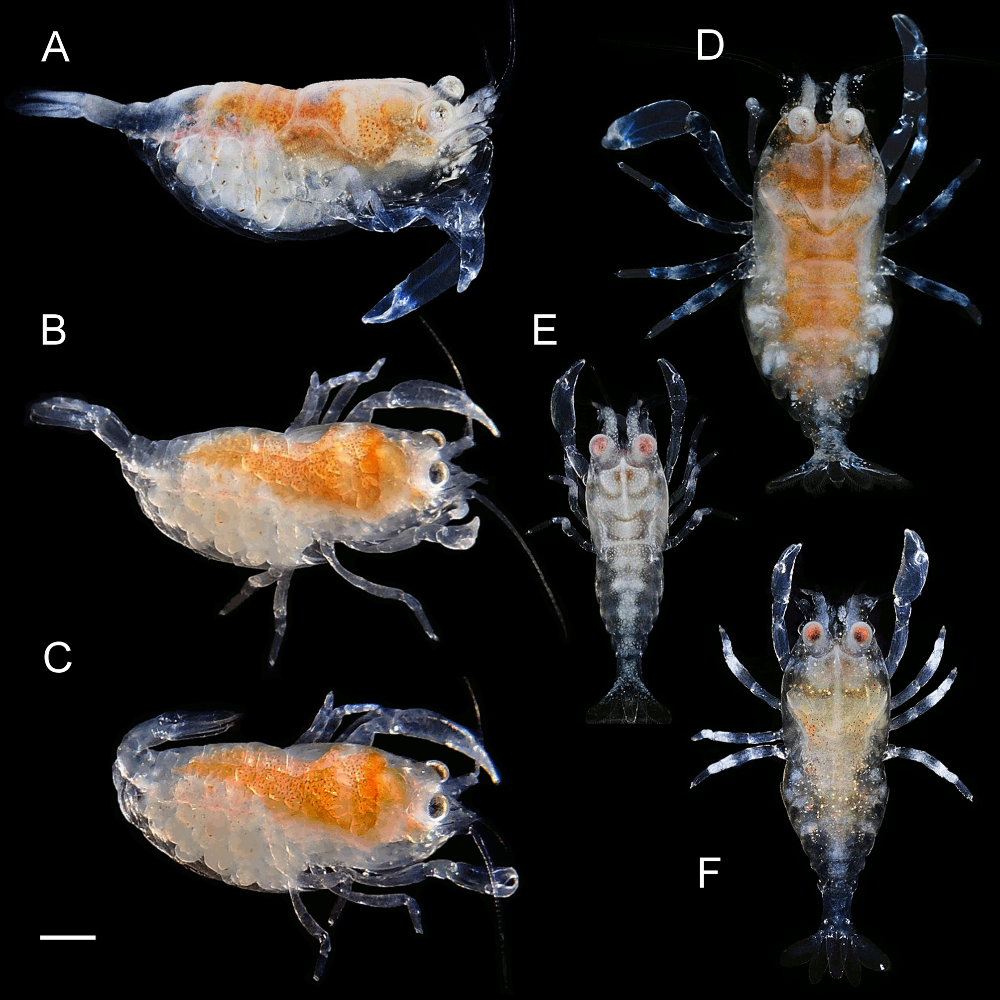
Etymology. From Latin scorpio (= scorpion) pointing on the unique among caridean shrimps ability of the new species to overturn the posterior pleonites in a manner resembling the typical body posture of scorpions ( Fig. 1 View FIGURE 1 C).
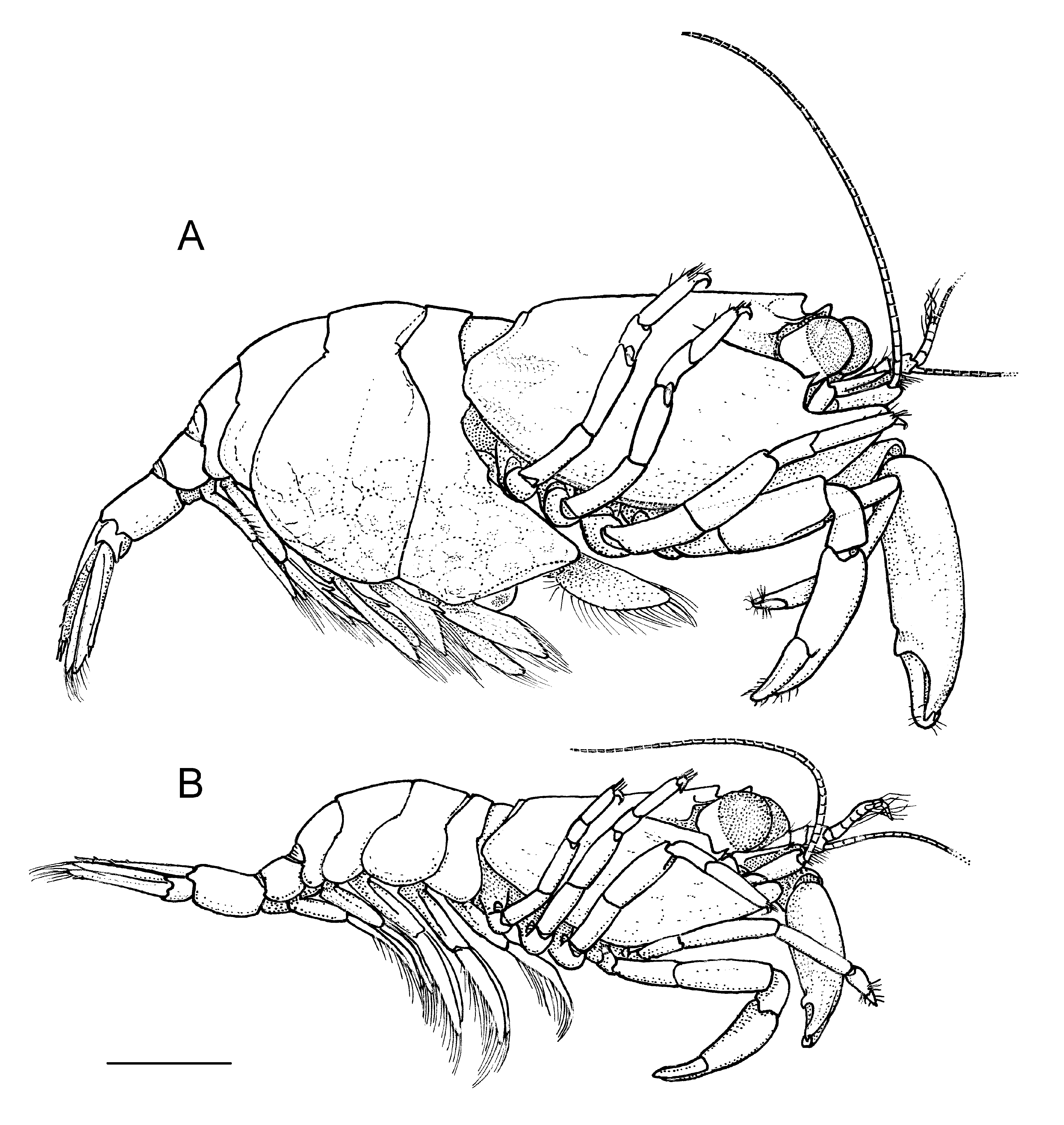
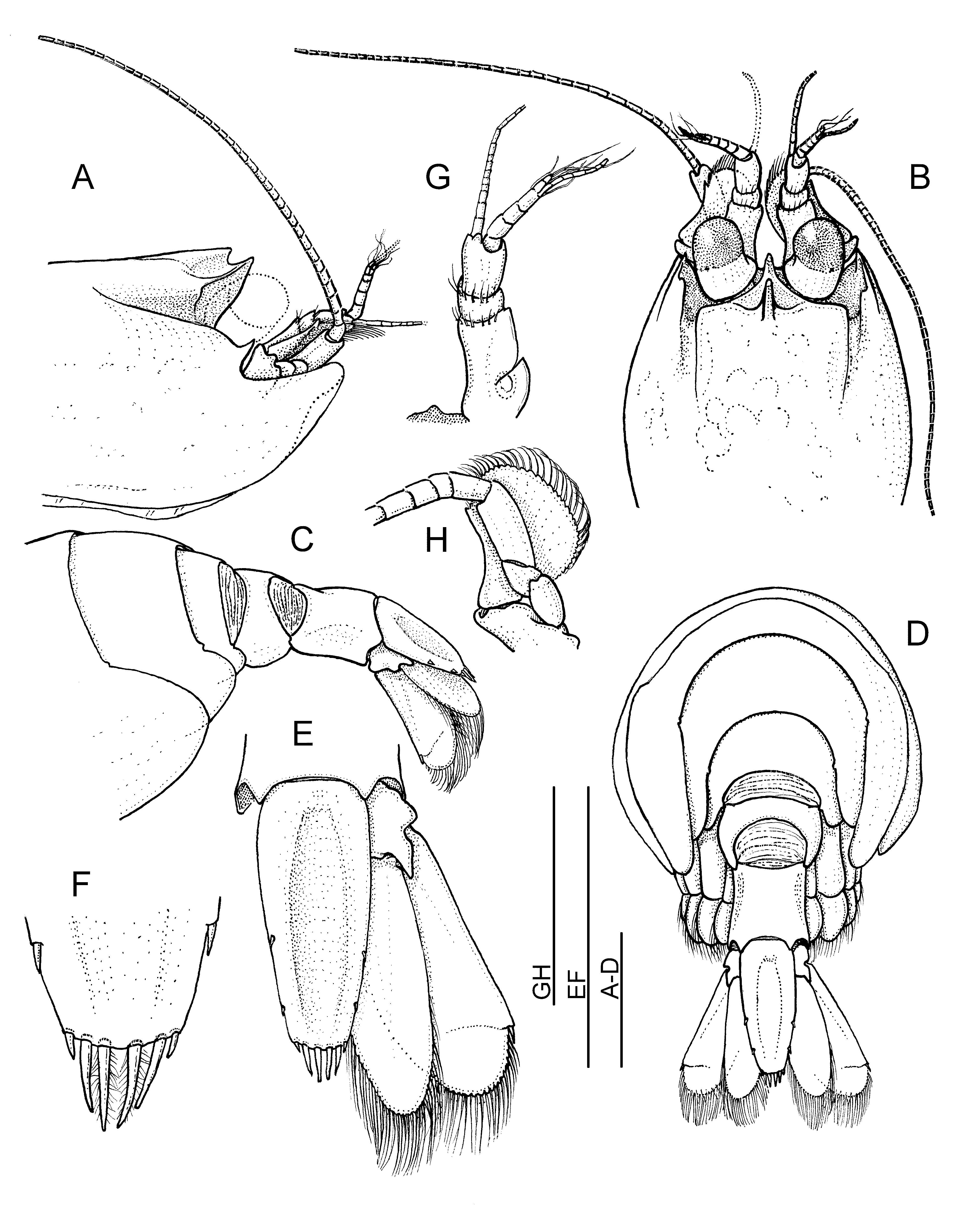
Description of female holotype ( Figs 1 View FIGURE 1 A–D, 2A, 3–5, 6A–E). Carapace ( Fig. 3 View FIGURE 3 A,B) smooth. Rostrum very short and strongly upward directed, compressed, not reaching half-length of basal segment of antennular peduncle; dorsal margin deeply concave anteriorly, with large subrectangular posterior tooth situated anterior to posterior orbital margin, and with widely oblique lateral margins overhanging orbits; pair of shallow depressions present anteriorly on sides of dorsal tooth; ventral margin of rostrum almost vertical, convex. Orbital margins sharply defined dorsally and ventrolaterally, but posteriorly interrupted; ventral margin continuing anteriorly to slightly upward directed antennal spine; shallow post-orbital furrow running slightly obliquely up; inferior orbital angle obsolete, situated medially to antennal spine. Supraorbital, epigastric and hepatic spines absent. Anterolateral angle of carapace produced anteromedially as elongate rounded lobe extending forwards beneath antennal peduncles to meet corresponding opposite lobe in midline enclosing thus all mouthparts. Ventral branchiostegite broadly rounded, with margin laminose, soft.
Thoracic sternum broad, unarmed, increasing in width posteriorly; fourth thoracic sternite with pair of anteriorly directed transverse ridges, widely separated in middle.
Pleon ( Fig. 3 View FIGURE 3 C,D) with all pleura rounded posteroventrally; first pleuron produced anteriorly as large triangular lobe. Posterior dorsal margins of fourth and fifth segments and anterior dorsal margins of fifth and sixth segments broadly concave, with wide soft membranous fields between corresponding segments’ tergites. Sixth segment slightly longer dorsally than fifth, about 0.6 of telson length.
Telson ( Fig. 3 View FIGURE 3 D–F) about 2.4 times longer than broad proximally, tapering posteriorly; dorsally flattened, with distinct broad median depression; lateral borders slightly convex, with 2 pairs of small dorsolateral spines situated at about 0.6 and 0.8 of telson length from anterior margin; posterior margin about 0.5 of anterior telson width, slightly convex, without median process, with 3 pairs of spines—outer pair short, similar to dorsolateral telson spines, intermediate pair about 2.5 times length of lateral spines, submedian spines setulose, long and slender, about 3 times length of lateral spines.
Eyes ( Figs 2 View FIGURE 2 , 3 View FIGURE 3 B), when directed forward, extending by length of cornea beyond tip of rostrum; eyestalks short and widest basally, cornea globular, subequal in length to eyestalk, with small accessory pigment spot incorporated dorsolaterally to posterior margin of cornea.
Basal segment of antennular peduncle ( Fig. 3 View FIGURE 3 G) reaching anterior margin of cornea of anteriorly extended yes; about 2 times longer than wide, medial margin distinctly convex, without ventral tooth; lateral margin convex, with broad and acutely pointed stylocerite attaining half-length of segment; anterolateral angle of segment obsolete, not noticeably produced. Intermediate segment short and unarmed; distal segment distinctly longer than intermediate segment and about half-length of basal segment. Antennular flagella short, lower flagellum with about eleven segments, subequal to peduncle in length; fused portion of upper flagellum consisting of 4 robust segments, shorter free ramus with 2 smaller segments, longer flagellum bearing about 9 slender segments.
Antenna ( Fig. 3 View FIGURE 3 H) with scaphocerite short and broad, not exceeding antennular peduncle; lamella 1.5 times as long as wide, with greatest width proximally to midlength, medial margin concave, lateral margin strongly arched, almost semicircular, distal margin extending beyond small distolateral tooth. Carpocerite robust and cylindrical, failing to reach distal end of scaphocerite. Antennal flagellum slender, exceeding post-orbital carapace length.
Epistome with obtuse median vertical keel anteriorly ( Fig. 3 View FIGURE 3 G).
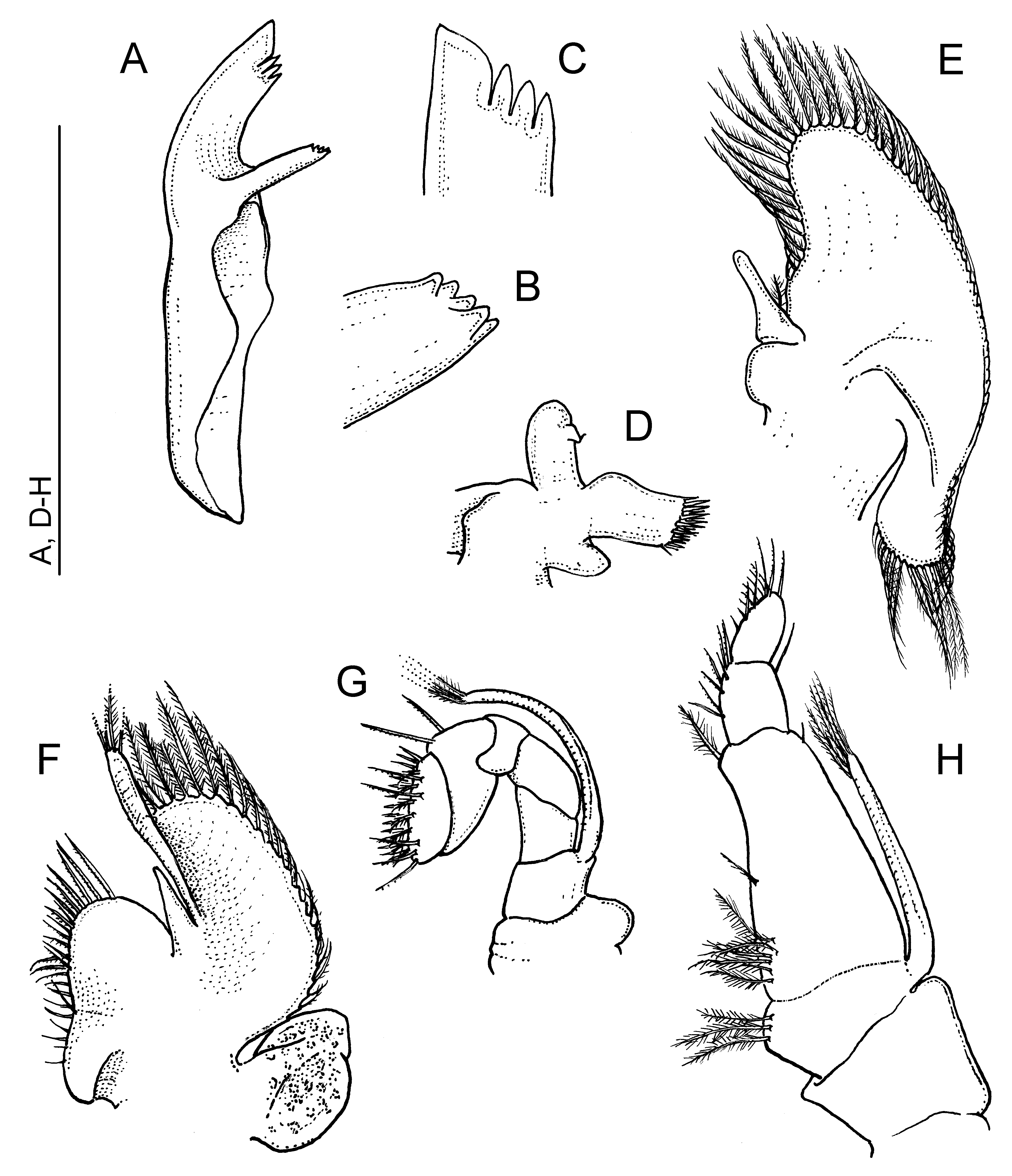
Mandible ( Fig. 4 View FIGURE 4 A–C) large, only slightly shorter than third maxilliped length; mandibular palp lacking; incisor process large and laminar, its distal border bearing one greatly enlarged truncate tooth and 3 smaller acute teeth; molar process reduced, slender, tapering to subacute process with group of low obtuse teeth terminally.
Maxillula ( Fig. 4 View FIGURE 4 D) small, with well-developed palp about twice longer than wide, with apex feebly bifid, medial lobe with small hooked seta; upper lacinia 1.6 times longer than broad, with row of stout spines distally; lower lacinia reduced to short rounded unsetose process.
Maxilla ( Fig. 4 View FIGURE 4 E) with endites reduced to single short rounded lobe; palp well developed, slender, with single basal seta laterally; scaphognathite also well developed, 2.2 times longer than wide in middle, broadly rounded distally and laterally, elongated posteriorly.
First maxilliped ( Fig. 4 View FIGURE 4 F) bearing short, elongate triangular, non-setose palp; endites of coxa and basis feebly separated, with serrulate setae medially and anteromedially; exopod reduced, short but functional, flagellum about 3 times longer than wide, compressed, with 4 terminal plumose setae, caridean lobe broadly rounded, 2.2 times longer than wide proximally, anteriorly reaching half-length of exopod; epipod robust, cordiform, feebly bilobed.
Second maxilliped ( Fig. 4 View FIGURE 4 G) normal in shape, very small, half size of third maxilliped; dactylar segment elongate ovoid in outer aspect; all segments well separated; exopod well developed, setose terminally; epipod lacking, coxa with rounded lateral lobe.
Third maxilliped ( Fig. 4 View FIGURE 4 H) short and broad; ischium slightly produced distomedially to small subtriangular tooth, lateral lobe shallow, with straight outer margin; basis short, convex and with 3 setulose setae medially; antepenultimate segment fused with merus and ischium, without subdivision, broad, about 1.7 times longer than basal breadth and 3 times longer than distal breadth; medial border bearing few slender setulose setae along proximal half and single similar distal seta; penultimate segment short, slightly longer than broad, about 0.3 of antepenultimate segment length, medial border bearing few serrulate setae; ultimate segment subequal to penultimate segment in length, stout, about twice as long as broad basally, medial border with few serrulate setae of which terminal ones longest. Exopod short, failing to reach end of antepenultimate segment of endopod, with 4 terminal and 2 subterminal plumose setae, lateral plate obsolete; no epipod nor arthrobranch present.
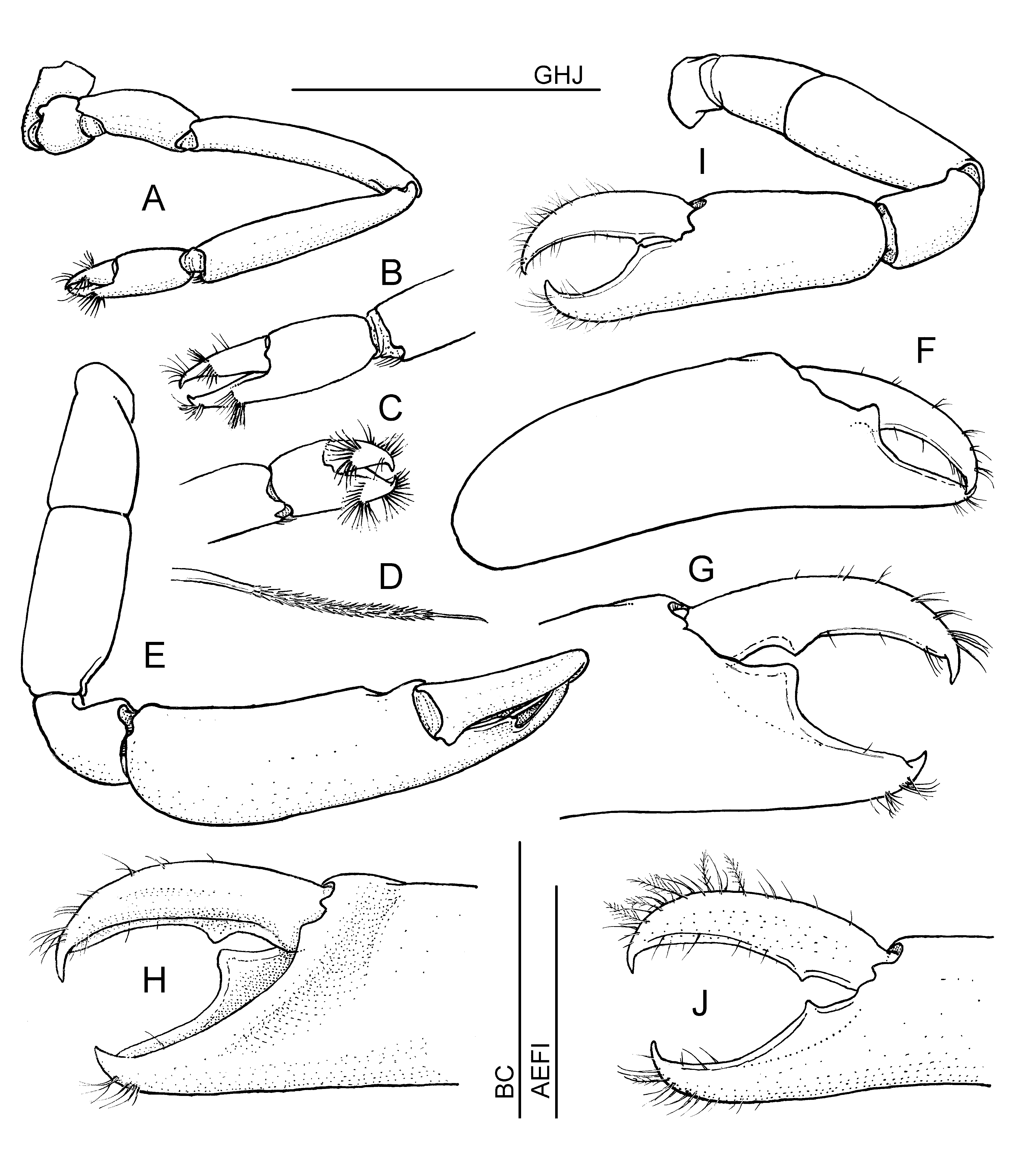
First pereiopods ( Fig. 5 View FIGURE 5 A–C) long and slender, with merus extending anteriorly almost to distal end of antennular peduncle. Fingers subequal to palm length, without distinct cutting edges, tips hooked; each finger with pair of transverse fan-shaped series of densely pappose setae with glabrous basal and terminal thirds ( Fig. 5 View FIGURE 5 D). Carpus almost twice as long as chela and about 5 times its maximum width distally, subequal to merus length. Ischium half length of carpus or merus. Basis and coxa short, stout, without special features.
Second pereiopods robust and similar in shape, but distinctly unequal ( Fig. 2 View FIGURE 2 A, 5E–J). Both chelipeds exceeding antennular peduncles by entire propodi, chelae held subparallel and bent down in front of body and facing by morphological ‘lateral side’ anteriad, with ‘ventral’ margins laterad; chelae distally incurved ( Fig. 2 View FIGURE 2 A), somewhat subspatulate. Major cheliped ( Fig. 5 View FIGURE 5 E–H) with fingers about 0.6 of palm length, subcylindrical, with opposing cutting edges laterally; dactylus slender, about 5 times longer than broad proximally, smoothly curved and tapering to slender apex; cutting edge lateral, shallowly laminar, with single triangular tooth on proximal third. Fixed finger cutting edge lateral, highly laminar, with large triangular compressed tooth proximally opposing smaller dactylar tooth; tips of fingers hooked and distal halves of cutting edges concave, widely gaping when fingers closed. Palm subcylindrical, more compressed distally, with shallow depression distomedially, proximally off dactylus and running obliquely anteroventrad. Carpus one third of palm length, twice as wide distally as proximally, unarmed. Merus 0.6 of palm length and twice as long as high, unarmed. Ischium 0.7 of merus length.
Minor cheliped ( Fig. 5 View FIGURE 5 I,J) with chela about 0.75 of major chela length. Fingers about 0.4 of palm length, similar to those of major chela, but with both cutting edges narrowly laminar and with single small tooth on proximal third. Carpus about one third of chela length and 0.6 of palm length, merus subequal to palm length, stout, ischium of similar width, about 0.7 of merus length.
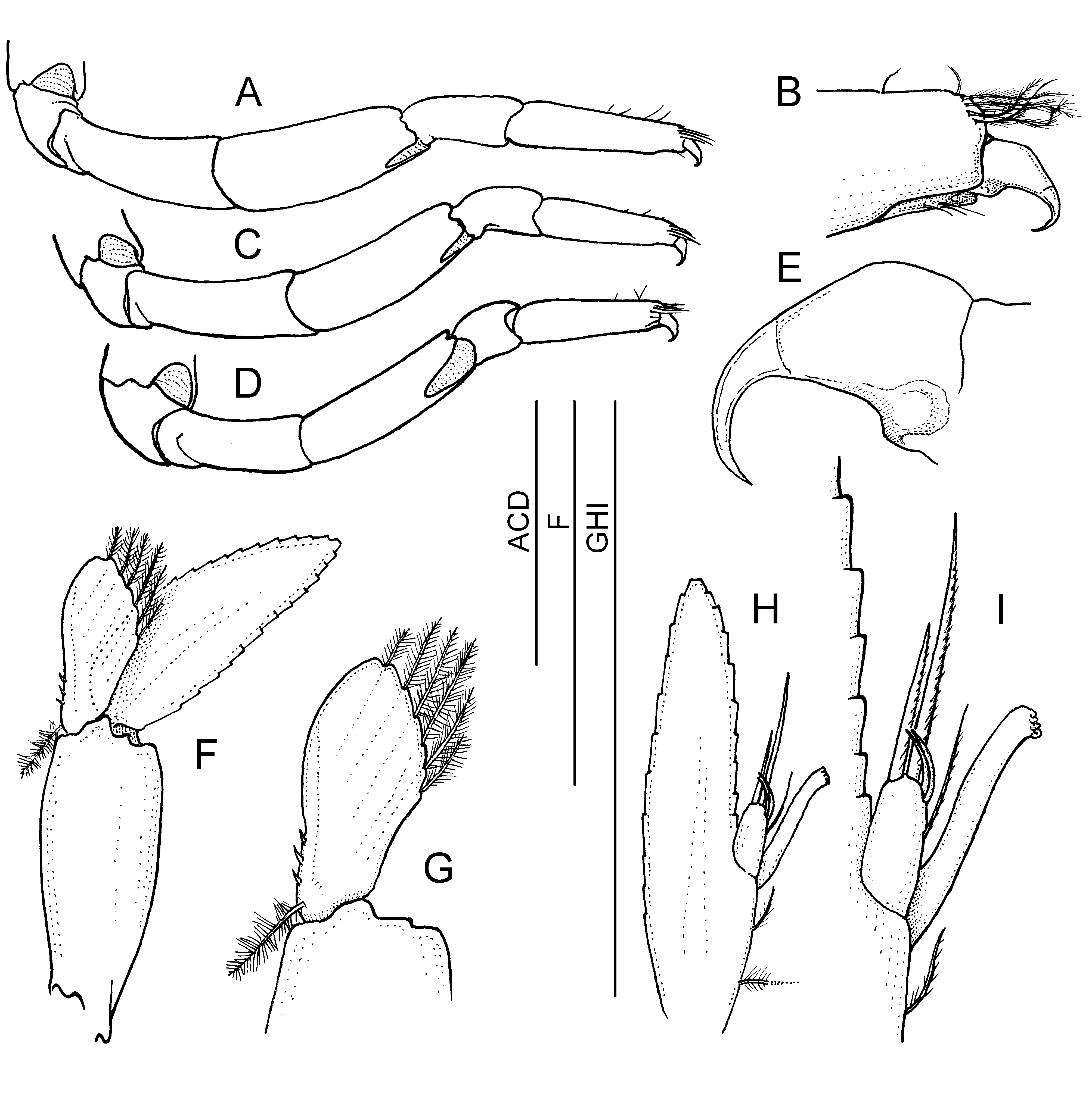
Third to fifth pereiopods ( Fig. 6 View FIGURE 6 A–E) similar and subequal, usually upturned over carapacial dorsum, with distal ends overreaching dorsum of cephalothorax, robust and similar, third being longest. All segments unarmed, lacking spines, but group of plumose setae present at distal end of propodi on each side dorsally over dactylar base. Third pereiopod ( Fig. 6 View FIGURE 6 A–B) exceeding antennular peduncle by dactylus when extended anteriorly. Dactylus small, with simple and strongly hooked unguis and with small basal protuberance retracting into propodus when dactylus flexed. Propodus 3.5 times longer than wide proximally, carpus slightly wider but shorter, 0.7 of propodus length; merus stout, slightly longer than propodus but almost twice as wide; ischium subequal in length to merus but slightly more slender.
Pleopods with depressed protopods and enlarged wide setose branches.
Uropods ( Fig. 3 View FIGURE 3 D–E) exceeding telson by almost distal third of their length. Exopod with lateral margin straight and bearing single small distolateral movable spinule but lacking acute fixed tooth. Diaeresis feebly demarcated.
Male allotype ( Figs 1 View FIGURE 1 E, 2B, 6F–I). Body size smaller than that of females examined; body shape generally similar, only pleonal pleurae short, rounded, with pleopodal protopods exposed. Endopod ( Fig. 6 View FIGURE 6 F,G) of first male pleopod about 0.6 of exopod length and 2.2 times longer than broad, with greatest width situated at half-length; medial border feebly concave with single setulose seta proximally and 3 short and stout simple spines along basal third of its length; distal margin rounded; lateral border slightly convex, oblique, with 6 plumose setae along distal half of margin. Endopod of second pleopod ( Fig. 6 View FIGURE 6 H,I) with slender appendix interna bearing several terminal cincinnuli; appendix masculina reaching 0.6 of appendix interna, short and stout, about twice as long as broad, with 5 distal setae—i.e. 2 long terminal serrulate setae 1.2 and 1.9 times longer than appendix masculina, 2 simple curved distomedial setae about 0.6 of appendix length medially, and single serrulate seta, subequal to appendix, more proximally.
Other specimens. Three remaining specimens, ovigerous females paratypes, are of almost the same size and morphologically closely similar to the holotype, showing no noticeable distinctions, except the NTOUM female paratype ( Fig.1 View FIGURE 1 F) which has the second pereiopods chelae smaller and less divergent in shape.
Measurements (in mm). Female holotype: PoCL, 2.3; TL, 6.8; dorsal length of 5th pleonal segment, 0.4 (sclerotized part, 0.2); length of 6th pleonal segment, 0.65; telson length, 1.0; telson width, 0.45; major chela length, 2.3; minor chela length, 1.65; propodus of third pereiopod length, 0.7. — Male allotype: PoCL, 1.5; TL, 5.5; dorsal length of 5th pleonal segment, 0.45 (sclerotized part, 0.2); length of 6th pleonal segment, 0.5; telson length, 0.85; telson width, 0.4; major chela length, 1.7; minor chela length, 1.35; propodus of third pereiopod length, 0.6. The female carried approx. 50 eggs of the size 0.60 x 0.43 mm (with early eyespots).
Remarks. The present new species is distinctly smaller than M. fungiacola , with the body and appendages stouter. It differs from the original description of M. fungiacola (cf., Bruce, 1967), and the present comparative material (below), mainly by the following characters: (1) the rostrum is short and stout, distinctly dorsad directed with almost vertical ventral margin and wide lateral carinae (vs. slender, directed anteriad or upturned distally, ventral margin almost horizontal, lateral carinae feeble), the rostrum reaches subequal height as the anterior median tooth of the carapace (vs. rostrum is lower-positioned, anteriad, with the anterior median tooth of the carapace distinctly higher); (2) the orbital margin widely interrupted posteriorly forming shallow but distinct longitudinal groove on the side of the carapace (vs. orbital margin well developed in holotype, or with only a slight depression there in some present Taiwanese specimens); (3) the basal segment of the antennular peduncle distinctly concave medially and without anterolateral tooth (vs. medial margins subparallel, anterolateral tooth small but distinct); and (4) the opposing dorsal margins of the 4th/5th and 5th/6th pleonal articulations deeply concave, with broad membranous fields between segments (vs. margins moderately convex).
In addition to those, the antennal scaphocerite in the new species is broad and short, with the length only slightly greater than the width, the lateral margin is distinctly concave, and the lamina is broadly semicircular medially (vs. scaphocerite distinctly longer than broad, with lateral margin almost straight, lamina moderately convex medially). The exopod of the third maxilliped is reported as reduced, terminally unsetose in M. fungiacola (cf. Bruce, 1967), while in the new species it is well developed, with terminal plumose setae, just falling short of the distal margin of the antepenultimate segment. The fourth thoracic sternite is wide and flattened, with distinct entire anterior ridge in M. fungiacola , while that of the new species is less prominent and developed only on sides and widely interrupted in the middle.
The molecular comparison of obtained sequences of the 16S rRNA gene (488 bp) for specimens of both species revealed 3.9 % distinction between species that also confirms a separate position of the new species.
Colour ( Fig. 1 View FIGURE 1 ). Body semitranslucent, adult females with orange ovaries with scattered dark dots extending from inside of carapace posteriorly to the fourth pleonite ( Fig. 1 View FIGURE 1 A–D), some specimens with diffused white and red dots ( Fig. 1 View FIGURE 1 A–C), others with symmetrical fields of densely set white spots dorsally on carapace, diffused white paths on antennules, dorsally on pleon and uropods; most remarkable being brightly white transverse line anteriorly across gastric region on carapace, with white median tooth ( Fig. 1 View FIGURE 1 E,F); eye cornea dark, reddish, or white; diffused whitish fields on walking legs.
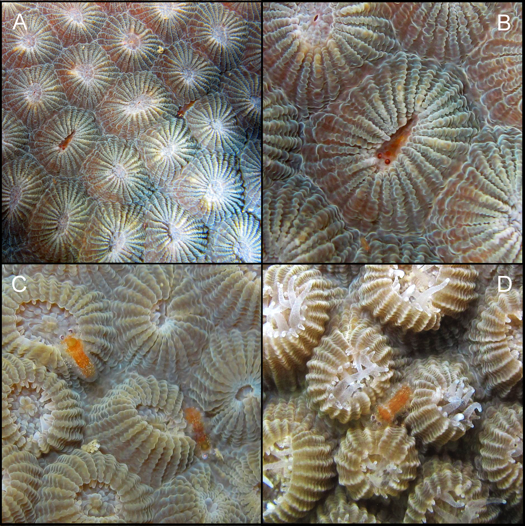
Behaviour. The new species is remarkable by its ability to overturn the posterior pleonites in a manner resembling typical body posture of scorpions ( Fig. 1 View FIGURE 1 C). Such manner of overturning the pleon by high dorsal bending of just the distal segments is unique among caridean shrimps. Some caridean shrimps like Phycocaris simulans Kemp, 1916 , or Neostylodactylus litoralis Okuno & Tachikawa, 2000 , held their whole pleon upturned highly dorsad almost touching the carapacial dorsum but posteriorly to the third segment the pleon is normally bent back down ( Kemp, 1916; Okuno & Tachikawa, 2000). The above mentioned behaviour of M. scorpio n. sp. most likely reflects the frequent position of the shrimp in grooves between closely set polyps of their coral host Diploastrea heliopora ( Fig. 7 View FIGURE 7 C,D), or directly in the ‘oral cavity’of its host ( Fig. 7 View FIGURE 7 A,B). In M. fungiacola , the pleon was also noted „to be frequently held in an elevated position, away from the substrate and with the caudal fan flexed” ( Bruce, 1972: 4). The morphology of the posterior pleonal segments of M. fungiacola allows the elevation of the pleon only in a lesser extent in comparison with M. scorpio n. sp.
Host. Coral Diploastrea heliopora (Lamarck) ( Scleractinia : Diploastreidae ) ( Fig. 7 View FIGURE 7 ).
Distribution. Southwestern Taiwan (type locality, present report).
GenBank accession number. KX169193 View Materials (see Material paragraph, above).
| RMNH |
National Museum of Natural History, Naturalis |
No known copyright restrictions apply. See Agosti, D., Egloff, W., 2009. Taxonomic information exchange and copyright: the Plazi approach. BMC Research Notes 2009, 2:53 for further explanation.