
Goniopsarites, Meng, Rui, Wang, Menglin & Wang, Yinglun, 2014
|
publication ID |
https://doi.org/ 10.11646/zootaxa.3866.1.4 |
|
publication LSID |
lsid:zoobank.org:pub:6202FA35-AF83-4F98-82EE-F089EA7ACC35 |
|
DOI |
https://doi.org/10.5281/zenodo.6143285 |
|
persistent identifier |
https://treatment.plazi.org/id/04DA0EC6-21BA-4B55-92D3-8669E981EBE8 |
|
taxon LSID |
lsid:zoobank.org:act:04DA0EC6-21BA-4B55-92D3-8669E981EBE8 |
|
treatment provided by |
Plazi |
|
scientific name |
Goniopsarites |
| status |
gen. nov. |
Goniopsarites View in CoL gen. nov.
Type species. Goniopsarites fronticonvexus sp. nov., here designated.
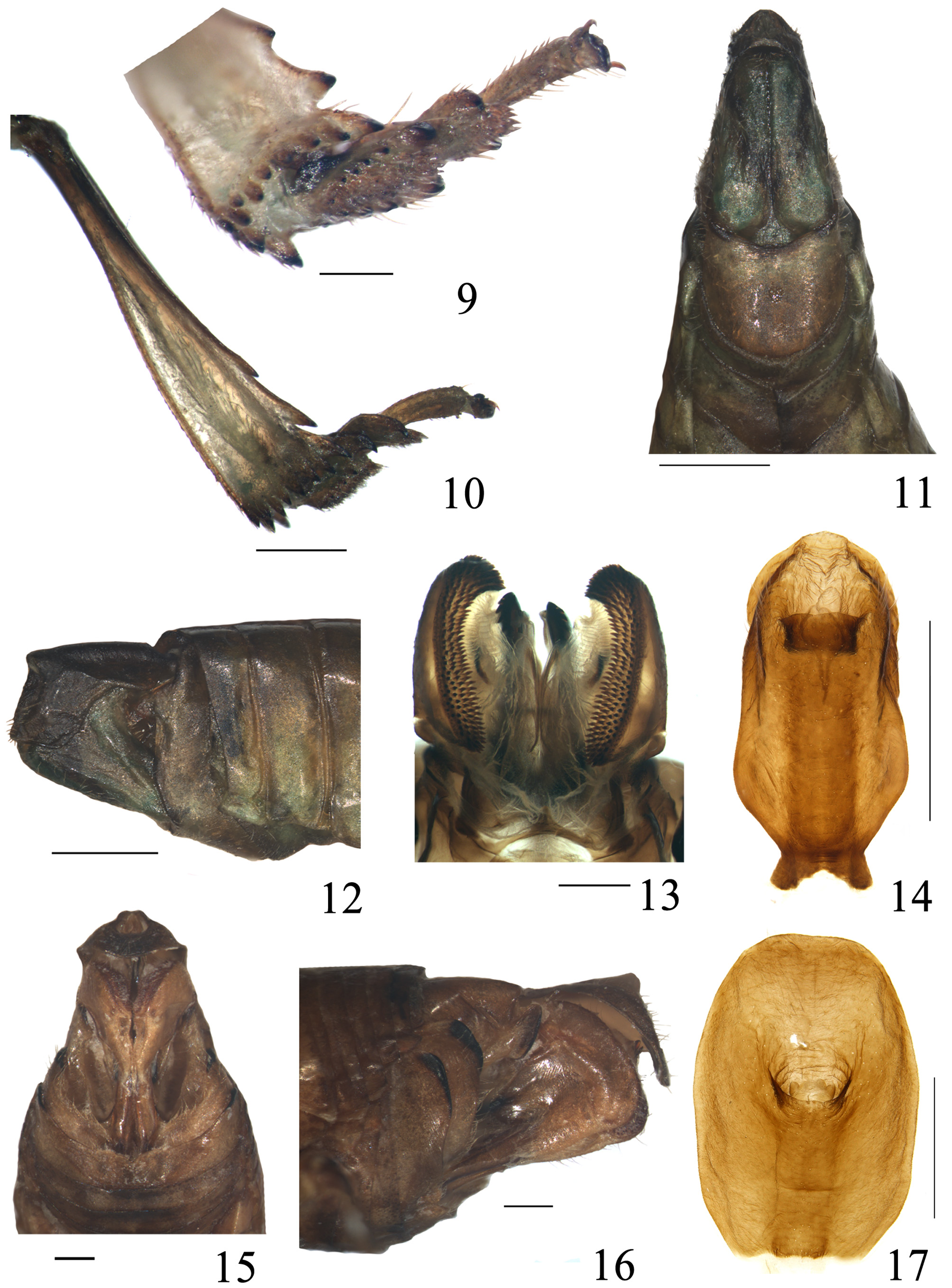
Description. Head with eyes slightly narrower than pronotum ( Figs 1, 4 View FIGURES 1 – 8 ). Vertex quite broad, 7.0 times wider than long in middle line, with disc depressed, lateral margins strongly keeled, anterior margin weakly concave and sinuate at middle, and posterior margin shallowly concave; median carina feeble, sublateral keels elevated ( Figs 1, 4 View FIGURES 1 – 8 ). Frons long, 1.7 times longer than wide in mid line, 1.5 times wider at widest upper margin than at base; upper margin deeply concave, lateral margin distinctly acutely elevated, slightly concave in middle; median carina distinct, disappeared near the lower third of frons, with Y-shaped keel and a pair of short keels at lower part; disc of frons depressed, abruptly forward protruding near frontoclypeal suture in lateral view ( Figs 3, 5, 6 View FIGURES 1 – 8 ). Clypeus narrow, triangular, with median carina, lateral margins extremely carinate, median area particularly convex in lateral view ( Figs 3, 5, 6 View FIGURES 1 – 8 ). Frontoclypeal suture acute ( Fig. 3 View FIGURES 1 – 8 ). Rostrum elongate surpassing post trochanters, apical segment short, subapical segment three times as long as apical segment ( Fig. 3 View FIGURES 1 – 8 ). Ocelli present. Eyes oval. Antennae with scape columned and short, pedicel subglobose. Pronotum about 3.0 times longer than vertex in middle line, with median carina ( Fig. 4 View FIGURES 1 – 8 ); anterior margin obtusely convex between eyes, posterior margin slightly concave, both distinctly carinate; lateral lobe distinctly broadened ( Fig. 5 View FIGURES 1 – 8 ). Mesonotum large, 2.5 times as long as vertex and pronotum in middle line, 1.5 times wider than long; disc slightly flat, with median carina and parallel lateral carinae, which are joined with arcuately transverse keel near anterior margin ( Fig. 4 View FIGURES 1 – 8 ).
Tegmina steeply tectiform ( Fig. 2 View FIGURES 1 – 8 ), much elongate and broad, costal margin strongly and convexly arched, but concave near apex, apical margin rounded obliquely, posterior margin straight, widest at basal half, distinctly narrow on apical third, about twice longer than wide at widest part; precostal area much narrower than costal cell (about 1:4) with no transverse veinlets. Sc+R and M veins emiting from the top of basal cell, Cu vein arising from lower part of basal cell, Sc+R forking very closed to basal cell, M forking slightly at one third from basal part, Cu forked a bit after middle of tegmen. Claval suture extraordinary distinct, clavus much elongated with apex reaching apical margin of tegmen and terminating with a distinct spine, two claval veins (A1 and A2) united at middle from base of clavus, with sparse transverse veinlets. Both longitudinal veins and transverse veinlets markedly prominent, transverse veinlets obviously dense and reticulated at apical part ( Fig. 7 View FIGURES 1 – 8 ). Wings well-developed and distinctly divided into three areas: preanal area, vanal region and anal area by two weak incisions of apical margin; costal margin weakly concave with small coupling lobe ( Fig. 8 View FIGURES 1 – 8 , indicated by arrow), apical margin with minute setae. The R vein bifurcated at one-quarter near apex; M vein basally fused with cubitus vein and simple; Cu vein tetrafurcate at middle. Both the CuA and Pcu postcubitus vein simple, almost closed to each other at apex, but not fused. First anal vein (A1) vein quadrifurcated, but second anal vein (A2) simple, fused with the fourth embranchment of A1 after basal part to terminal. The longitudinal vein R, M, and Cu separately branched apically. About 6–7 transverse veinlets on apical region. Veins as in fig. 8.
Fore femora and tibiae moderately broad, hind tibia widened at apical half and with two lateral spines ( Fig. 10 View FIGURES 9 – 17 ). Spinal formula of hind leg 12–11–2, indicating number of spines at apex of hind tibia and hind tarsomeres I and II ( Fig. 9 View FIGURES 9 – 17 ).
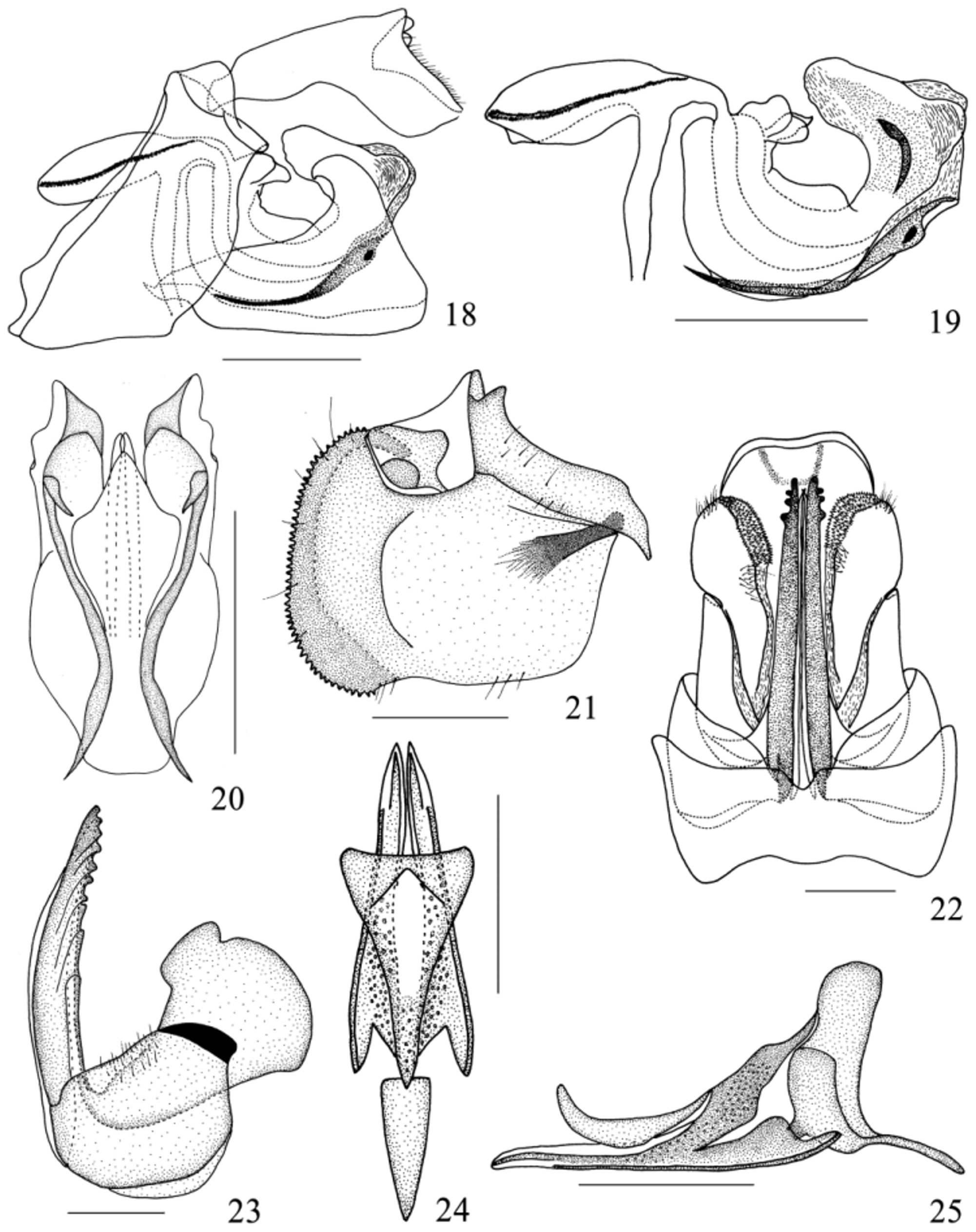
Male terminalia. Anal tube in lateral view large and long, widest subapically, bent down at midlength. Anal column short, located at middle ( Figs 12, 14 View FIGURES 9 – 17 , 18 View FIGURES 18 – 25 ). Genital styles expanding distally, subtriangular in lateral view. Capitulum short, without teeth. Aedeagus moderately dumpy, apical portion curved cephalad in lateral view.
Female terminalia. Anal tube relatively large and broad, widest medially, bent down at midlength in lateral view. Anal column very short, situated at point of flexure ( Fig. 17 View FIGURES 9 – 17 ). Gonoplac minutely denticulate in posterior view, basal part bearing a black sclerous clavate structure close to base of gonapophyses IX ( Figs 13 View FIGURES 9 – 17 , 21 View FIGURES 18 – 25 ). Gonapophyses IX elongated, triangular-shaped ( Fig. 24 View FIGURES 18 – 25 ). Gonapophyses VIII narrow and long, tapering distally, with teeth along dorsal margin ( Fig. 23 View FIGURES 18 – 25 ).
Diagnosis. The new genus resembles Goniopsara Metcalf ( Melichar, 1899) , but can be distinguished from the latter by: 1) frons with upper margin deeply concave, lateral keels from lateral margin, disc forward protruding at lower part (in Goniopsara , frons with upper margin almost straight, lateral keels from upper margin, disc flat); 2) mesonotum with anterior margin slightly arced, apical point in the same level of the lower edge of eyes (in Goniopsara , mesonotum with anterior margin acutely convex medially surpassing the lower edge of eyes obviously); 3) tegmen with costal margin strongly convex near basal one third, clavus nearly reaching apical margin, with apex terminating in a distinct spine (in Goniopsara , costal margin slightly convex, clavus with apex about one forth from apical margin, and not terminating in a spine).
Etymology. Goniopsarites refers to the resemblance of this genus to Goniopsara Metcalf. The gender is masculine.

No known copyright restrictions apply. See Agosti, D., Egloff, W., 2009. Taxonomic information exchange and copyright: the Plazi approach. BMC Research Notes 2009, 2:53 for further explanation.