
Confractamnis johnjelli, Young, Gavin C., 2005
|
publication ID |
https://doi.org/ 10.3853/j.0067-1975.57.2005.1443 |
|
persistent identifier |
https://treatment.plazi.org/id/8F2287C9-FFFE-FFF8-C7E3-2F18FB85F9D9 |
|
treatment provided by |
Felipe |
|
scientific name |
Confractamnis johnjelli |
| status |
n.gen. and n.sp. |
Confractamnis johnjelli n.gen. and n.sp.
1992 eubrachythoracid nov.–Young et al., 1993: 247 (pars).
Name. From the Latin confractus (broken) and amnis (stream or river), with reference to the Broken River. The species name recognizes the collector, Prof. John Jell, University of Queensland, who has conducted research in the Devonian of the Broken River area over many decades.
Diagnosis. A large brachythoracid attaining a length of at least 2 m; trunk armour high, with anterior lateral, posterior dorsolateral, and posterior lateral plates all dorsoventrally elongated. Median dorsal plate enclosing a midline angle of about 150° between left and right laminae. Anterior dorsolateral plate crossed by single sensory canal groove close and subparallel to the lateral margin of the median dorsal plate. Dorsal corner of anterior lateral plate narrow, rounded, and bilobed, with 25–30° angle between main margins. Posterior dorsolateral plate extensively overlapped by anterior dorsolateral plate; posterior lateral plate with high and narrow exposed part, and elongate anterior overlap for the anterior dorsolateral plate. External surface of dermal bones smooth, or with very fine, closely spaced tuberculation of low relief.
Remarks. Because the skull roof is unknown in this new taxon, only trunk armour characters are available for assessing its affinities. It is clearly not a coccosteid, in which the ADL plate is crossed by an additional ventral sensory canal groove. The absence of the ventral sensory groove is one of four characters by which Carr (1991: 382) characterized the most derived pachyosteomorph subgroup within the Brachythoraci (comprising the Dinichthyidae and Aspinothoracidi). Within the former family, Eastmanosteus differs from the new taxon in possessing tubercular ornament, and both Eastmanosteus and Dunkleosteus have an extensive exposed part of the ADL plate above the sensory groove. Levisosteus Otto, 1999 is a poorly known brachythoracid showing possible affinity with Dunkleosteus , and indicating that ornament reduction may have occurred by the Eifelian, but its trunk armour is completely unknown, so no other comparisons are possible with the specimens described here. The aspinothoracids are characterized by various skull features in addition to the absence of a spinal plate in the trunk armour ( Carr, 1991). There is no information on whether this small bone was present in Confractamnis n.gen., but it is likely that the spinal was lost independently in several groups, as suggested by Denison (1984). One group in which this had occurred by the Middle Devonian is the Heterostiidae . Heterostius resembles Confractamnis n.gen. in large size, reduction of ornament, and position of the lateral line groove near the dorsal exposed edge of the ADL plate. Heterostius , however, differs in extreme trunk armour reduction, with the AL plate fused to the ADL plate, whereas the AL was clearly a separate bone in Confractamnis n.gen. The Emsian forms Tityosteus , Taemasosteus and Antineosteus also resemble Confractamnis n.gen. in the dorsal position of the sensory groove on the ADL plate, but differ in the dorsal configuration of the AL plate (as indicated by the shape of its overlap on the ADL), in the shape of the PDL and PL plates, in the much flatter MD plate ( Tityosteus and Antineosteus ), and in the transversely elongate articular condyle attached along its length to the ADL plate ( Antineosteus ; a character defining the Homostiidae ). Close affinity with Atlantidosteus (known from two species, one in Morocco and one in the Broken River sequence) can be excluded on the assumption that this taxon was a homostiid, with a dorsoventrally compressed trunk armour. One isolated ADL plate from the Emsian of Morocco resembles Confractamnis n.gen. in its pointed articular condyle, the narrow rounded dorsal angle of the overlap area for the AL plate, and the position of the sensory groove close and subparallel to the lateral margin of the MD. This specimen may represent a closely related taxon, but it differs from Confractamnis n.gen. in its smaller size, coarse tubercular ornament, more acute angle between the dorsal lamina of the ADL plate and the long (transverse) axis of the articular condyle, and the lack of a bilobed dorsal corner on the AL plate.
Material. ANU V 1028 (holotype), a large left ADL plate with part of the MD plate attached, associated with left PDL and PL plates (all incomplete); ANU V 1031, a very incomplete MD plate .
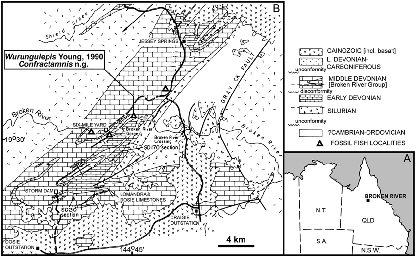
Locality. University of Queensland locality L4399, north bank of the Broken River, Grid Reference 640 460 on the Burges 1:100 000 sheet (see Fig. 1 View Fig ); field numbers 58/L1 ( V 1028) and 58/L2 ( V 1031).
Horizon and age. According to information provided by Prof. J.A. Talent to A. Basden (pers. comm., 28 August 1995), the holotype of Wurungulepis , from the same locality as the material described here, came from strata that were pre-Dosey Limestone in the sequence, and equivalent to the Bracteata Formation and Lomandra Limestone. Outcrop in this vicinity is referred to as “undifferentiated Broken River Group” on the most recent published geological map ( Sloan et al., 1995: fig. 2). Prof. J.S. Jell (letter of 17 April, 1980) suggested a Middle Devonian (?Eifelian) age for this locality, and the sequence is shown spanning the Emsian- Eifelian boundary by Sloan et al. (1995: fig. 3).
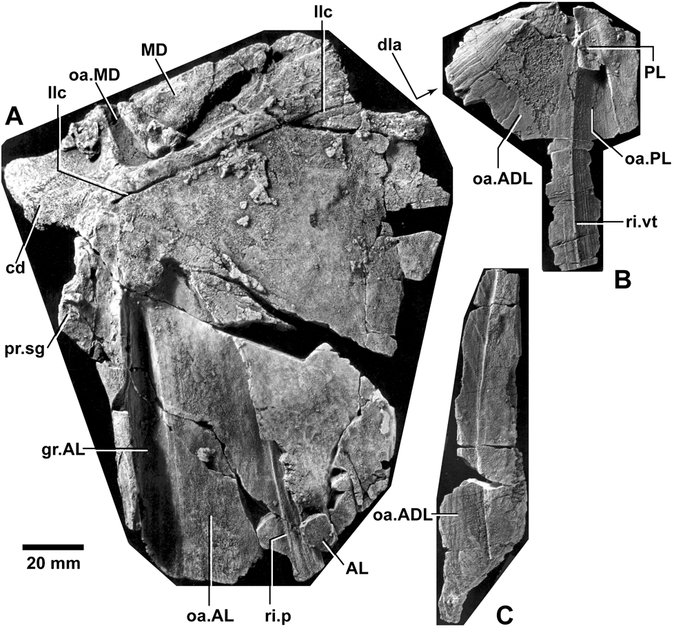
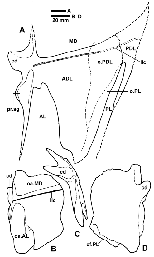
Description. The holotype ( ANU V 1028) includes remains of four bones from the trunk armour of a very large brachythoracid. The left anterior dorsolateral (ADL) plate is particularly massive ( Figs. 2A View Fig , 3 View Fig ), with the bone some 35 mm thick at the base of the articular condyle (cd) for the dermal neck-joint. The ventral part of the ADL is missing, and the posterior margin is broken and incomplete, but the fragmented posterior parts reduce in thickness to a little more than 1 mm, with part of the actual margin preserved. Together with the evidence of adjacent bones it is possible to determine the general shape of the ADL plate, except for the extent of the ventral margin.
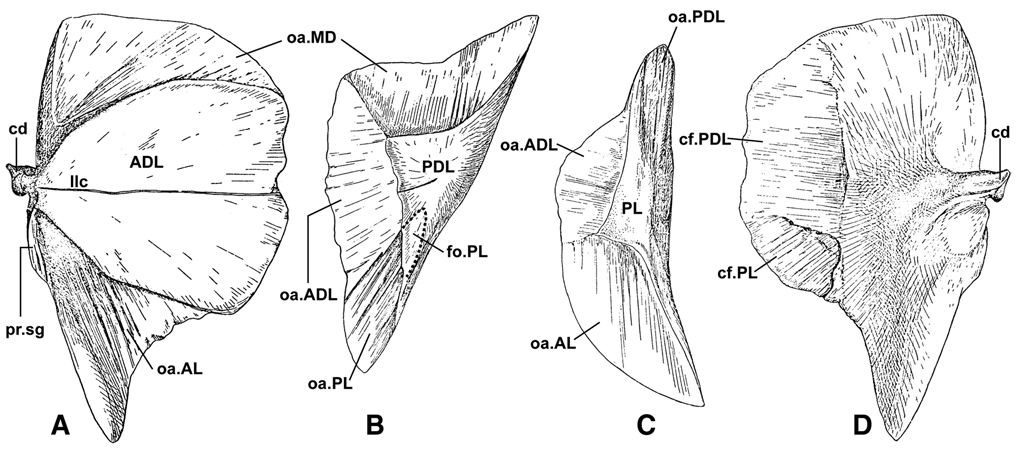
The external surface of the ADL ( Fig. 2A View Fig ) shows two deeply incised overlap areas for the median dorsal (MD) and anterior lateral (AL) plates (oa.MD, oa.AL). A broken fragment of the left anterolateral corner of the MD plate is still attached to the specimen, but slightly displaced. Its thickness (13 mm), so close to the lateral margin, gives some indication of the large size of this bone (cf. ANU V 1031 below). The rest of this normally massive bone was either weathered away, or perhaps was contained in another uncollected limestone block. The external surface of the MD fragment appears smooth, as does the exposed surface of the ADL. On close examination, some parts show a faint tuberculation of closely spaced fine tubercles (about 15 per crosses the ADL plate from the region of the articular condyle, as in all arthrodires with a developed dermal neckjoint. It runs, however, close and subparallel to the edge of the overlap for the MD, which is a point of difference to many other brachythoracids. The overlap area for the AL plate is set in about 10 mm along a deep anterior groove, which braced the AL against the anterior edge of the ADL plate (gr.AL, Fig. 2A View Fig ). The overlap shows that the AL had a narrow rounded bilobed dorsal corner, which forms a much more acute angle (about 25–30°), and is quite different in shape to the triangular overlap of Eastmanosteus or Dunkleosteus (dorsal angle 45–50°). Near the ventral preserved edge the overlap area expands as a posterior embayment, in which a fragment of the overlying AL plate is preserved (AL, Fig. 2A View Fig ). At its margin, this embayment slopes gradually to the external surface, in contrast to the thick and undercut margins of the deeply incised main part of the overlap area (oa.AL). These show that immediately dorsal to the embayment the posterior edge of the AL was enclosed by the ADL to a depth of about 6 mm. The undercut surface is exposed by the broken margin of the overlap area, to reveal a strong ridge (ri.p, Fig. 2A View Fig ).
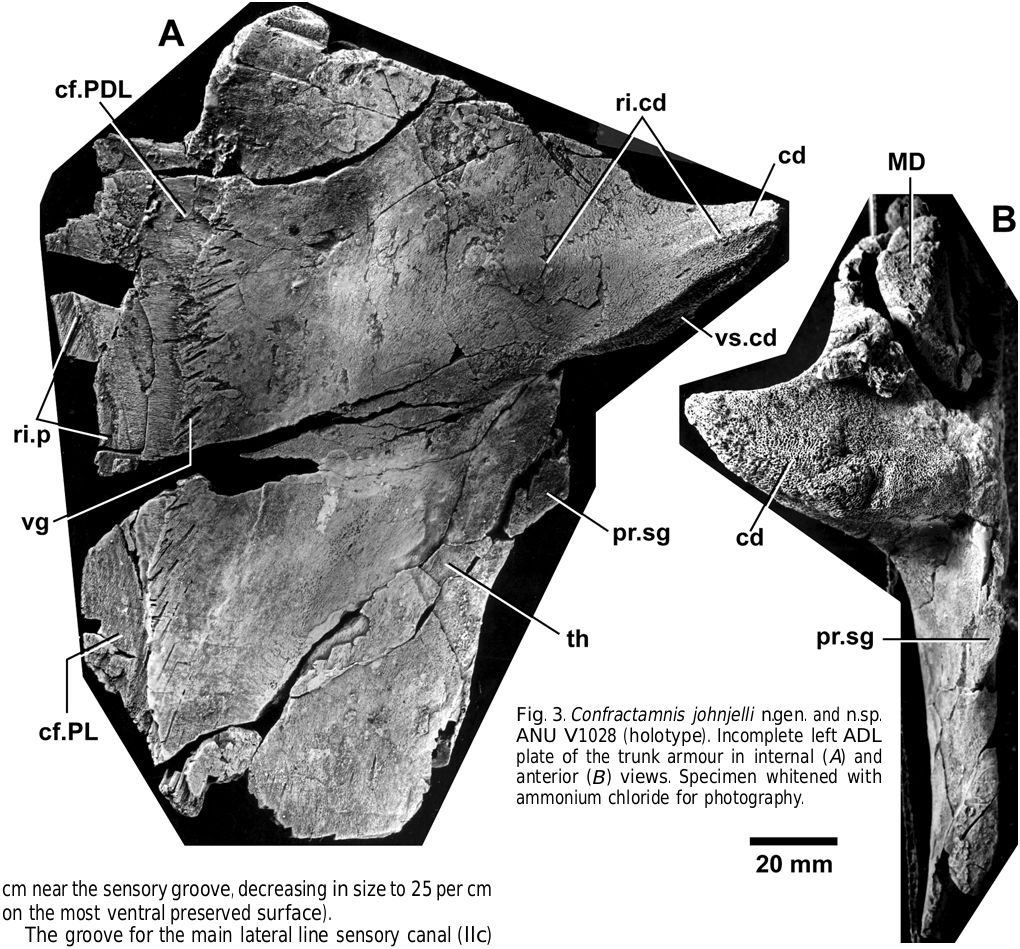
The articular condyle for the dermal neck-joint in Confractamnis n.gen. is a very strongly developed projection from the anterior margin of the ADL (cd, Figs. 2 View Fig , 3 View Fig ). In mesial view, the condyle shows a triangular crosssection. The ventral side of the triangle, with typical “siebknochen” texture of spongy bone (invested with articular cartilage in life), is partly visible in internal view (vs.cd, Fig. 3A View Fig ). The posterior side of the triangle forms the smooth inner surface, which is slightly convex about a low ridge crossing the condyle from near the mesial termination to the thickened part of the ADL (ri.cd). An anterior view of the ADL plate ( Fig. 3B View Fig ) shows the strong dorsal support for the condyle (the “condylus ridge” of Heintz, 1934: 73), and the mesial termination of the condyle as a rounded point, with its anterodorsal surface (anterior side of triangle) being flat to slightly concave, and also with “siebknochen” texture (sieve-like bone). The actual articular surface is higher laterally than mesially (see Fig. 5C View Fig ), a condition also noted in both Dunkleosteus and Homostius by Heintz (1934: 73). Lelièvre (1995) used this as a character to group some primitive brachythoracids from Morocco. Beneath the condyle is a prominent subglenoid process (pr.sg), which would have articulated against the paraarticular process of the skull roof.
The inner surface of the ADL plate shows extensive posterior contact faces for the PDL and PL plates (cf.PDL, cf.PL, Fig. 3A View Fig ). The distinct anterior margin of the very extensive PDL contact face is marked by vascular grooves (vg) with radiating orientation from the ossification centre of the bone (situated anteriorly in the region of the articular condyle attachment). Ventrally this margin becomes the anterior margin for the PL contact face. A fainter ridge near the posterior border of the ADL plate (ri.p) represents the anterior margin for the more dorsal part of the PL contact face. The anterior half of the inner ADL surface is convex, where the bone is massively thickened (th) to form a broad dorsoventral ridge, decreasing ventrally where the internal convexity lies beneath the deeply incised overlap for the AL plate on the external surface. The posterior part of the inner surface in ANU V 1028 is flat to concave, and inflected dorsoventrally at an angle of about 140° about the dorsolateral ridge, at the level of the sensory groove on the external surface. The PDL plate has a similar inflection (“dla” arrow, Fig. 2B View Fig ).
Associated left PDL and PL plates of Confractamnis n.gen. are also partly preserved in the holotype. They can be placed against the ADL plate to confirm their life position. The PDL ( Fig. 2B View Fig ) is missing much of its dorsal part, which was overlapped by the MD plate. Only a narrow posterior strip of the external bone surface is preserved, and most of the bone comprises an internal lamina overlapped extensively by both ADL and PL plates. A thin vertical ridge (ri.vt), which was entirely internal, separates the two overlaps (oa.ADL, oa.PL). The edges of the bone are extremely thin, with many fine fractures which collapsed during acid preparation, and could be only partly reconstructed from fragments. The margins of the bone, however, are indicated by the extent of the contact face inside the ADL plate (cf.PDL, Fig. 3A View Fig ), which shows that the degree of overlap was more extensive than in many other brachythoracids. The external surface of the PDL plate may have expanded dorsally (PDL, Fig. 4A View Fig ), and presumably was crossed by a posterior continuation of the lateral line groove (llc). Its distinctive shape can be inferred from the marked dorsoventral elongation of the internal ridge (ri.vt, Fig. 2B View Fig ), and the extensive overlap surfaces (o.PDL, Fig. 4A View Fig ). The narrow dorsal corner of the PL plate is still attached to the PDL (PL, Fig. 2B View Fig ). There is no sign of the internal fossa which received this corner in Dunkleosteus (fo.PL, Fig. 6B View Fig ).
The inner surface of the PDL plate is smooth and concave, inflected about the dorsolateral angle at a level that corresponds to that on the ADL (dla, Fig. 2B View Fig ). The bone margins show inner vascular grooves like those on the inside of the ADL (vg, Fig. 3A View Fig ), which may indicate increased blood supply adjacent to the bone margins during periods of growth. The PDL fits inside the ADL such that the vertical ridge (ri.vt, Fig. 2B View Fig ) abuts against a slight ridge inside the posterior margin of the ADL plate (ri.p, Fig. 3A View Fig ).
The incomplete PL plate of Confractamnis n.gen. ( Fig. 2C View Fig ) is a remarkably high and narrow splint of bone, comprising an anterior overlap area, and high and narrow posterior exposed part. The posterior margin of the preserved part is partly broken, but the form of the inner surface shows that not much is missing. The PL plate slots into the grooved overlap behind the vertical ridge on the PDL (where part of the PL is still attached; oa.PL, Fig. 2B View Fig ). With both bones in place inside the ADL, the convex internal surface of the PL forms a thickened posterior border to the articulated armour. Its overlap area is sandwiched between the PDL and ADL plates, with its anterior edge against the posterior ridge dorsally (ri.p, Fig. 3A View Fig ), and ventrally extending past the PDL to fit into a more deeply incised contact face (cf.PL). This arrangement is summarized in Fig. 4A View Fig . The ventral part of the PL plate is unknown.
One small fragment of the AL plate is also preserved attached to the external surface of the ADL in ANU V 1028. Otherwise this bone is unknown, except for the distinctive shape of its dorsal part (AL, Fig. 4A View Fig ), as indicated by the overlap area on the ADL.
The only other arthrodire specimen from locality UQL 4399 also belonged to a brachythoracid, and is provisionally referred to Confractamnis n.gen. ANU V 1031 is an incomplete MD plate that lacks most diagnostic characters. However the external surface is largely smooth, with similar texture to the small portion of MD attached to the ADL plate in ANU V 1028, which is consistent with it belonging to a smaller individual of the same taxon. The more complete left lamina is gently curved, 95 mm from the midline to the preserved lateral edge, and 50 mm across between broken anterior and posterior margins. As all margins are incomplete, and no contact faces for the ADL and PDL plates can be discerned on the internal surface, there is insufficient information for a reasonable estimate of total length and overall shape. The bone may have been slightly broader than long, consistent with the shape of the MD overlap area on the ADL plate of the holotype ( MD, Fig. 4A View Fig ). This suggests a somewhat angular shape, and a straight to gently curved lateral margin .
The MD plate of ANU V 1031 evidently came from an individual only about half the size of the holotype, but the bone reaches about 10 mm thickness close to the midline. The incomplete carinal process is 70 mm long and 50 mm deep as preserved. Its anteroventral edge thins to about 1 mm, and probably not much is missing. The posteroventral edge is expanded to a thickness of about 8 mm, but is incomplete in lacking the knob-like or grooved termination that is normally developed on the carinal process. The angle between the carinal process and the left lamina is about 75 °.
Reconstruction
Arriving at a reliable three-dimensional reconstruction of the trunk armour of a brachythoracid from isolated bones is difficult. It took over 120 years from the first attempts ( Miller, 1841) for a reliable reconstruction of the armour of Coccosteus (Miles & Westoll, 1968: fig. 44), this reconstruction depending on earlier attempts for other forms (e.g., Dunkleosteus Heintz, 1932 : fig. 68; Homostius Heintz, 1934 : fig. 45).
With the discovery of exceptional three-dimensional preservation in the acid-prepared arthrodires from Gogo, Western Australia, it was possible to check the reliability of such restorations with actual specimens. Trial and error in the Natural History Museum , London , showed that if bones were glued together with a tight fit on the clearly defined overlap areas, the left and right sides did not join up—only by leaving a small space around the edges of bone overlaps could a symmetric reconstruction be achieved ( Dr R. S. Miles, pers. comm.). The first illustration of an actual reconstructed specimen from the uniquely preserved Gogo fauna was the holotype of Harrytoombsia by Miles & Dennis (1979: fig. 9) .
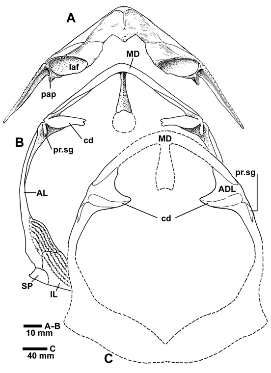
The ADL plate on its own is one of the most informative in attempting a reconstruction from isolated bones, because the axis of articulation on the condyle must have been horizontal for the dermal neck-joint to function in the living animal. This provides some constraint on the cross-sectional shape of the trunk armour, a point first exploited by Heintz (1934: fig. 47) in comparing Dunkleosteus with the dorsoventrally compressed armour of Homostius . There is, however, always a degree of uncertainty regarding the precise orientation of the axis of articulation, because the condyle in life was invested with articular cartilage, so its true shape is not preserved. Thus, it is possible that the ADL may have been slightly more steeply inclined (implying a higher trunk armour) than depicted in the reconstruction ( Fig. 5C View Fig ), although the preserved angles of the MD plate in ANU V 1031 are generally consistent with this reconstruction.
Using the trunk armour in the holotype of Harrytoombsia as a model (Miles & Dennis, 1979: fig. 9), the armour of Confractamnis n.gen. may have been some 37 cm in height, and over 30 cm across. As there is no control of the width across the articular condyles of the ADL, the armour could have been considerably larger by comparison with Harrytoombsia (which has proportionately smaller condyles), but it is unlikely to have been any smaller. Such a fish would have been at least 206 cm long, judging by proportions in Coccosteus cuspidatus , where whole animals including the tail are preserved, and total length of the fish is some 5.5 times trunk armour height (see Miles & Westoll, 1968: figs. 44, 48).
| MD |
Museum Donaueschingen |
| PL |
Západoceské muzeum v Plzni |
| V |
Royal British Columbia Museum - Herbarium |
| R |
Departamento de Geologia, Universidad de Chile |
No known copyright restrictions apply. See Agosti, D., Egloff, W., 2009. Taxonomic information exchange and copyright: the Plazi approach. BMC Research Notes 2009, 2:53 for further explanation.
|
Kingdom |
|
|
Phylum |
|
|
Class |
|
|
Order |
|
|
Genus |