
Euricania paraclara, Ren, Lan-Lan, Stroiński, Adam & Qin, Dao-Zheng, 2015
|
publication ID |
https://doi.org/ 10.11646/zootaxa.4033.1.8 |
|
publication LSID |
lsid:zoobank.org:pub:8A95CF0B-BCD9-4D34-AD74-F1C3DA4616D6 |
|
DOI |
https://doi.org/10.5281/zenodo.6092285 |
|
persistent identifier |
https://treatment.plazi.org/id/8F2DD502-C26F-FF9F-61E5-A899FDB03613 |
|
treatment provided by |
Plazi |
|
scientific name |
Euricania paraclara |
| status |
sp. nov. |
Euricania paraclara View in CoL sp. nov.
( Figs 1–17 View FIGURES 1 – 13 View FIGURES 14 – 17 )
Etymology. The specific epithet refers to the similarity of this new species to Euricania clara Kato, 1932 .
Diagnosis. Euricania paraclara sp. nov. is similar to Euricania clara Kato , but differs from the latter by having long apical spinose process of aedeagus surpassing half length of periandrium in midline (apical spinose process of aedeagus short, not surpassing half length of periandrium in E. clara ).
Description. Length (including tegmina): male 9.5–11.0 mm, female 8.5–9.5 mm.
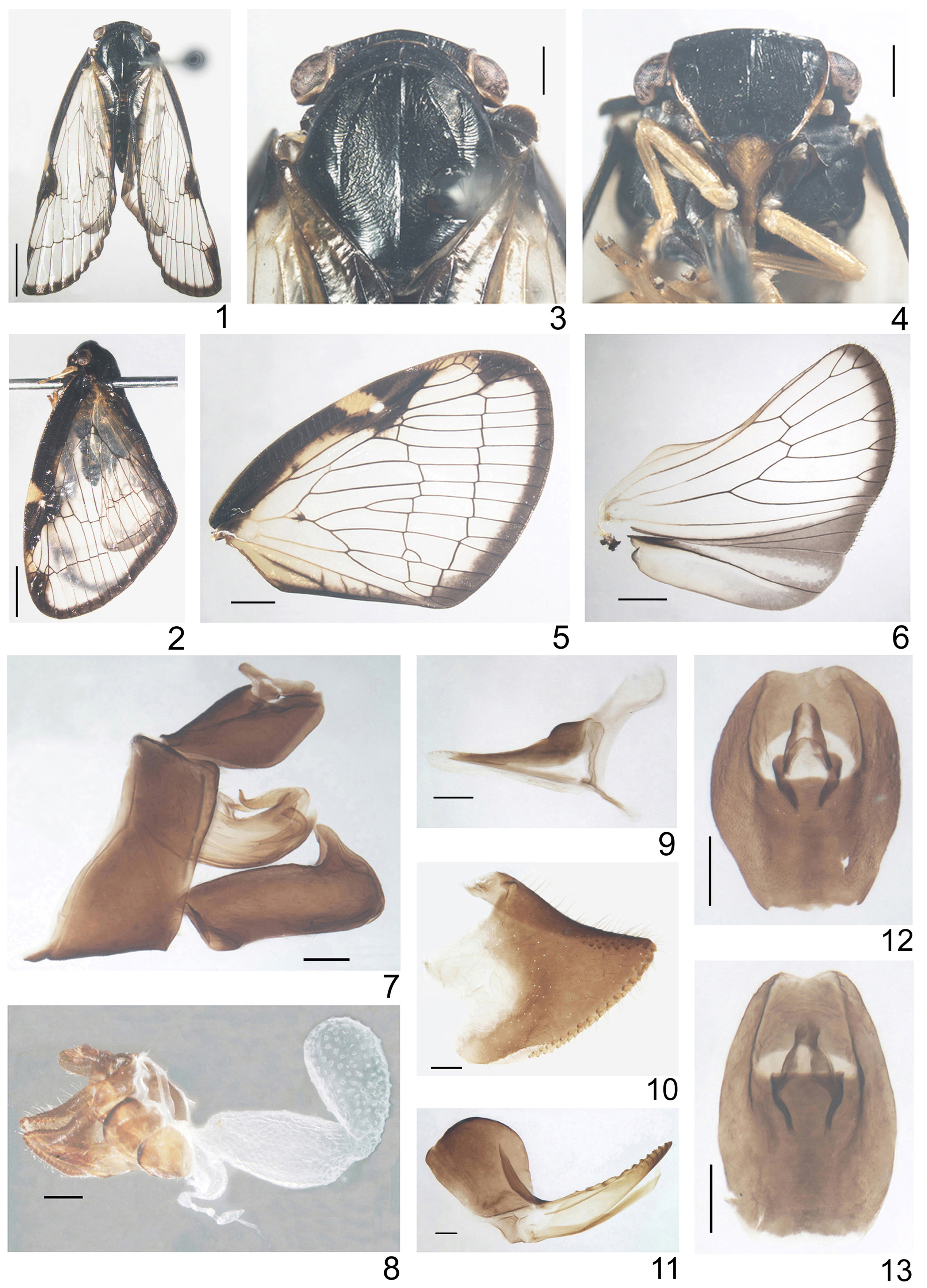
HEAD. Head with compound eyes (in dorsal view) about as wide as mesonotum ( Figs 1, 3 View FIGURES 1 – 13 ). Vertex transverse, 9 times wider at the anterior margin than long in midline, with all margins strongly carinate, anterior and posterior margins arcuate, almost parallel; disc of vertex without median carina ( Fig. 3 View FIGURES 1 – 13 ).
Frons at upper margin 1.35 times as wide as tall in midline; frons widest at lower part of compound eyes shorter than long in midline about 1.45: 1; upper margin slightly convex, lateral margins arcuate, not incised near ocelli, in lower part slightly curved to frontoclypeal suture; frontal disc with 3 carinae separated basally and delicately rugose vertically, median carina extending the midlength of frons, lateral carinae arcuate, shorter than median carina about 1: 1.6, reaching the level of ocelli ( Fig. 4 View FIGURES 1 – 13 ).
Compound eyes oval, with small callus at lower margin. Pedicel elongate, barrel-shaped, with plate organs located apically. Ocelli present.
Frontoclypeal suture arcuate. Clypeus without carinae, with median portion convex ( Fig. 4 View FIGURES 1 – 13 ).
Rostrum reaching mesotrochanters, apical segment shorter than subapical.
THORAX. Pronotum distinctly longer in midline than vertex; disc of pronotum with median carina and two lateral impressions, anterior and posterior margins in median portion almost parallel ( Fig. 3 View FIGURES 1 – 13 ).
Mesonotum elongate, distinctly longer than cumulative length of vertex and pronotum in midline; median carina distinctly visible, keel-shaped and reaching almost scutellum; lateral carinae connected basally, reaching posterior margins; anterolateral carinae not surpassing the lateral angles of mesonotum, connected with lateral carinae ( Figs 1–3 View FIGURES 1 – 13 ).
Tegmina ( Figs 1, 2 & 5 View FIGURES 1 – 13 ) membranous, elongately-triangular; costal margin weakly arcuate, anterior angle broadly rounded, placed distad to claval angle, posterior margin angulate. Costal area of tegmen with transverse veinlets, a little wider than postcostal cell and widened apically; postcostal cell narrower than costal area with a few incomplete transverse veinlets, basal cell widely rounded; veins ScP+RA, MP and CuA separated at base. ScP+RA vein forked a bit distad than MP fork, CuA stem distinctly longer than other stems, forked after MP1+2 and MP3+4. Tegmen with 2 lines of transverse veinlets, apical and subapical cells longer than wide. Cubital cell without transverse veinlets, icu veinlet present. Claval veins Pcu and A1 fused about midlength of clavus, transverse veinlets present between CuP-Pcu and Pcu+A1-CuP.
Wings small, with precostal cell longer than wide, and 2 transverse veinlets r-m and m-cu ( Fig. 6 View FIGURES 1 – 13 ).
Metatibia longer than metafemur, with 2 lateral spines placed distally and with 6 apical teeth; basitarsomere as long as cumulative length of meta- and hind tarsomere, with 8 apical teeth. Metatibiotarsal formula 2/6/8.
Coloration. General colour of body dark brown to dark ( Figs 1–2 View FIGURES 1 – 13 ). Posterolateral corner of vertex has a visible brown macula on each side ( Fig. 3 View FIGURES 1 – 13 ). Lateral margins of vertex, frons in apical ⅔, clypeus and rostrum yellowish brown ( Figs 1–3 View FIGURES 1 – 13 ). Eyes sordid brownish ornamented with irregular black patches ( Figs 1–4 View FIGURES 1 – 13 ). Tegmina with costal marginal fascia dark brown, with a yellow, rhomboid patch near middle, beneath with a white small spot; apical and inner margins with brown fasciae ( Figs. 1, 4 & 5 View FIGURES 1 – 13 ). Costal and inner margins of wings with narrow, brown fasciae; apical marginal fascia brown and broad, vannal region brownish except for a grayish longitudinal band between veins A1 and A2 ( Fig. 6 View FIGURES 1 – 13 ). Abdomen dark brown, each tergite with a narrow brown stripe posteriorly ( Fig. 1 View FIGURES 1 – 13 ). Legs yellow ( Fig. 3 View FIGURES 1 – 13 ).
MALE TERMINALIA. Anal tube oval in dorsal view, widest in middle, length at midline: maximum width ratio =1.44: 1, posterior margin slightly concave, basal margin almost straight, lateral margins strongly convex, anus placed a bit after midlength ( Figs 7 & 12 View FIGURES 1 – 13 ). Anal tube in lateral view ( Fig. 7 View FIGURES 1 – 13 ) not extending the end of the genital styles; ventral margin strongly arcuate.
Pygofer, in lateral view ( Fig. 7 View FIGURES 1 – 13 ), taller than wide; dorsal part narrower than ventral, posterior margin almost straight; posterior-dorsal angle without process, caudoventral angle obtuse ( Fig. 7 View FIGURES 1 – 13 ).
Genital styles (in lateral view, Fig. 7 View FIGURES 1 – 13 ), distinctly longer than wide and bearing distinct spine-like process at the end of dorsal margin; lower and upper margin weakly arcuate, almost parallel; hind margin of caudo-dorsal angle widely rounded, surpassing the posterior margin of process.
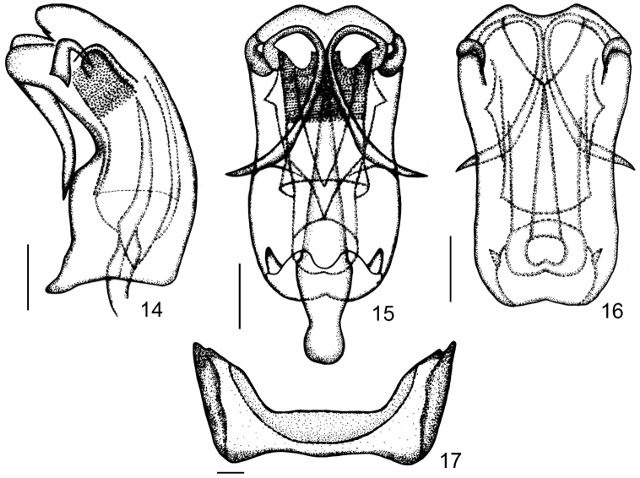
Phallic complex ( Figs 7 View FIGURES 1 – 13 , 14–16 View FIGURES 14 – 17 ). Periandrium with lateral split shorter than the half of its length. Basal part of periandrium elevated without any additional structures; dorsal periandrium shorter than ventral one, upper median lateral fold of periandrium well developed, narrow and smooth.
Aedeagus a bit longer than periandrium, with pair of well sclerotized, smooth, spinose processes. Each process with a single apex, base of the process placed under the lateral lobe of dorsal periandrium. Apical process distinctly longer than subapical one, surpassing the midlength of periandrium, oriented cephalad and laterad; subapical lateral process distinctly shorter than apical, strongly curved, with apex oriented basad.
FEMALE TERMINALIA. Pregenital sternite with median portion distinctly narrower, comparing to well developed lateral lobes; anterior margin weakly convex medially, posterior margin almost straight ( Fig. 17 View FIGURES 14 – 17 ).
Anal tube in lateral view with ventral margin convex ( Fig. 8 View FIGURES 1 – 13 ).
Anal tube in dorsal view egg-shaped, widest at basal third, length at midline: maximum width ratio = 1.67: 1, lateral margins convex, basal margin nearly straight, posterior margin concave, anus placed after midlength ( Fig. 13 View FIGURES 1 – 13 ).
Gonoplac unilobate, triangular, with posterior margins bearing 2 rows of blunt and short teeth, posteroventral part partly membranous ( Fig. 10 View FIGURES 1 – 13 ).
Gonapophysis VIII partly laterally flattened, tapering apicad; dorsal margin shallowly concave with sharp apex and well visible teeth at the postero-dorsal margin, near apex with spiniferous microsculpture; endogonocoxal process narrower and shorter than gonaphophysis VIII ( Fig. 11 View FIGURES 1 – 13 ).
Gonapophysis IX with posterior connective lamina sclerotized, gonospiculum bridge finger-like dorsobasally, needle-like ventrobasally ( Fig. 9 View FIGURES 1 – 13 ).
Bursa copulatrix with widely connected 2 pouches; first pouch with well visible cells and sclerotized ornamentation, the second one without cells but with well visible numerous superficial pores ( Fig. 8 View FIGURES 1 – 13 ).
Spermatheca well developed; ductus receptaculi wrinkled, longer than diverticulum ductus.
Type materials. Holotype, male: [ China: Guizhou, Dabanshui, 1000m, coll. Lifang Zheng, 26 Aug. 2012]. Paratypes: 12 males and 40 females, same data as holotype.
Distribution. China (Guizhou).
No known copyright restrictions apply. See Agosti, D., Egloff, W., 2009. Taxonomic information exchange and copyright: the Plazi approach. BMC Research Notes 2009, 2:53 for further explanation.