
Gordonopsis, Guinot & Richer de Forges, 1995
|
publication ID |
https://doi.org/10.26107/RBZ-2020-0023 |
|
publication LSID |
lsid:zoobank.org:pub:057B08EE-1006-43E8-AE20-0AF8F224BA2D |
|
DOI |
https://doi.org/10.5281/zenodo.4576678 |
|
persistent identifier |
https://treatment.plazi.org/id/8F3E87B9-A805-BA0B-71AB-FBC1D9F14FB1 |
|
treatment provided by |
Carolina (2021-03-03 14:08:27, last updated by Plazi 2023-11-01 22:38:20) |
|
scientific name |
Gordonopsis |
| status |
|
Key to species of Gordonopsis View in CoL
1. Carapace and pereiopods covered with long dense setae which obscures surfaces and margins ( Fig. 27 View Fig A–C); distal edge of dorsal margin of cheliped merus with 1 spine ( Fig. 28E, G, H View Fig ) ..... Gordonopsis velutina View in CoL , new species (Admiralty Islands)
– Carapace and pereiopods glabrous, covered with short setae that may obscure surface or longer sparser setae that does not obscure surface (e.g., Figs. 2 View Fig A–C, 14A–C, 17A–C); distal edge of dorsal margin of cheliped merus with 2 spines (e.g., Figs. 3F, G View Fig , 15G View Fig ) .............................................................................2
2. Latero-posterior margin of carapace with distinct tubercle or spine (e.g., Fig. 2A View Fig ); outer margin of P2–P4 coxae unarmed, smooth .....................................................................................3
– Latero-posterior margin of carapace with very low tubercle, barely visible (e.g., Fig. 17A View Fig ); outer margin of P2–P4 coxae with a spine or tubercle (may be reduced or absent on some of articles) (e.g., Figs. 18J, K View Fig , 22I, J View Fig ) ....................................5
3. Carapace relatively narrower with lateral margins straighter, less convex ( Fig. 9B View Fig ); carpus of cheliped with 1 spine on each of outer and inner margins, dorsal ridge smooth, unarmed ( Fig. 10F, G View Fig ) ......... Gordonopsis alaini View in CoL , new species ( Madagascar)
– Carapace longitudinally ovate, with posterior part more globose, lateral margins distinctly convex (e.g., Fig. 2A View Fig ); carpus of cheliped with 3 or more spines and spinules on each of outer and inner margins, dorsal ridge not armed with spines or spinules (e.g., Fig. 3F, G View Fig ) .......................................................4
4. Ambulatory legs long ( Figs. 2A View Fig , 6A View Fig ); distal part of P5 merus extending beyond gastric groove when folded anteriorly ( Figs. 4A View Fig , 8A View Fig ); dorsal ridge of carpus of cheliped with row of low granules ( Figs. 3F, G View Fig , 7G View Fig ); tip of G1 subtruncate ( Figs. 4H View Fig , 8K View Fig ) ... Gordonopsis profundarum ( Alcock & Anderson, 1899) (western Indian Ocean)
– Ambulatory legs short ( Fig. 12A, B View Fig ); distal part of P5 merus just reaching gastric groove when folded anteriorly ( Fig. 12D View Fig ); dorsal ridge of carpus of cheliped with row of low sharp tubercles ( Fig. 13F View Fig ); tip of G1 appears bifurcated ( Fig. 13K View Fig ) ... Gordonopsis robusta Ng, Padate & Saravanane, 2019 View in CoL (Andaman Sea)
5. Supraorbital margin proportionately wider, pseudorostral spines relatively short, directed slightly obliquely laterally at angle of 20–30° ( Figs. 14B View Fig , 15D View Fig , 17B View Fig )................................................6
– Supraorbital margin C-shaped, less wide, pseudorostral spines long, directed almost 45° obliquely laterally ( Figs. 21B View Fig , 24B View Fig ) .........................................................................................7
6. Carapace appears inflated when viewed laterally and frontally ( Fig. 15A, B View Fig ); P2–P4, particularly merus, proportionately shorter ( Fig. 16 View Fig E–G, K, L); outer surface of P4 merus with 1 tubercle on proximal part ( Fig. 16G, I View Fig )............... Gordonopsis pacifica Takeda & Suyama, 2019 View in CoL (southern Japan)
– Carapace appears inflated when viewed laterally and frontally ( Fig. 18A, B View Fig ); P2–P4, particularly merus, proportionately longer ( Fig. 19 View Fig D–F); outer surface of P4 merus unarmed, completely smooth ( Fig. 19F View Fig )...... Gordonopsis hera View in CoL , new species ( Papua New Guinea)
7. Outer margin of each P3 and P4 coxa with 2 long spines each ( Figs. 22I, J View Fig , 23B, H View Fig ); outer surface of P4 merus with 1 perpendicular spine on proximal part... Gordonopsis phorcys View in CoL , new species ( Solomon Islands)
– Outer margin of each P3 and P4 coxa with 1 spine each ( Fig. 26 View Fig B–D); outer surface of P4 merus unarmed, completely smooth ..... Gordonopsis ceto View in CoL , new species (South China Sea)
Alcock A & Anderson ARS (1899) Natural history notes from H. M. royal Indian marine survey ship ' Investigator, ' Commander T. H. Heming, R. N., commanding. - Series III., No. 2. An account of the deep-sea Crustacea dredged during the surveying-season of 1897 - 98. The Annals and Magazine of Natural History, Including Zoology, Botany, and Geology, Series 7, 3: 1 - 27.
Ng PKL, Padate VP & Saravanane N (2019) Gordonopsis robusta, a new species of deep-sea porter crab (Crustacea: Brachyura: Homolidae) from the Andaman Sea, India. Raffles Bulletin of Zoology, 67: 510 - 516.
Takeda M & Suyama N (2019) Five species of crabs (Crustacea: Decapoda: Brachyura) from the depths off Okino-Torishima, the southernmost island in Japan. Bulletin of the National Museum of Nature and Science, Tokyo, Series A (Zoology), 45: 45 - 59.

Fig. 27. Gordonopsis velutina, new species, holotype female (20.8 × 27.8 mm) (MNHN-IU-2013-2269), Admiralty Islands. A, overall habitus; B, dorsal view of carapace; C, dorso-frontal view of carapace.

Fig. 28. Gordonopsis velutina, new species, holotype female (20.8 × 27.8 mm) (MNHN-IU-2013-2269), Admiralty Islands. A, lateral view of cephalothorax; B, frontal view of cephalothorax; C, left third maxilliped; D, ventral view showing buccal cavity, epistome, antennae, and antennules; E, outer view of merus and carpus of right cheliped; F, outer view of right carpus and chela; G, dorsal view of left cheliped; H, dorsal view of right cheliped.
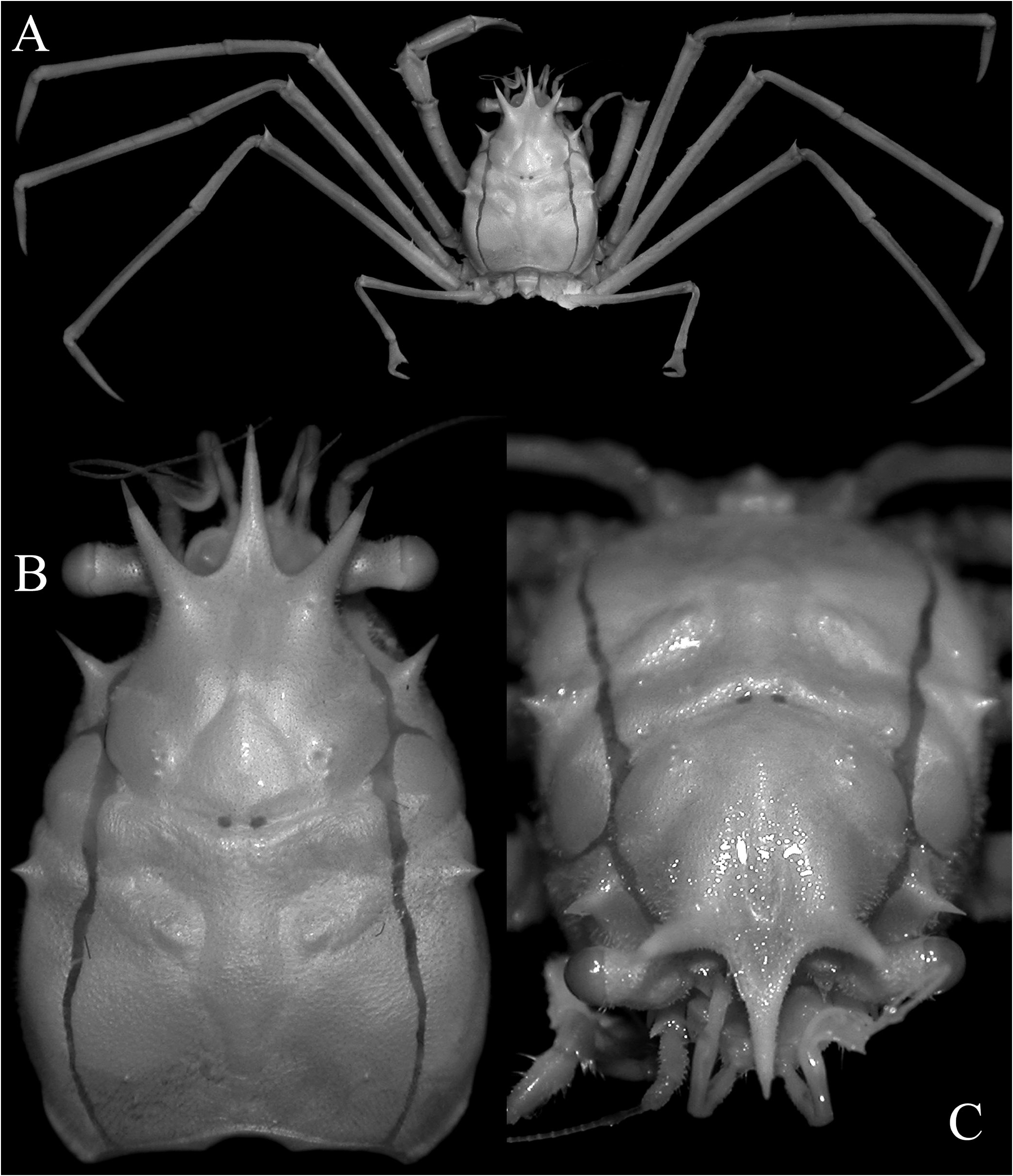
Fig. 2. Gordonopsis profundarum (Alcock & Anderson, 1899), neotype male (16.0 × 11.0 mm) (NHM 1948.9.7.28), Maldives. A, overall habitus; B, dorsal view of carapace; C, dorso-frontal view of carapace.
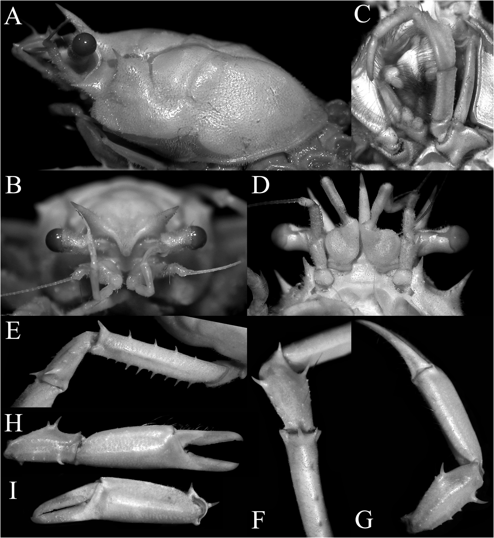
Fig. 3. Gordonopsis profundarum (Alcock & Anderson, 1899), neotype male (16.0 × 11.0 mm) (NHM 1948.9.7.28), Maldives. A, lateral view of cephalothorax; B, frontal view of cephalothorax; C, left third maxilliped; D, ventral view showing buccal cavity, epistome, antennae, and antennules; E, outer view of merus and carpus of left cheliped; F, dorsal view of left merus, carpus, and chela; G, dorsal view of right carpus and chela; H, outer view of right chela; I, outer view of left chela.

Fig. 15. Gordonopsis pacifica Takeda & Suyama, 2019, holotype male (31.0 × 43.0 mm) (NSMT-Cr 26053), Okino-Torishima Island, Japan. A, lateral view of cephalothorax; B, frontal view of cephalothorax; C, left third maxilliped; D, dorsal view of front; E, outer view of merus and carpus of right cheloped; F, outer view of right chela; G, dorsal view of carpus of right cheliped.

Fig. 17. Gordonopsis hera, new species, holotype ovigerous female (21.8 × 31.2 mm) (MNHN-IU-2013-2245), Papua New Guinea. A, overall habitus; B, dorsal view of carapace; C, dorso-frontal view of carapace.

Fig. 18. Gordonopsis hera, new species, holotype ovigerous female (21.8 × 31.2 mm) (MNHN-IU-2013-2245), Papua New Guinea. A, lateral view of cephalothorax; B, frontal view of cephalothorax; C, left third maxilliped; D, ventral view showing buccal cavity, epistome, antennae, and antennules; E, dorsal view of carpus of left cheliped (denuded); F, dorsal view of carpus of right cheliped (denuded); G, outer view of right chela; H, inner view of right chela; I, dorsal view of right cheliped; J, right P2–P4 coxae and basis-ischia (denuded); K, left P2–P4 coxae (denuded).

Fig. 22. Gordonopsis phorcys, new species, holotype female (10.9 × 16.8 mm) (MNHN-IU-2008-12211), Solomon Islands. A, lateral view of cephalothorax; B, frontal view of cephalothorax; C, left third maxilliped; D, ventral view showing buccal cavity, epistome, antennae, and antennules; E, outer view of merus and carpus of right cheliped; F, outer view of right carpus and chela; G, dorsal view of left cheliped; H, dorsal view of right cheliped; I, right P2–P4 coxae and basis-ischia (denuded); J, left P2–P4 coxae and basis-ischia (denuded).
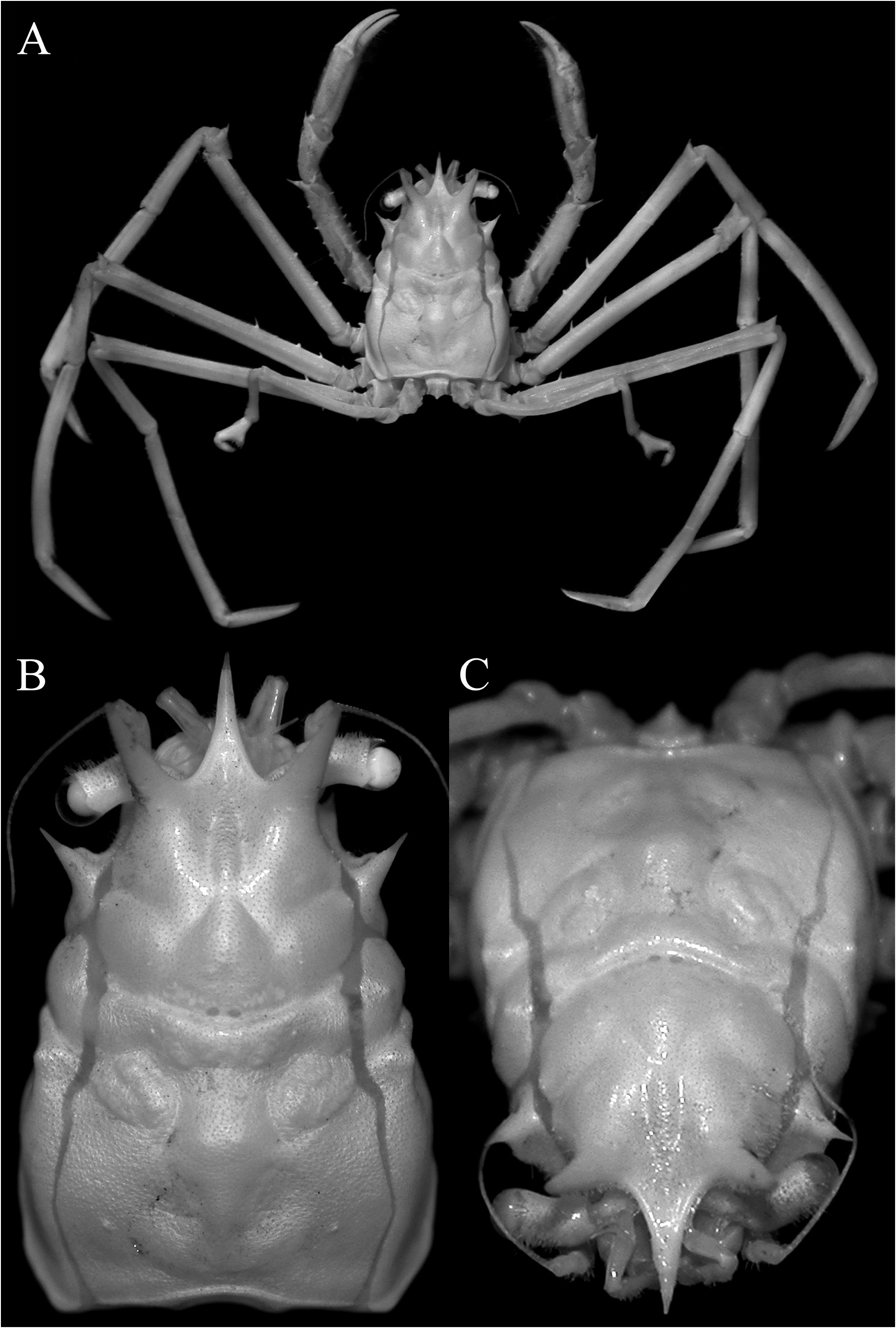
Fig. 9. Gordonopsis alaini, new species, holotype male (15.4 × 10.0 mm) (MNHN-IU-2017-9049), Madagascar. A, overall habitus; B, dorsal view of carapace; C, dorso-frontal view of carapace.

Fig. 10. Gordonopsis alaini, new species, holotype male (15.4 × 10.0 mm) (MNHN-IU-2017-9049), Madagascar. A, lateral view of cephalothorax; B, frontal view of cephalothorax; C, left third maxilliped; D, ventral view showing buccal cavity, epistome, antennae, and antennules; E, outer view of right carpus and chela; F, dorsal view of left cheliped; G, dorsal view of right cheliped; H, ventral view of right G1; I, ventral view of right G2.

Fig. 6. Gordonopsis profundarum (Alcock & Anderson, 1899), male (16.3 × 24.1 mm) (ZMB 13645), East Africa. A, overall habitus; B, dorsal view of carapace; C, dorso-frontal view of carapace.
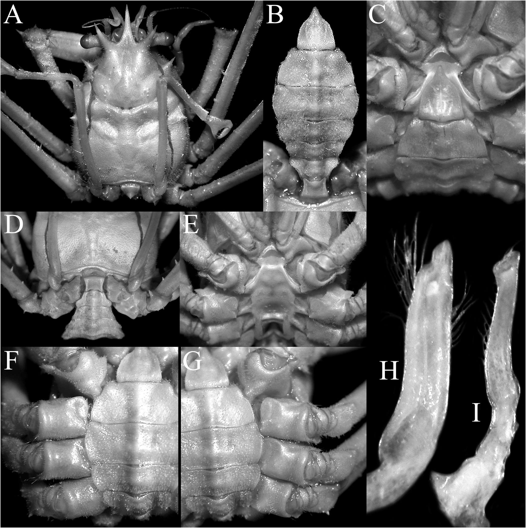
Fig. 4. Gordonopsis profundarum (Alcock & Anderson, 1899), neotype male (16.0 × 11.0 mm) (NHM 1948.9.7.28), Maldives. A, dorsal view of carapace showing relative P5 length; B, pleon; C, telson and pleonal somite 6; D, posterior part of carapace and pleon; E, sternopleonal cavity; F, right P2–P4 coxae and basis-ischia (denuded); G, left P2–P4 coxae and basis-ischia (denuded); H, ventral view of left G1; I, ventral view of left G2.
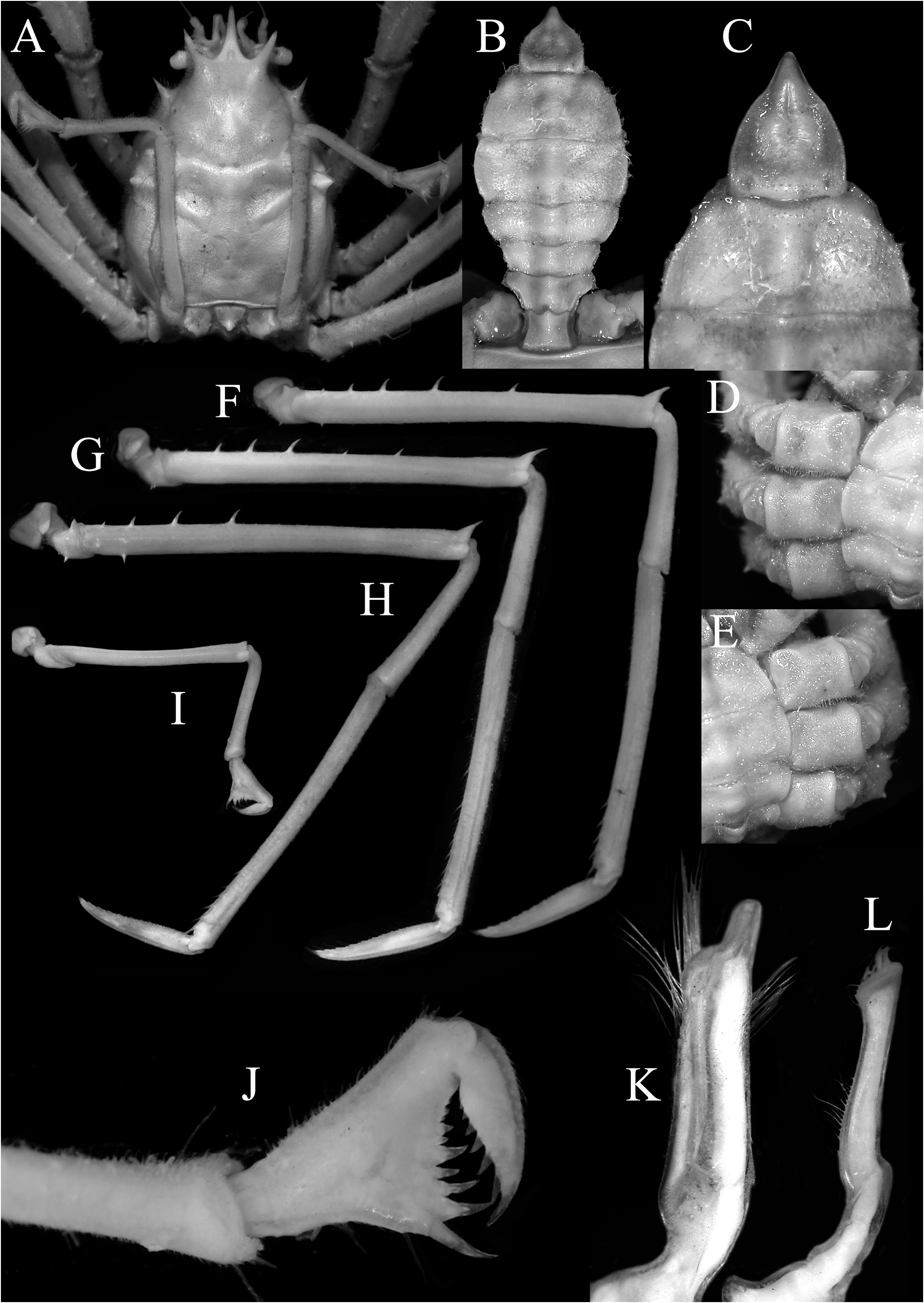
Fig. 8. Gordonopsis profundarum (Alcock & Anderson, 1899), male (16.3 × 24.1 mm) (ZMB 13645), East Africa. A, dorsal view of carapace showing relative P5 length; B, pleon; C, telson and pleonal somite 6; D, right P2–P4 coxae and basis-ischia (denuded); E, left P2–P4 coxae and basis-ischia (denuded); F–I, right P2–P5, respectively (all to same scale); J, right P5 pseudochela; K, ventral view of left G1; L, ventral view of left G2.
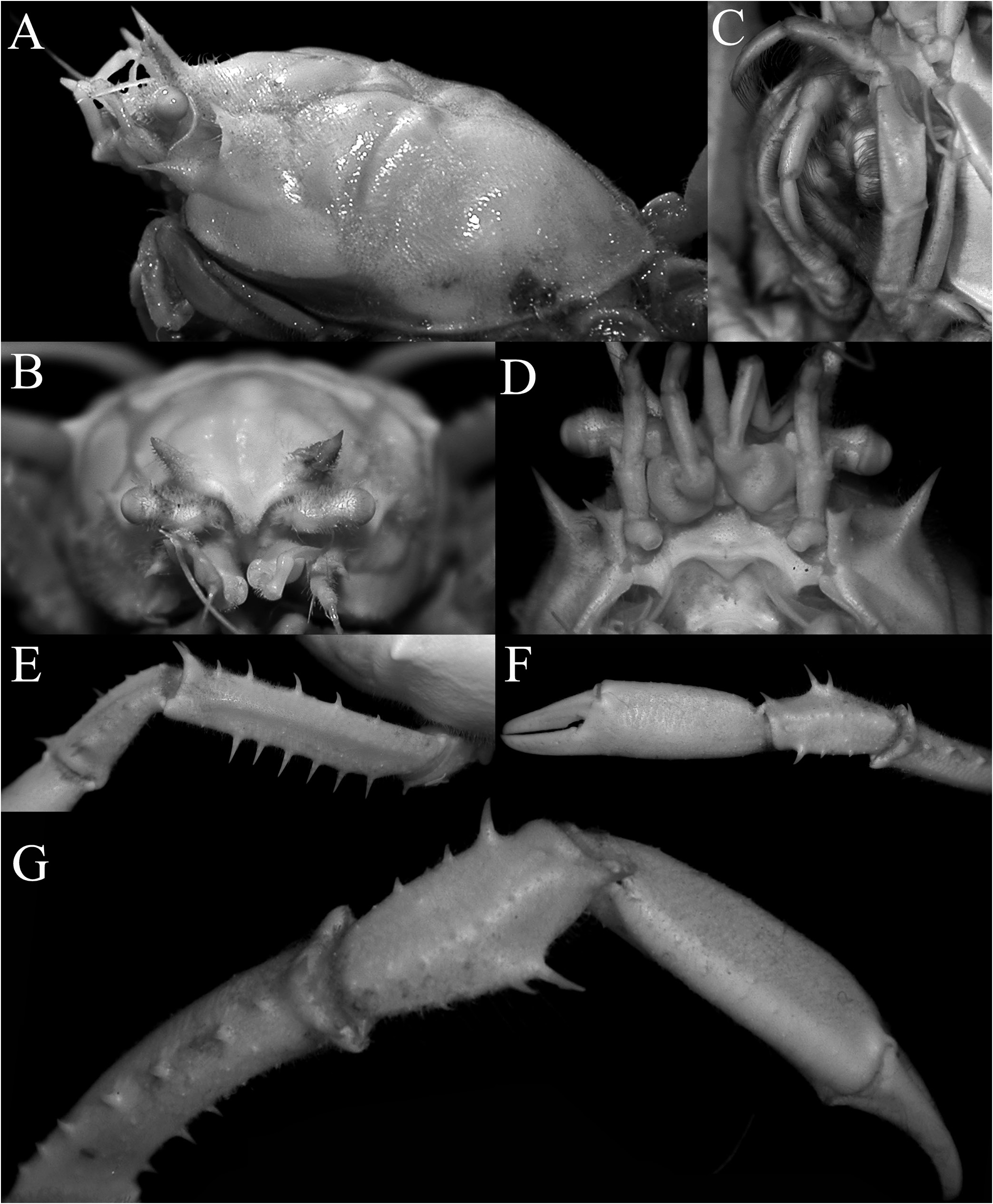
Fig. 7. Gordonopsis profundarum (Alcock & Anderson, 1899), male (16.3 × 24.1 mm) (ZMB 13645), East Africa. A, lateral view of cephalothorax; B, frontal view of cephalothorax; C, left third maxilliped; D, ventral view showing buccal cavity, epistome, antennae, and antennules; E, outer view of merus and carpus of left cheliped; F, outer view of left carpus and chela; G, dorsal view of left cheliped.

Fig. 12. Gordonopsis robusta Ng, Padate & Saravanane, 2019. A, C, E, holotype male (33.5 × 44.9 mm) (CMLRE), Andaman Sea; B, D, paratype female (15.6 × 20.8 mm) (CMLRE), Andaman Sea. A, B, overall habitus; C, dorsal view of carapace; D, dorsal view of carapace showing relative P5 length; E, outer view of right chela.
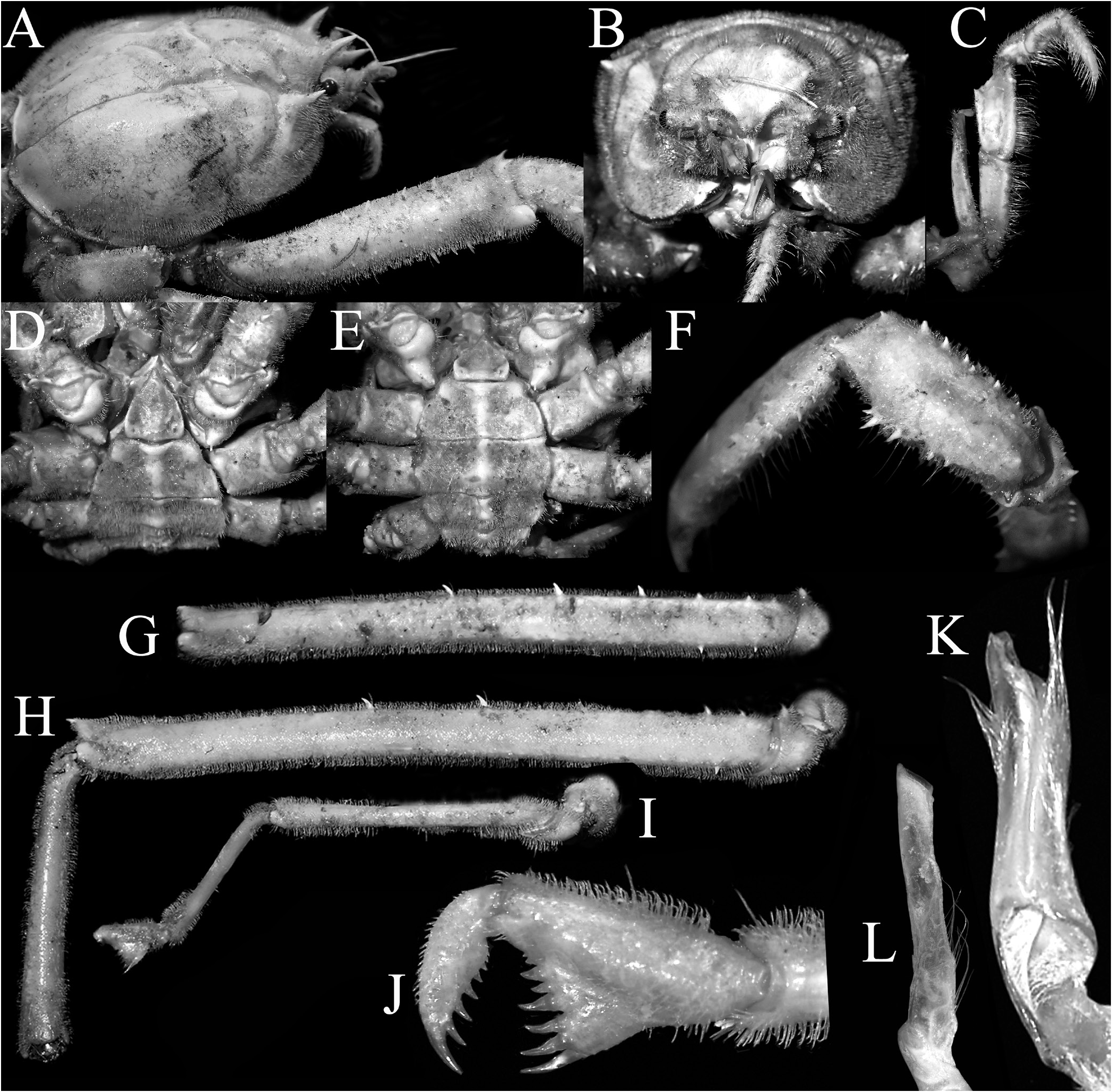
Fig. 13. Gordonopsis robusta Ng, Padate & Saravanane, 2019, holotype male (33.5 × 44.9 mm) (CMLRE), Andaman Sea. A, dorso-lateral view of cephalothorax; B, frontal view of cephalothorax; C, right third maxilliped; D, E, ventral view of cephalothorax showing pleon and coxae; F, dorsal view of carpus of right cheliped; G–I, left P3–P5 meri and leg, respectively (all to same scale); J, left P5 pseudochela; K, ventral view of right G1; L, ventral view of right G2.
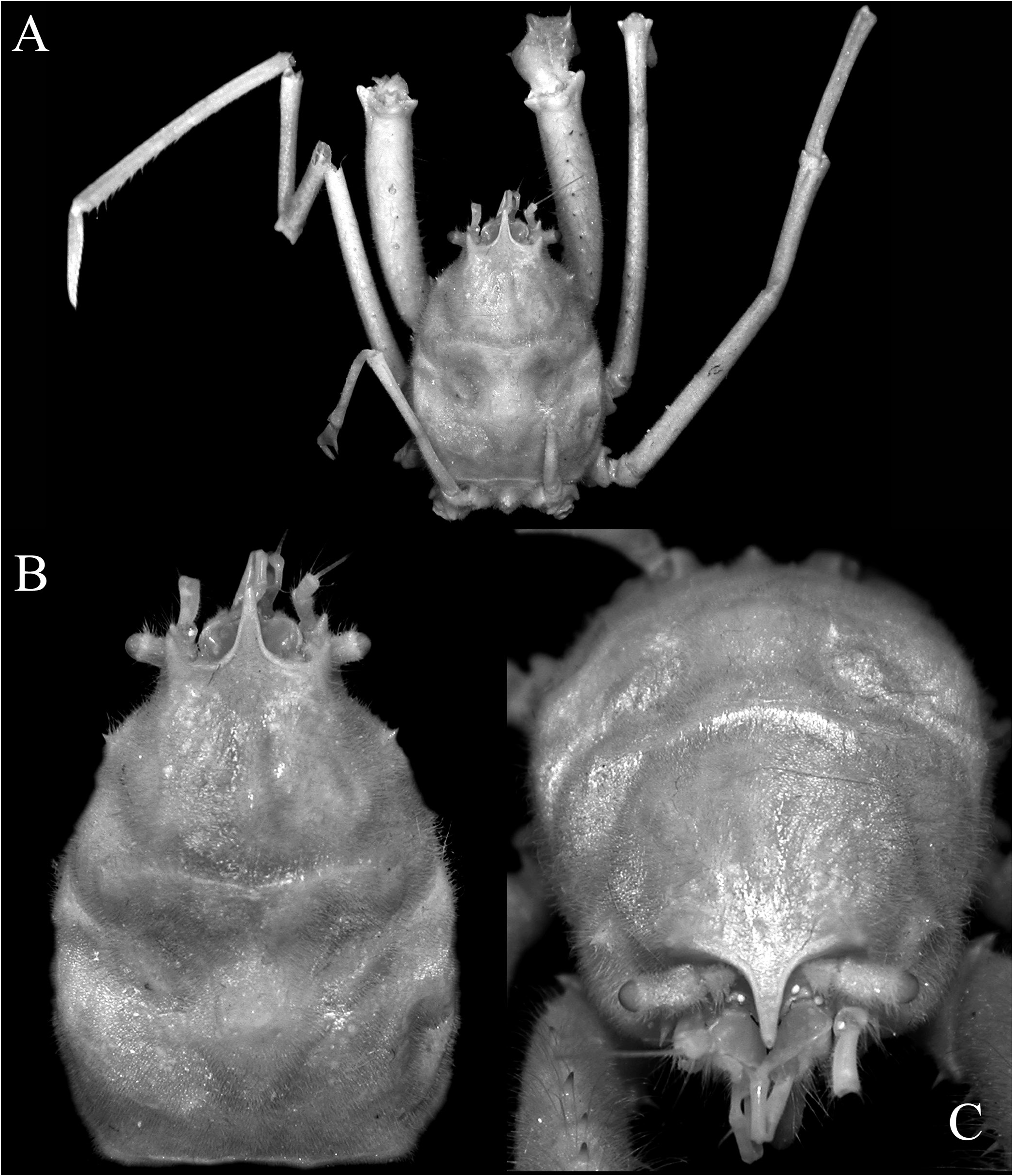
Fig. 14. Gordonopsis pacifica Takeda & Suyama, 2019, holotype male (31.0 × 43.0 mm) (NSMT-Cr 26053), Okino-Torishima Island, Japan. A, overall habitus; B, dorsal view of carapace; C, dorso-frontal view of carapace.
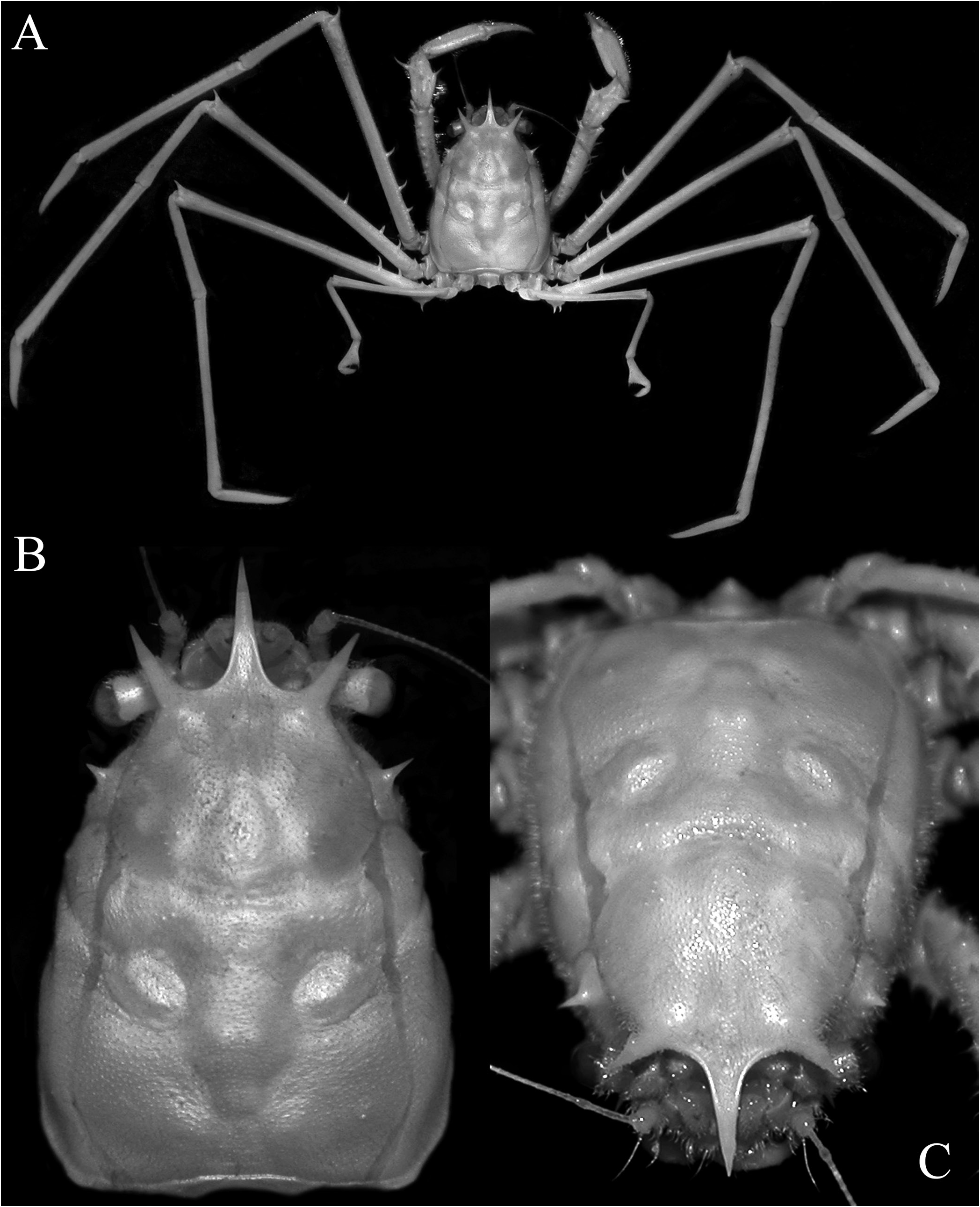
Fig. 21. Gordonopsis phorcys, new species, holotype female (10.9 × 16.8 mm) (MNHN-IU-2008-12211), Solomon Islands. A, overall habitus; B, dorsal view of carapace; C, dorso-frontal view of carapace.
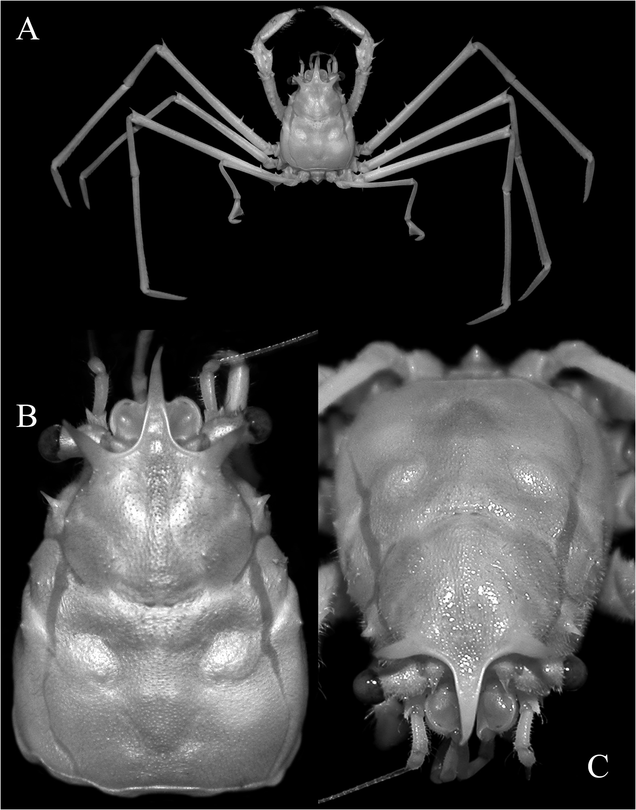
Fig. 24. Gordonopsis ceto, new species, holotype female (9.3 × 13.6 mm) (NTOU), South China Sea. A, overall habitus; B, dorsal view of carapace; C, dorso-frontal view of carapace.
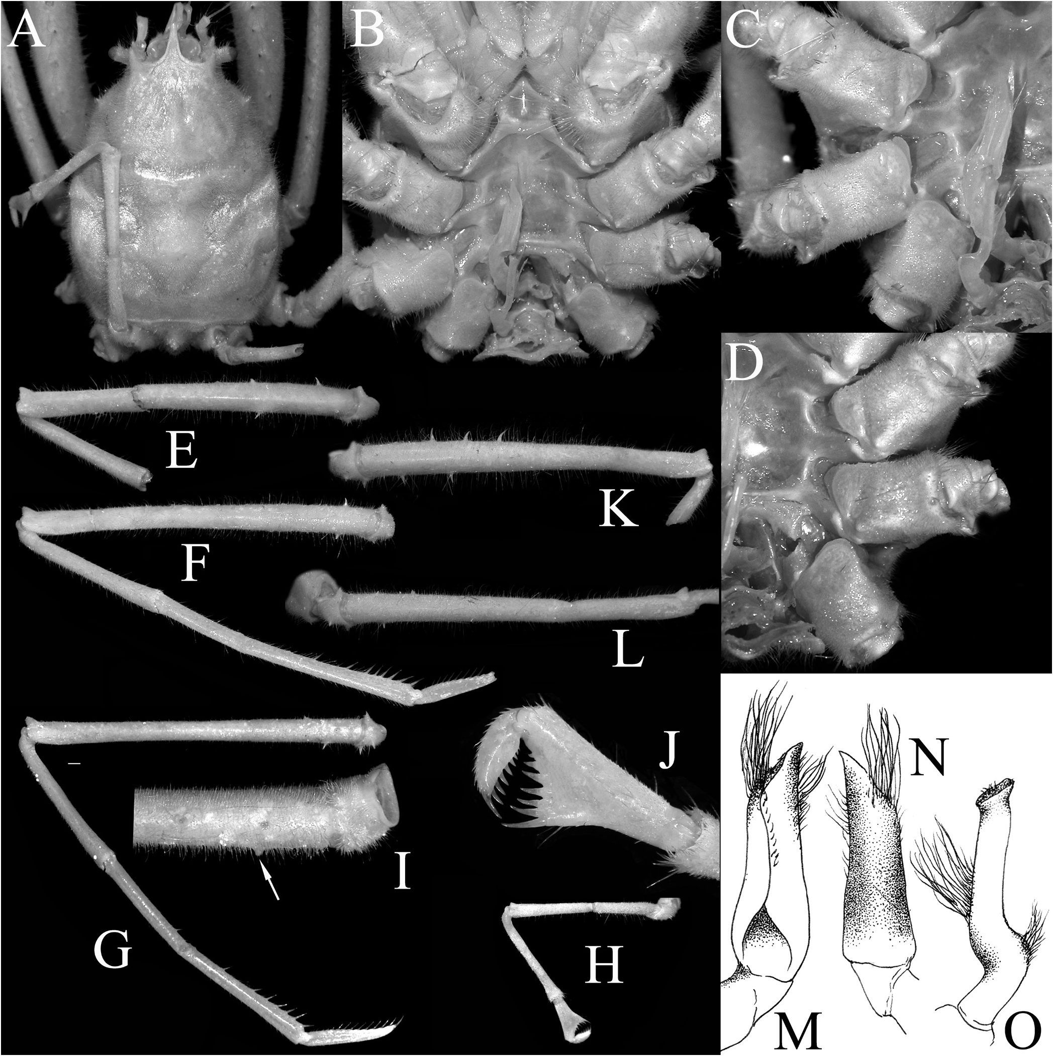
Fig. 16. Gordonopsis pacifica Takeda & Suyama, 2019, holotype male (31.0 × 43.0 mm) (NSMT-Cr 26053), Okino-Torishima Island, Japan. A, dorsal view of carapace showing relative P5 length; B, sternopleonal cavity; C, right P2–P4 coxae and basis-ischia; D, left P2–P4 coxae; E–H, left P2–P5, respectively; I, dorsomesial view of proximal part of merus of P4 showing tubercle on outer surface (arrow); J, left P5 pseudochela; K, L, right P2 and P3, respectively; M, N, left G1; O, left G2. All ambulatory legs shown to same scale. M–O after Takeda & Suyama (2019: fig. 4B–D).
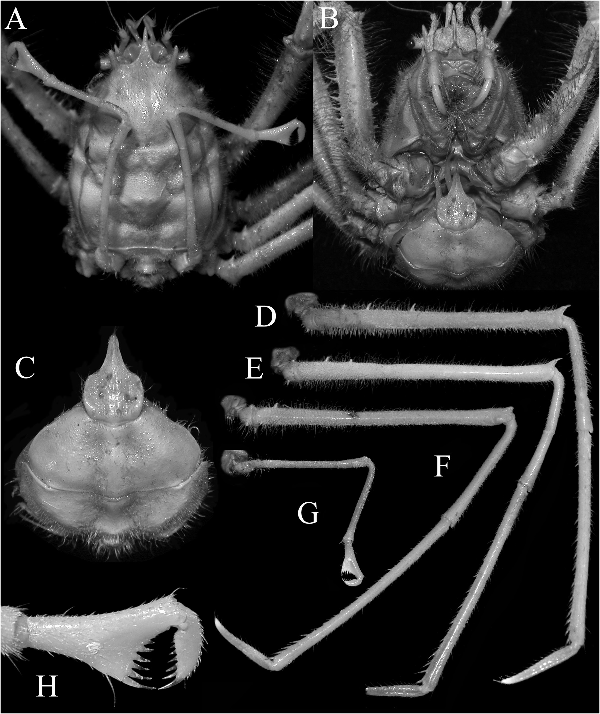
Fig. 19. Gordonopsis hera, new species, holotype ovigerous female (21.8 × 31.2 mm) (MNHN-IU-2013-2245), Papua New Guinea. A, dorsal view of carapace showing relative P5 length; B, ventral view of cephalothorax showing pleon; C, telson and pleon somites 5 and 6; D–G, right P2–P5, respectively (all to same scale); H, right P5 pseudochela.

Fig. 23. Gordonopsis phorcys, new species, holotype female (10.9 × 16.8 mm) (MNHN-IU-2008-12211), Solomon Islands. A, dorsal view of carapace showing relative P5 length; B, ventral view of cephalothorax showing coxae, basis-ischia, and pleon; C–F, right P2–P5, respectively (all to same scale); G, right P5 pseudochela; H, ventral view of cephalothorax showing sternopleonal cavity, coxae, and basis-ischia.

Fig. 26. Gordonopsis ceto, new species, holotype female (9.3 × 13.6 mm) (NTOU), South China Sea. A, dorsal view of carapace showing relative P5 length; B, ventral view of cephalothorax showing coxae, basis-ischia, and pleon; C, right P2–P4 coxae and basis-ischia (denuded); D, left P2–P4 coxae and basis-ischia (denuded); E–H, right P2–P5, respectively (all to same scale); I, right P5 pseudochela.
No known copyright restrictions apply. See Agosti, D., Egloff, W., 2009. Taxonomic information exchange and copyright: the Plazi approach. BMC Research Notes 2009, 2:53 for further explanation.
|
Kingdom |
|
|
Phylum |
|
|
Class |
|
|
Order |
|
|
Family |