
Balala Distant, 1908, 1907
|
publication ID |
https://doi.org/10.11646/zootaxa.4731.1.2 |
|
publication LSID |
lsid:zoobank.org:pub:FC77AA02-EE32-4BDE-8A8A-6AE9B8A8D3AA |
|
persistent identifier |
https://treatment.plazi.org/id/8F4E87BD-FFBF-FFEA-D0BC-FCE57CD7FB9B |
|
treatment provided by |
Plazi |
|
scientific name |
Balala Distant, 1908 |
| status |
|
Genus Balala Distant, 1908 View in CoL new records to Malaysia and Thailand
Balala Distant 1908: 250 View in CoL ; Schmidt 1909: 262; Schmidt 1911: 229; Jacobi 1914: 380; Schumacher 1915: 97; Schmidt 1920a: 117, 118; Schmidt 1920b: 127; Evans 1946: 46; Metcalf 1962: 13. Type species: Penthimia fulviventris Walker, 1851 View in CoL , by original designation.
Wania Liu 1939: 297 View in CoL . Type species: Wania membracioidea Liu, 1939 , by original designation. Synonymised by China 1941: 255.
Diagnosis. This genus can be distinguished from other genera of Hylicinae by the very robust body; the short, round and deflected head; and the strongly dilated, compressed front tibia.
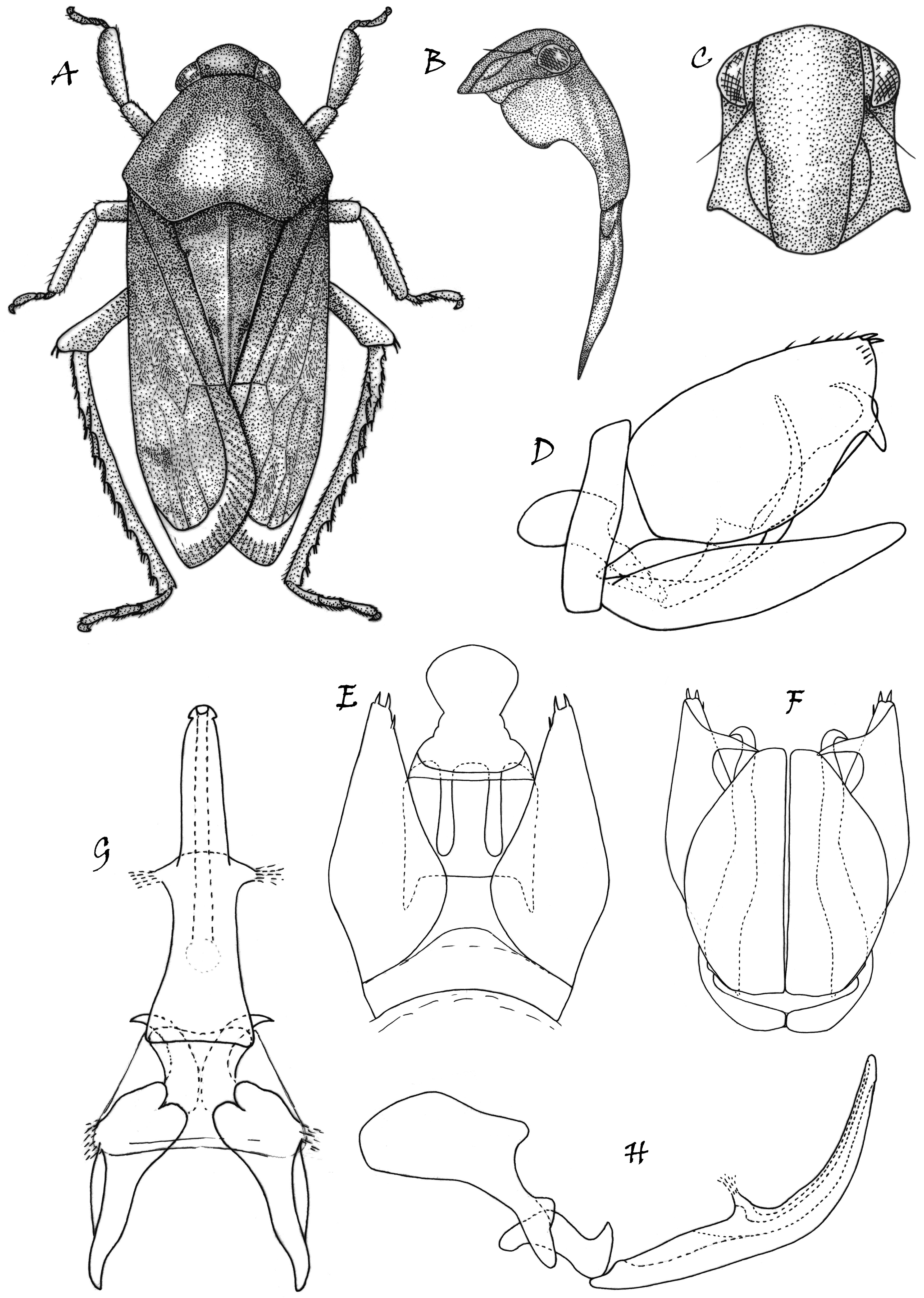
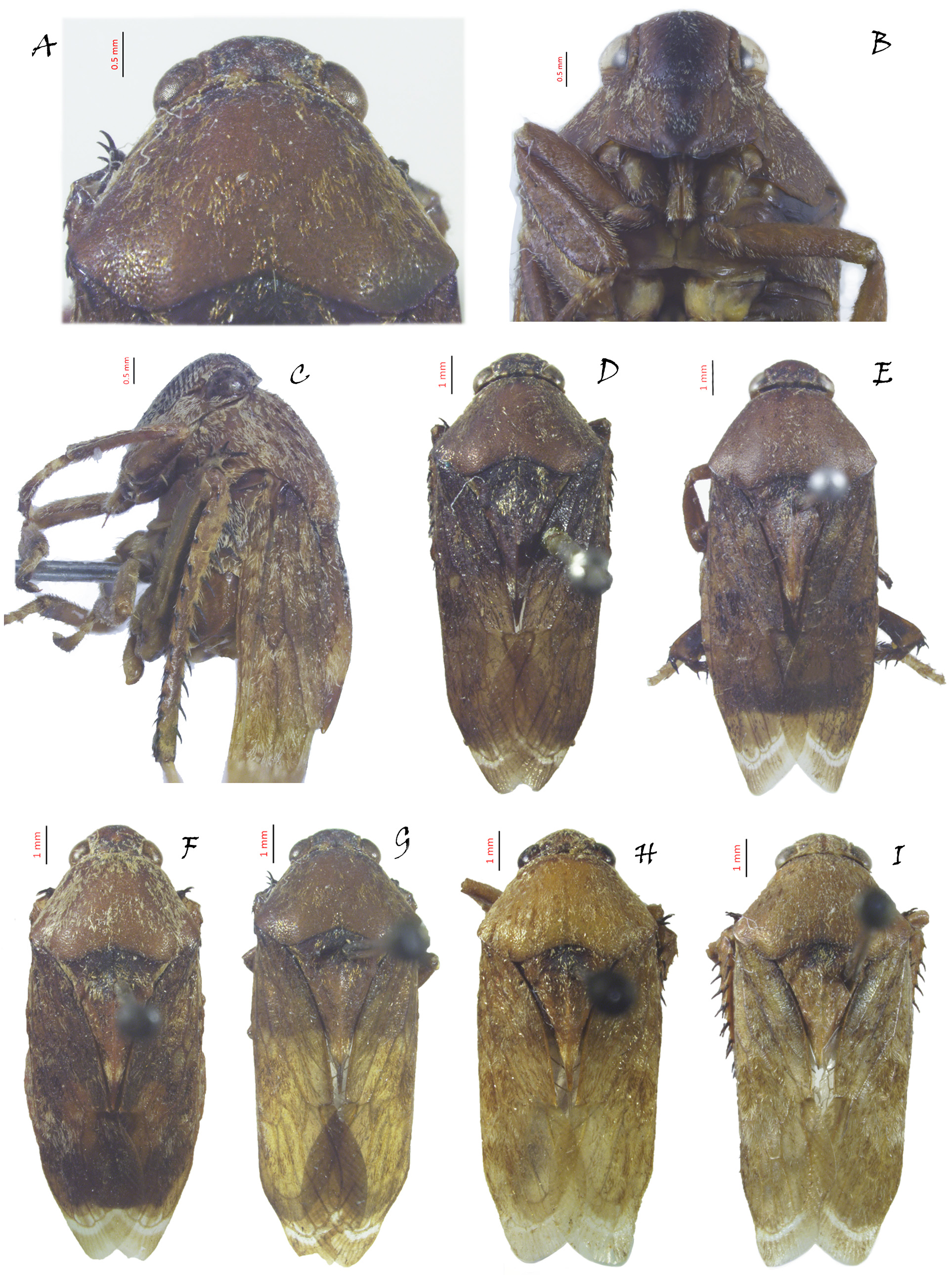
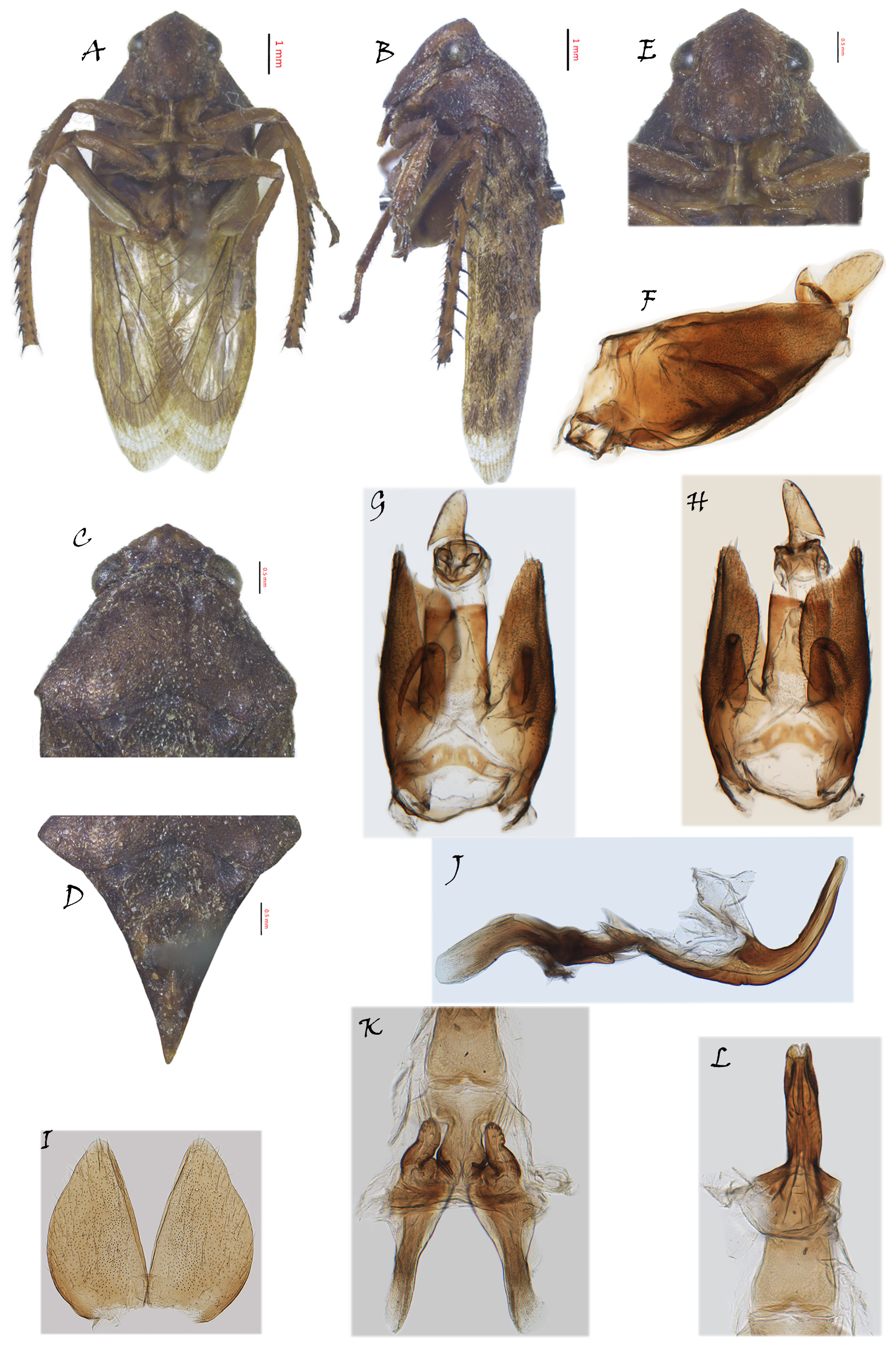
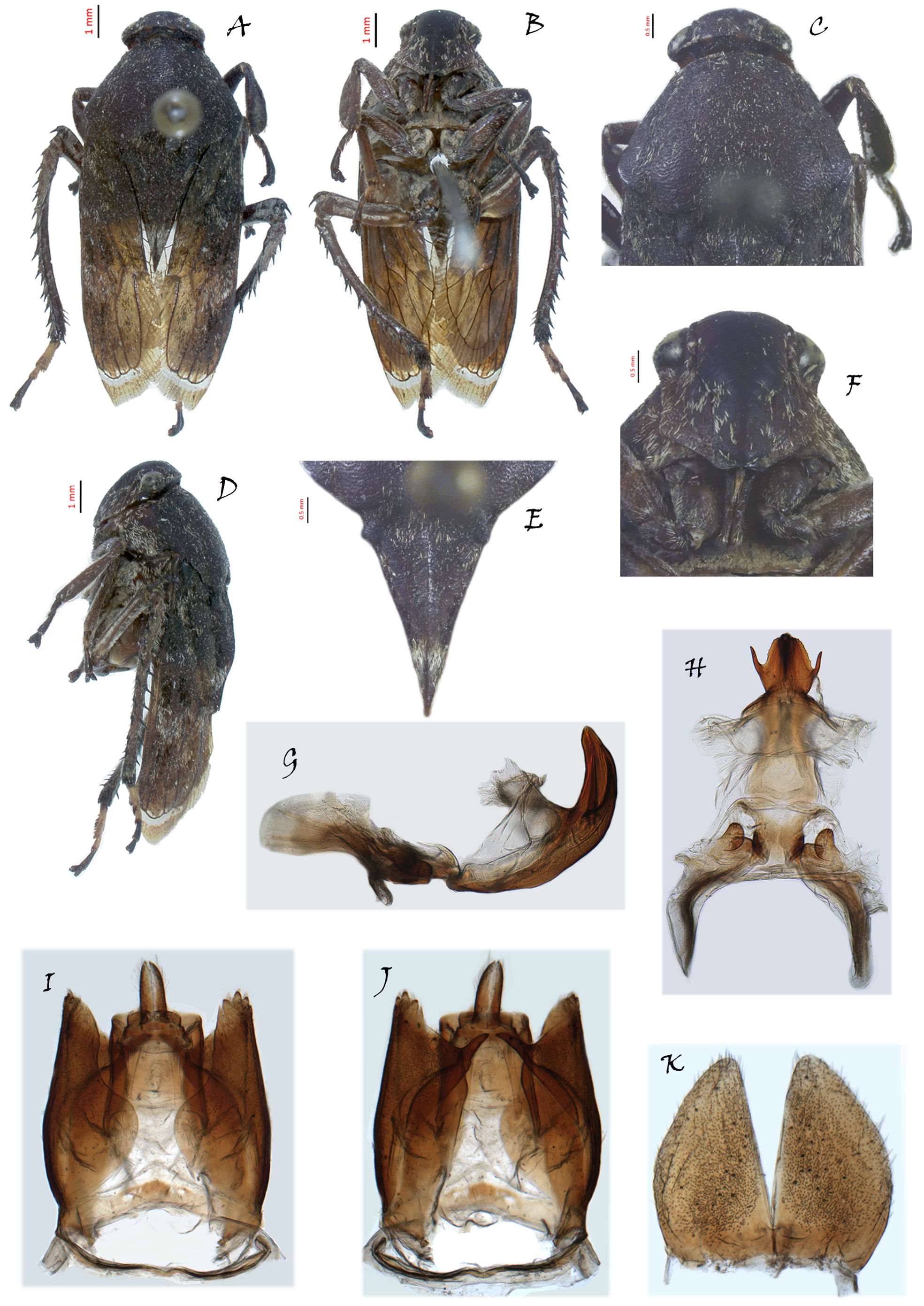
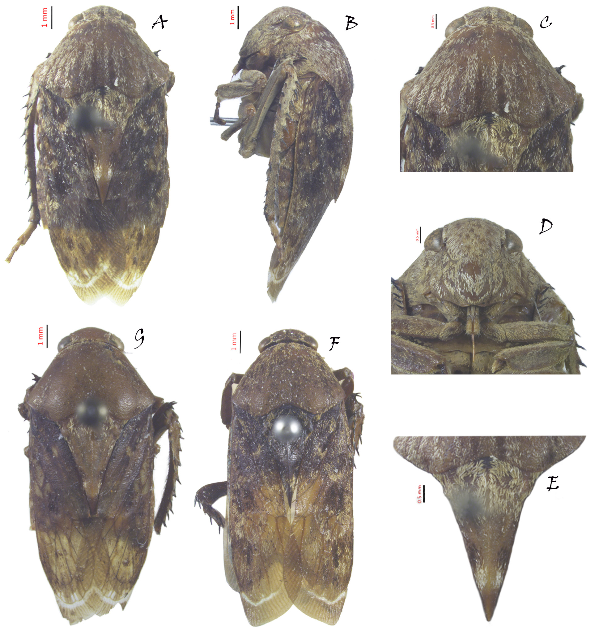
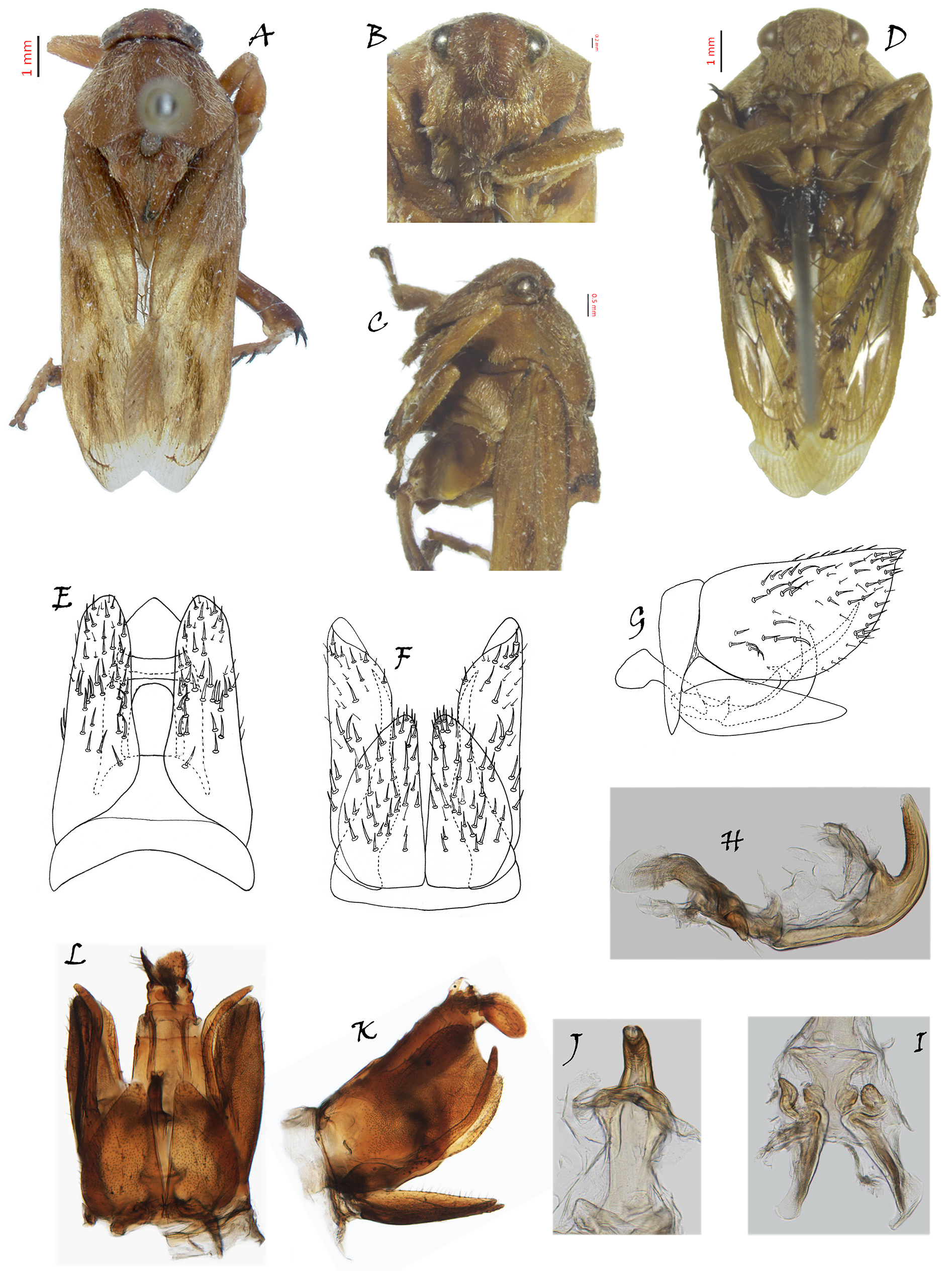
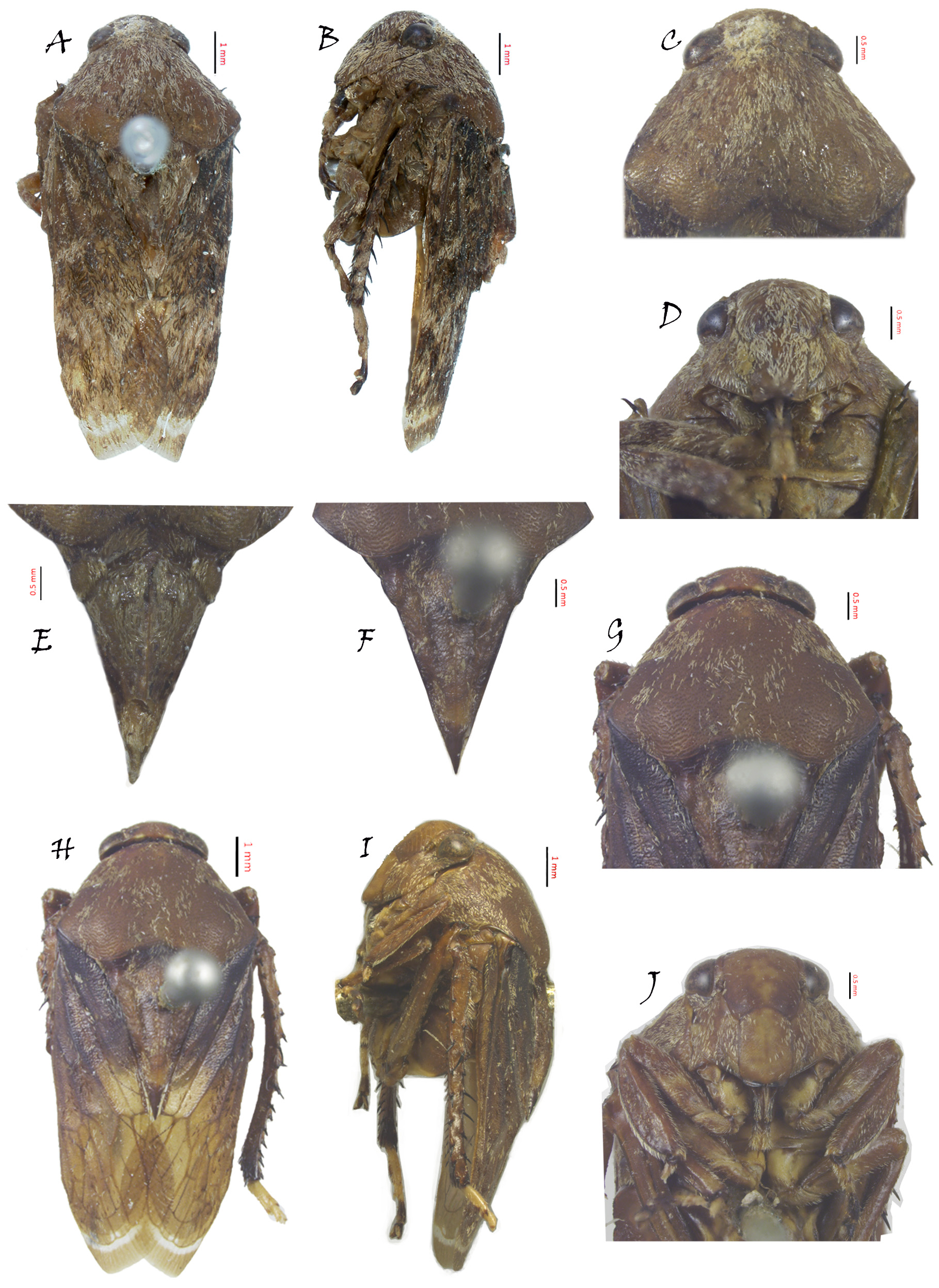
Description. Large leafhoppers (body length ♂ 10. 86–14.19 mm, ♀ 13.12–14.84 mm), body very robust. Yel- low brown to dark brown ( Figs. 2D View FIGURE 2 , 4B View FIGURE 4 , 5A View FIGURE 5 , 6A View FIGURE 6 , 8A View FIGURE 8 , 9A, H View FIGURE 9 ). Whole body densely or loosely covered with blackish and whitish scale-shaped setae. Thorax and forewings usually granulose or transversely rugulose. Female ( Fig. 12A View FIGURE 12 ) usually larger than male, with longer crown and paler in coloration.
Crown ( Figs. 1A View FIGURE 1 , 2A View FIGURE 2 , 4C View FIGURE 4 , 5C View FIGURE 5 , 6C View FIGURE 6 , 8A View FIGURE 8 , 9C, G View FIGURE 9 ) very short and declined toward apex, length shorter than width, anterior margin usually round in front of eyes, without median longitudinal carina, one pair of very small submedial semiovoid spots without setae between eyes on posterior margin, posterior margin slightly concave forward, nearly as wide as and slightly higher than anterior margin of pronotum, eyes reniform, distance between eyes about or slightly longer than 2x width of eye, ocelli located between inside margin of eyes, much nearer to eyes than to each other.
Face ( Figs. 1C View FIGURE 1 , 2B View FIGURE 2 , 4E View FIGURE 4 , 5F View FIGURE 5 , 6D View FIGURE 6 , 8B View FIGURE 8 , 9D, J View FIGURE 9 ) usually short and broad, lateral frontal suture extending onto crown in front of ocelli, antennal ridge underdeveloped, frontoclypeus and anteclypeus both convex and parallel-sided, frontoclypeus longer and broader than anteclypeus, with muscle impressions on each side extending onto crown, clypeal sulcus arcuate or weak, anteclypeus extended somewhat beyond gena, lateral margin of gena and apical margin of anteclypeus carinate, lorum narrow and short, dorsal end slightly beyond clypeal sulcus, well-separated from gena, gena exposing part of proepisternum, rostrum short, usually just passing front coxae.
Pronotum ( Figs. 1A View FIGURE 1 , 2A View FIGURE 2 , 4C View FIGURE 4 , 5C View FIGURE 5 , 6C View FIGURE 6 , 8A View FIGURE 8 , 9C, G View FIGURE 9 ) elevated posterad and sloping laterally, anterior margin protruded forward, lateral margin oblique and divergent posterad, posterior margin carinate and strongly excavated before base of mesonotum and formed into W, some small irregular depressions without setae located anterolaterally, lateral surface narrowed downward and concealing most of mesepisternum, with many longitudinal setae strips on dorsal surface or not.
Exposed part of mesonotum and scutellum ( Figs. 1A View FIGURE 1 , 2C View FIGURE 2 , 4D View FIGURE 4 , 5E View FIGURE 5 , 6E View FIGURE 6 , 8C View FIGURE 8 , 9E, F View FIGURE 9 ) triangular, longer than crown and pronotum together, often reaching apex of clavus, exposed part of mesonotum very short, with center region sunken, anterolateral regions swollen, posterolateral regions formed into small tubercles, scutellum elevated and tapering to tip, with median longitudinal ridge.
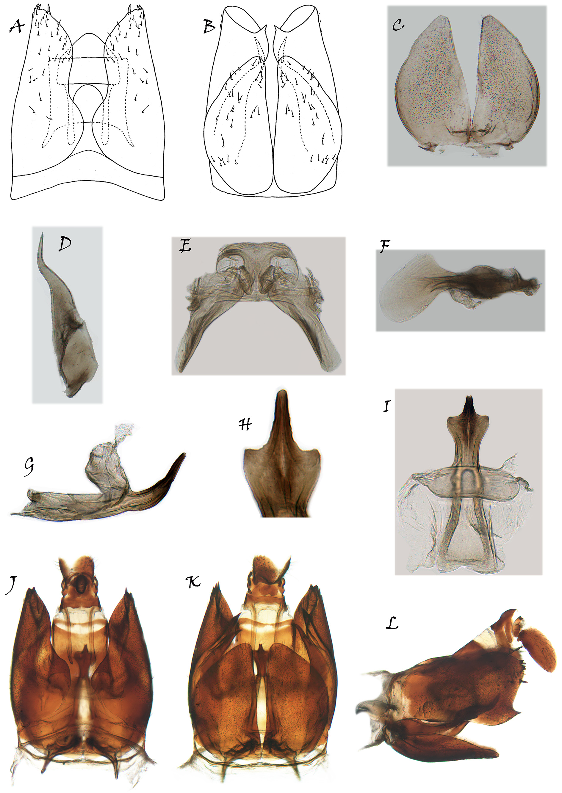
Forewings ( Fig. 12B View FIGURE 12 ) generally covered with several hair clusters or not, valvate behind clavus which’s apex truncate, claval suture obviously folded downward, venation somewhat prominent, largely obscured by opaque sclerotization and bushy hair cluster in basal 2/3, vein RA1 somewhat weak or absent, veins Pcu and A1 separated from each other, three anteapical cells, central anteapical cell slightly longer than outer and inner anteapical cells, four to five apical cells, 3rd apical cell usually shortest, appendix well developed and with numerous longitudinal crenulae, extended around wing apex from claval suture to 4th apical cell.
Abdomen ( Figs. 2E, F View FIGURE 2 , 6A View FIGURE 6 ) oval, slightly broader than tegmina in middle and gradually narrowed posteriorly at rest, abdominal tergites of male generally with meristic yellow patches.
Front tibia strongly dilated and compressed, hind femur with 5–7 macrosetae at apex and most basal one usually poorly developed, hind tibia slightly curved and strongly spinose, hind tarsi with first and second tarsi yellowish, claw and even third tarsi blackish.
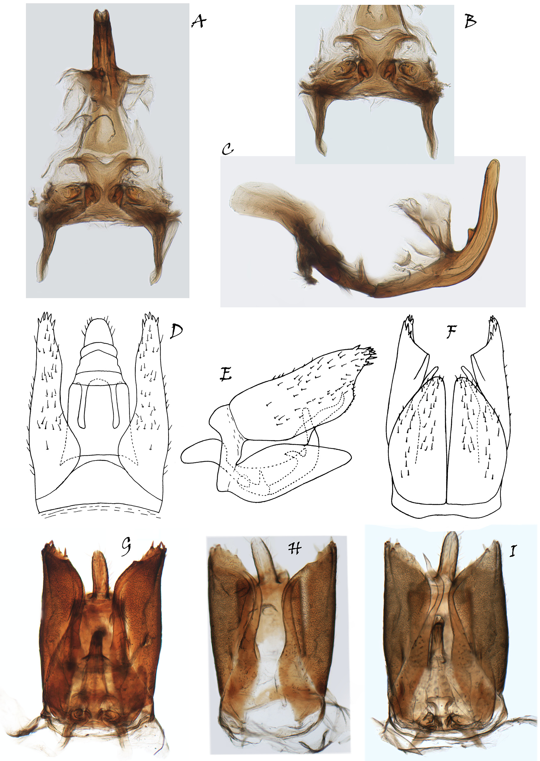
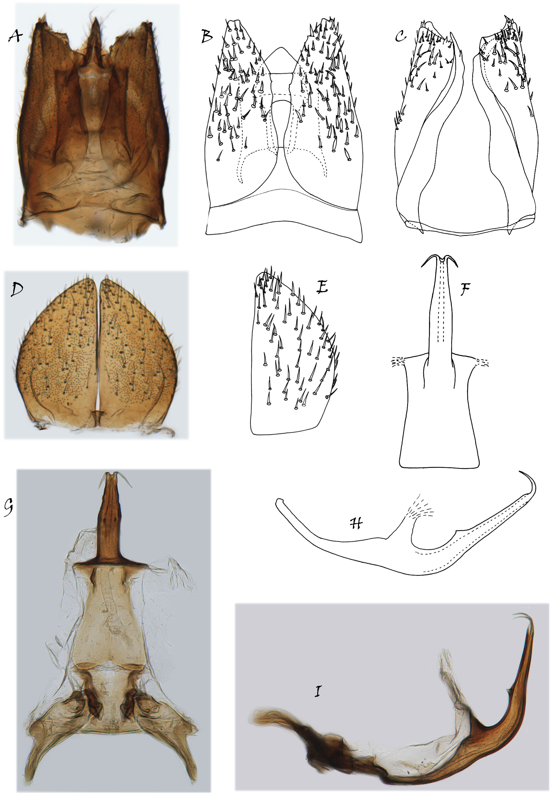
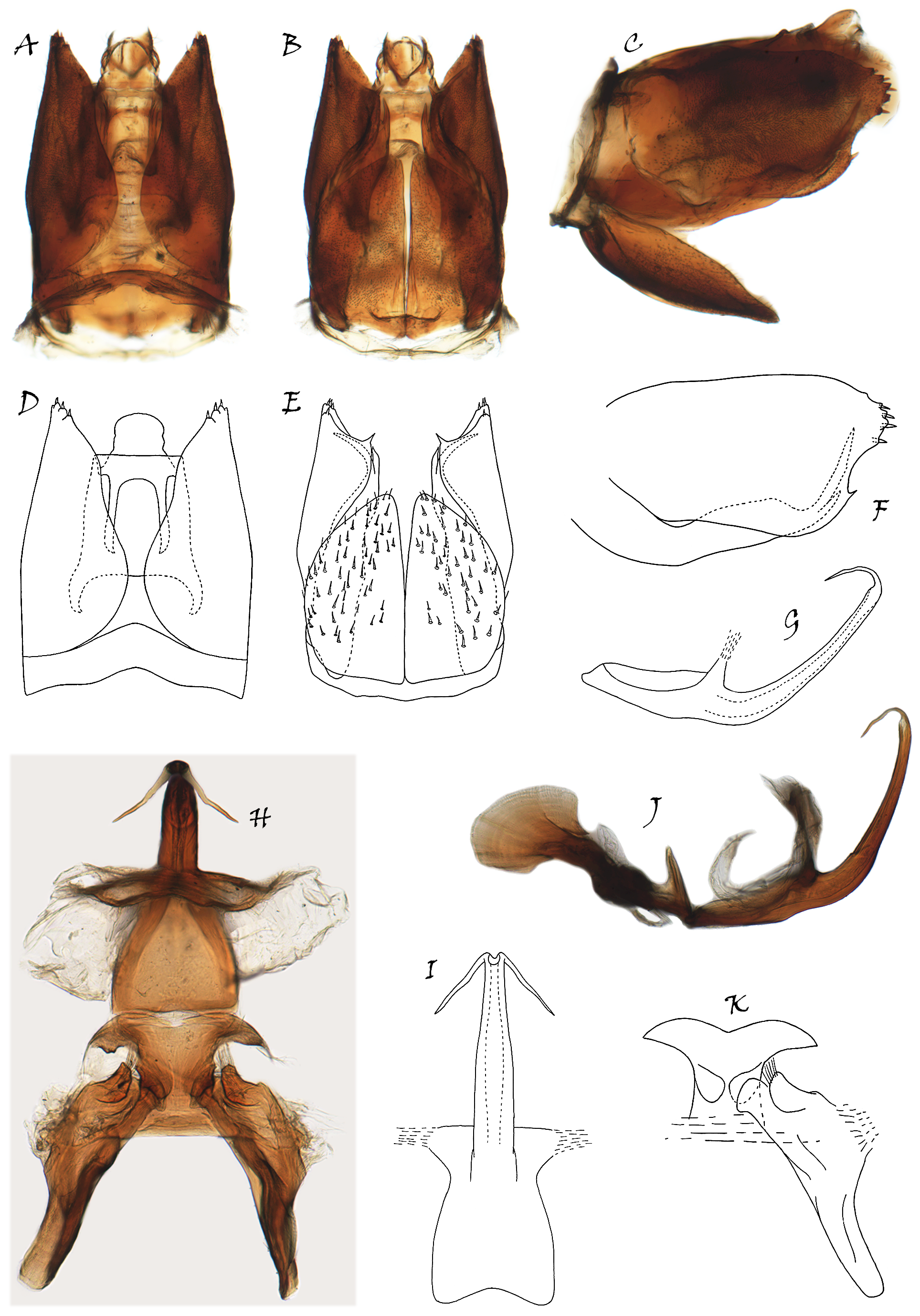
Male genitalia. Pygofer ( Figs. 1 View FIGURE 1 D–F, 3D–I, 4F–I, 5I–K, 7J–L, 8E–G, K, L, 10A–E, 11A–F) base very short, separated from lobe by vertical membranous cleft, lobe granulose and covered with many scales, deeply bifid in dorsal view, apical margin slightly produced and with about 4–8 stout setae with spine-like setal bases; ventral appendage partly separated from base of pygofer by membrane and extended to inner surface of lobe, base broad and flat, apex tapered. Subgenital plate with base fused, inner margins nearly straight, outer margins evenly convex, gradually narrowing posteriorly, ventral surface with scattered macrosetae; valve reduced, fused with pygofer and base of subgenital plate. Connective ( Figs. 1G View FIGURE 1 , 3B View FIGURE 3 , 4K View FIGURE 4 , 5H View FIGURE 5 , 7E View FIGURE 7 , 8I View FIGURE 8 , 10G View FIGURE 10 , 11K View FIGURE 11 ) approximately quadrangular, longer than wide, two sides of apex extended laterally into lamelliform lobes with apices tapered and curved dorsad in lateral view, basal margin below styles and connecting them; style with apodeme long, apophysis reduced and connected with lower middle part of connective, apex of apophysis thumb-like, stretched ventrad and with several slender setae; aedeagus ( Figs. 1G View FIGURE 1 , 3A View FIGURE 3 , 4L View FIGURE 4 , 5H View FIGURE 5 , 7I View FIGURE 7 , 8J View FIGURE 8 , 10F View FIGURE 10 , 11I View FIGURE 11 ) symmetrical and curved dorsad, preatrium well-developed. Segment X with pair of long, slender apodemes.
Female. See the species B. nigrifrons .
Distribution. Borneo, China, India, Japan, Malaysia, Myanmar, Sumatra, Thailand, Vietnam.

No known copyright restrictions apply. See Agosti, D., Egloff, W., 2009. Taxonomic information exchange and copyright: the Plazi approach. BMC Research Notes 2009, 2:53 for further explanation.
|
Kingdom |
|
|
Phylum |
|
|
Class |
|
|
Order |
|
|
Family |
|
|
SubFamily |
Hylicinae |
Balala Distant, 1908
| Tang, Jiu & Zhang, Yalin 2020 |
Wania
| China, W. E. 1941: 255 |
| Liu, G. Z. 1939: 297 |
Balala
| Metcalf, Z. P. 1962: 13 |
| Evans, J. W. 1946: 46 |
| Schmidt, E. 1920: 117 |
| Schmidt, E. 1920: 127 |
| Schumacher, F. 1915: 97 |
| Jacobi, A. 1914: 380 |
| Schmidt, E. 1911: 229 |
| Schmidt, E. 1909: 262 |
| Distant, W. L. 1908: 250 |